Commun.Fac.Sci.Univ.Ank.Series A2-A3 Volume 60, Number 1, Pages 21-30(2018) DOI: 10.1501/commua1-2_0000000108 ISSN 1303-6009 E-ISSN 2618-6462
http://communications.science.ankara.edu.tr/index.php?series=A2A3
Received by the editors: November 10, 2017; Accepted: January 27, 2018.
Key word and phrases: Neuroinformatics, Hodgkin-Huxley neuron, speed gradient feedback, collective bursting control, epileptiform dynamics.
This work has been supported by the TÜBİTAK project 116F049 “Controlling Spiking and Bursting Dynamics in Hodgkin-Huxley Neurons”.
© 2018 Ankara University Communications Faculty of Sciences University of Ankara Series A2-A3: Physical Sciences and Engineering
CONTROL OF COLLECTIVE BURSTING IN SMALL
HODGKIN-HUXLEY NEURON CLUSTERS
SERGEY BORISENOK, ÖNDER ÇATMABACAK, AND ZEYNEP ÜNAL
Abstract.The speed gradient-based control algorithm for tracking the membrane potential of Hodgkin-Huxley neurons is applied to their small clusters modeling the basic features of an epileptiform dynamics. One of the neurons plays a role of control element detecting the temporal hyper-synchronization among its network companions and switching their bursting behavior to resting. The ‘toy’ model proposed in the paper can serve as an algorithmic basement for developing special control elements at the scale of one or few cells that may work autonomously and are able to detect and suppress epileptic behavior in the networks of real biological neurons.
1. Introduction
Epilepsy is a disease involving changes at multiple different spatial and temporal scales and, therefore, demands for its modeling such a basic neuron mathematical element that possesses many degrees of freedom, like Hodgkin-Huxley (HH) neurons [1]. The chains of many HH neurons in the epileptiform regime demonstrate good agreement with animal in vivo recordings [2,3]. The synchronization/desynchronization of the cell behavior in the neural networks is the sufficient phenomenon for the understanding the mechanism of epilepsy [4,5]. Modern neuroscience demonstrates a great progress in study of the collective chaotic regimes of biological neurons, but its mathematical modeling still needs a sufficient improvement [6]. The Hodgkin-Huxley’s system covers some possible scenarios of the appearance of the collective bursting: ion channel mutations and fluctuations in concentration gradient of ions from inside to outside the axon [7].
Recently we’ve developed the efficient algorithm to track an arbitrary dynamical regime in a single HH neuron controlled by an external electrical signal [8]. Here Fradkov’s speed gradient feedback [9] is applied to suppress the collective bursting
in the small clusters of Hodgkin-Huxley neurons via the driving action potentials in the neural axons. Our tracking algorithm allows to detect the hyper-synchronized dynamics in the cluster and to transfer the behavior of some selected neurons from the collective bursting to the resting.
The control is performed with the single element of the cluster via the feedback to its bursting companions. The proposed algorithm can be used efficiently for studying, detecting and suppressing the epileptiform behavior [7] of spiking and bursting in the models for biological neuronal networks.
2. Speed gradient feedback control for hodgkin-huxley neurons To model the basic element of the neural cluster we use here the Hodkin-Huxley (HH) model proposed in [1]. The k-th neuron in the population is described by the dynamical set of ordinary differential equations:
3 ( ) 4 ( ) ( ) ( ) ; ( ) (1 ) ( ) ; ( ) (1 ) ( ) ; ( ) (1 ) ( ) . k M Na k k k Na K k k K Cl k Cl k k m k k m k k k n k k n k k k h k k h k k dv C g m h v E g n v E g v E I t dt dm v m v m dt dn v n v n dt dh v h v h dt (2.1)
Here vk(t) stands for the action potential of the axon, mk(t), nk(t), hk(t) are its membrane gate variables. The summary current Ik(t) entering the k-th cell plays a role of an external signal stimulating spiking or bursting dynamics of the neuron. αm,n,h, βm,n,h are fenomenologically found functions related to the membrane gate probabilities and given by [1]:
(2.2)
The set of constants in (2.1) includes the potentials ENa (equilibrium potential at which the net flow of Na ions is zero), EK (equilibrium potential at which the net flow of K ions is zero), ECl (equilibrium potential at which leakage is zero) in mV, the membrane capacitance CM and the conductivities gNa (sodium channel conductivity), gK (potassium channel conductivity), gCl (leakage channel conductivity) in mS/cm2: . 36 . 10 ; 3 . 0 ; 12 ; 36 ; 115 ; 120 Cl Cl K K Na Na E g E g E g
(2.3)
The link element transfers the electrical stimulation from the axon of (k –1)-th neuron to the input of k-th neuron via synapses, dendrites and soma of the k-th cell. We use the gain model:
1 rest
( ) [ ( ) ] ; const 0,
k k
I t v t v (2.4) where the reference rest potential of an HH neuron is given by [1]:
. 45 . 0 ; 3 ; 1 ; 150 ; 560 ; 50 ; 440 ; 400 ; 20 ; log 58 int ext int ext int ext int int int ext ext rest Cl Na K Cl Na K ext Cl Na K P P P Cl Cl Na Na K K Cl P Na P K P Cl P Na P K P v (2.5) 0.1 (25 ) ( ) ; ( ) 4 exp ; 25 18 exp 1 10 0.01 (10 ) ( ) ; ( ) 0.125 exp ; 10 80 exp 1 10 1 ( ) 0.07 exp ; ( ) . 30 20 exp 1 10 m m n n h h v v v v v v v v v v v v v v
The algorithm for tracking the membrane potential v(t) in a single neuron have been developed in [8]. For a single element tracking provides the reproduction of an arbitrary target function v*(t) by the potential v(t) via the designing the control current ISG(t). To do it, let’s define the scalar target (goal) function of the HH neuron as:
2 * 1 ( ) ( ) . 2 G v t v t (2.6)The speed gradient algorithm [9] defines the gradient control in the space of the control signal. In the case of single neuron it is reduced to the partial derivative:
. ) ( SG dt dG I t I (2.7)
Here is a positive constant. For the HH model (2.1) the algorithm (2.7) implies [8]: . )] ( ) ( [ ) ( * SG v t v t C t I M (2.8) Together with the dynamical set (2.1)-(2.4) Eq.(2.8) forms the control model for the HH cluster.
3. Control model of the epileptiform suppression
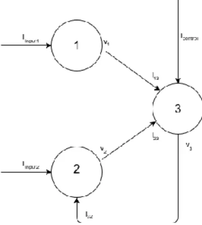
In this paper we introduce a basic ‘toy’ model for the epileptiform suppression. Let’s consider the sub-cluster of three HH neurons, see the configuration on Figure 1.
Figure 1. Basic model for an epileptiform suppression in the cluster of three Hodgkin-Huxley neurons.
Here the neurons 1 and 2 are involved into the collective bursting stimulated by the currents Iinput1 and Iinput 2 coming from other companion cells in the neural population. The neuron 3 is a monitoring element providing the switch on and off for the algorithm of suppression. It plays two roles. First, it detects the over-synchronization of the signals coming from the neurons 1 and 2 through the input currents I13 and I23 (sure, the neurons 1 and 2 may also stimulate other neurons in the bigger cluster, they are not shown on Figure 1). Second, if the neuron 3 observes the over-synchronization in a certain interval of time, it triggers the control algorithm of the suppression through the feedback loop to the neuron 2 by the current I32. The control current Icontrol reflects the inner degree of freedom for the neuron 3. Thus, this element works as an automat driving the neuron 2 from the bursting regime to the resting if and only if it detects its over-synchronization with the neuron 1. The basic cluster configuration on Figure 1 can be written in the form of coupled differential equations of (2.1)-type:
, 3 , 2 , 1 ; ) ( ) 1 ( ) ( ; ) ( ) 1 ( ) ( ; ) ( ) 1 ( ) ( ; ) ( ) ( ) ( ; ) ( ) ( ) ( ; ) ( ) ( ) ( 3 control 23 13 3 3 4 3 3 3 3 3 3 31 input2 2 2 4 2 2 2 3 2 2 input1 1 1 4 1 1 1 3 1 1 k h v h v dt dh n v n v dt dn m v m v dt dm I I I E v g E v n g E v h m g dt dv C I I E v g E v n g E v h m g dt dv C I E v g E v n g E v h m g dt dv C k h k k h k k k n k k n k k k m k i m k Cl Cl K K Na Na M Cl Cl K K Na Na M Cl Cl K K Na Na M (3.1) with the synaptic links:
. ] ) ( [ ) ( ; ] ) ( [ ) ( ; ] ) ( [ ) ( rest 3 31 rest 2 23 rest 1 13 v t v t I v t v t I v t v t I (3.2)
Here we use our method of ‘back spread’ algorithmic goal: the real control signal is passing from the neuron 3 to the neuron 2, while the algorithmic definition of the goal follows the opposite direction, from 2 to 3, see Eqs.(3.5)-(3.7) below.
First, we apply SG algorithm (2.8) to the neuron 3: . )] ( ) ( [ ) ( 3 3* control t v t v t I (3.3) The goal v3* of the tracking potential in the neuron 3 is defined as the inverse function to (2.4): . ) ( ) ( 31* rest * 3 v t I t v (3.4) The control current Icontrol entering the neuron 3 is given also in the SG form (2.8):
() ()
[ () ], ) ( 13 23 2 * 31 t I t I t v t vrest I (3.5) where ∆ stands for the smooth model of delta-function:. 0 const ; exp 1 ) ( 2 2 d d x d x (3.6)
The factor ∆ in (3.5) switches on the control algorithm only for the synchronized currents I13 and I23, and in the case of their time over-synchronization, i.e. only in the period of their epileptiform dynamics, leads the neuron 2 to the stabilization at the rest membrane potential.
This algorithm can be easily extended for a larger number of collective bursting neurons and their feedback links in the population.
4. Numerical simulations
For the purpose of numerical simulations the following set of parameters has been chosen: . 1 . 0 ; 1 ; 50 ; 10 ; 50 ; 50 input2 1 input I C d I M (4.1)
The results of the simulation are presented on Figure 2.
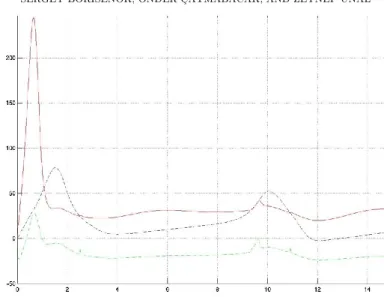
Figure 2. The neuron membrane potentials v1 (blue dashed line), v2 (red solid line) and v3 (green dash-dotted line) vs time.
On the Figure 2 one can see that after the beginning instability at the scale t = 2 the potential v2 is suppressed approximately in two times to compare with the bursting potential v1. This result seems to be very good for such a simple control model. When the collectively synchronized bursting is starting to growth (closed to t = 10), the control mechanism is switching on to drive the potential of the neuron 2 far away from the synchronization. The same is happen when t = 12.
Sure, the presented algorithm reflects only the basic features of the bursting suppression. The ‘toy’ control (3.5)-(3.6) needs to be sufficiently improved for the better detecting the chaotic hyper-synchronization in the clusters and feating more flexible details of the neuron dynamics.
5. Conclusions
The control algorithm developed in [8] for tracking the membrane potential of a single Hodgkin-Huxley neuron can be applied to a small configuration of HH elements modeling the basic features of an epileptiform dynamics. In this population one of the neurons plays a role of control element detecting the temporal hyper-synchronization among its network companions and switching on the feedback signal
that it sends to some selected neurons in the population to drive them off the epileptiform regime.
The ‘toy’ model proposed in the paper can serve as an algorithmic basement for developing special control elements at the scale of one or few cells that may work autonomously and are able to detect and suppress epileptic behavior in the networks of real biological neurons.
Acknowledgments
This work has been supported by the TÜBİTAK project 116F049 “Controlling Spiking and Bursting Dynamics in Hodgkin-Huxley Neurons”.
References
[1] Hodgkin and A.L. Huxley, A.A., Journal of Physiology, 117 (1952), 500-544. [2] Rubio, C, Rubio-Osornio, M., Retana-Márquez, S., López, M., Custodio, V. and
Paz, C., Central Nervous System Agents in Medicinal Chemistry, 10/4 (2010) 298-309.
[3] Naze, S. Bernard and Ch. Jirsa, V., PLOS Computational Biology, 1 (2015) 1-21.
[4] Reynolds, E.H., Epilepsy: The disorder, (Epilepst Atlas, WHO, 2005, 16-27). [5] Eftaxias, K. Minadakis, G. Athanasopoulou, L. Kalimeri, M. Potirakis, S.M.
and Balasis, G., Are epileptic seizures quakes of the brain? An approach by means of nonextensive Tsallis statistics, 2011, arXiv preprint arXiv:1110.2169. [6] Petkov, G., Goodfellow, M., Richardson, M. P. and Terry, J., Frontiers in
Neurology, 5 (2014) 261.
[7] Wendling, F., Benquet, P., Bartolomei and F. Jirsa, V., Journal of Neuroscience
Methods, 260 (2016) 233-251.
[8] Borisenok, S. and Ünal, Z., MATTER: International Journal of Science and
Technology, 3/2 (2017) 560-576.
[9] Fradkov, A.L., Cybernetical physics: From control of chaos to quantum control, Springer, Berlin, 2007.
Current Address: Sergey BORISENOK: Department of Electrical and Electronics Engineering, Faculty of Engineering, Abdullah Gül University, Kayseri, Turkey Sergey BORISENOK: Feza Gürsey Center for Physics and Mathematics, Boğaziçi University, Istanbul, Turkey
E-mail: [email protected], [email protected] ORCID: https://orcid.org/0000-0002-1992-628X
Current Address: Önder ÇATMABACAK: Faculty of Engineering and Natural
Sciences, Sabancı University, Istanbul, Turkey E-mail Address: [email protected] ORCID:https://orcid.org/0000-0002-6419-0363
Current Address: Zeynep ÜNAL: Department of Electrical and Electronics
Engineering, School of Engineering, Abdullah Gül University, Kayseri, Turkey E-mail Address: [email protected]