http://journals.tubitak.gov.tr/biology/ © TÜBİTAK
doi:10.3906/biy-1703-104
The MTT viability assay yields strikingly false-positive viabilities although the cells are
killed by some plant extracts
Didem KARAKAŞ1,*, Ferda ARI 2,*, Engin ULUKAYA1,**
1Department of Medical Biochemistry, Faculty of Medicine, İstinye University, İstanbul, Turkey 2Department of Biology, Faculty of Arts and Sciences, Uludağ University, Bursa, Turkey
1. Introduction
In the past years, several methods have been developed to determine cell viability in cell culture (Cook and Mitchell, 1989). Among these methods tetrazolium salt-based assays are widely used in order to measure cytotoxicity or cell proliferation (Mosmann, 1983; Berridge et al., 1996).
The principle of the MTT [3-(4,5-dimethyl-2-thiazolyl)-2,5-diphenyl-2H-tetrazolium bromide] assay is the reduction of water-soluble yellow tetrazolium salt by the dehydrogenase system of metabolically active/live cells into water-insoluble blue/magenta (MTT) formazan crystals (Morgan et al., 1998). In this way, the concentration of dissolved formazan crystals can be quantified using a spectrophotometer and it is in direct correlation to the number of metabolically active cells (Gabrielson et al., 2002; Tunney et al., 2004; Wang et al., 2010). The MTT assay represents a simple and rapid colorimetric assay and yields quantitative data (Alley et al., 1988). This assay is carried out entirely in 96-well microtiter plates; thus, large
numbers of experiments examining a number of variables can be readily performed (Cole, 1986).
On the other hand, the MTT assay has some disadvantages that are dependent on the cell ability to overcome cell death. One remarkable disadvantage is that damaged mitochondria may be still able to reduce MTT to formazan crystals (Mosmann, 1983; Page et al., 1988; Sieuwerts et al., 1995). Loveland et al. (1992) showed that cells with inactivated mitochondria were also able to produce formazan crystals as well as cells with active mitochondria. Furthermore, many nonmitochondrial dehydrogenases and flavin oxidases are able to reduce MTT (Altman, 1976; Burdon et al., 1993). Besides, different conditions and some chemicals/phytochemicals can also lead to changes in metabolic activity (Plumb et al., 1989; Hsu et al., 2003). The MTT compound may interact with some chemicals/phytochemicals, resulting in false results in viability (Hsu et al., 2003; Ulukaya et al., 2004; Devika and Stanely Mainzen Prince, 2008).
Abstract: The MTT assay is one of the often used cell viability/cytotoxicity assays. However, when the methanol extracts of plants are used to test their cytotoxic potential, interference may occur, resulting in false-positive viability results. Therefore, in this study, the reliability of the MTT assay was investigated in the case of plant use. The methanol extracts of three different plants (Hypericum adenotrichum, Salvia kronenburgii, and Pelargonium quercetorum) were tested in breast cancer cell lines (MCF-7 and MDA-MB-231) using the MTT assay and the results were compared to the ATP assay, which is a much more sensitive and reliable assay due to its interference-free feature. Additionally, decreased cell density was confirmed with phase-contrast microscopy and fluorescence staining (Hoechst 33342 dye). Although both of the viability/cytotoxicity assays are considered as metabolic assays, viabilities (in %) in the MTT assay were found to be strikingly higher when compared to the results with the ATP assay. Even in the case of total death, the MTT assay still produced artificial/false increases in viability. The morphology-based evaluation of viability/cytotoxicity by phase-contrast microscopy and Hoechst 33342 staining were greatly compatible with the ATP assay results. Overestimated (false) viabilities in the MTT assay suggests a serious interference between the MTT assay itself and the extracts used. Some ingredients of plants may have reducing activity (like the dehydrogenase activity of the cells) that converts the MTT compound into the colored formazan that is the principle of the assay. Therefore, the MTT assay may not be a suitable assay for some plant extracts, urging great caution when plants are used. Key words: MTT assay, ATP assay, interference, cytotoxicity, plant extract, breast cancer
Received: 30.03.2017 Accepted/Published Online: 16.09.2017 Final Version: 18.12.2017
Research Article
* These authors contributed equally to this work. ** Correspondence: [email protected]
More importantly, investigated agents may interfere with mitochondrial dehydrogenase activity, resulting in activation or inhibition of mitochondrial dehydrogenases and thus over/underestimation of the MTT assay results (Jaszczyszyn and Gąsiorowski, 2008).
The ATP assay is also used to determine cell viability. It is the fastest and the most sensitive cell viability assay, and it is less prone to artifacts compared to other viability assay methods (Riss et al., 2013). Therefore, it may be considered as a kind of reference assay for cell viability measurement. The ATP assay was first developed by Lundin et al. as a somatic cell viability assay (Lundin et al., 1986). The principle of this method is conversion of luciferin to oxyluciferin by the luciferase enzyme in the presence of Mg2+ ions and ATP. The ATP assay yields a luminescent signal and a linear relationship exists between the intensity of the luminescent signal and the ATP concentration, and therefore the number of cells (Mueller et al., 2004).
Plant extracts contain diverse chemicals and some of them could reduce the activity of the MTT compound, giving false results. If this occurs, then the MTT assay has to be expected to result in false-positive results for viability, regardless of any dehydrogenase activity in the cell. Therefore, in this study, we aimed to investigate how reliable the MTT assay is in the testing of the cytotoxic activity of plant extracts (Hypericum adenotrichum, Salvia kronenburgii, and Pelargonium quercetorum) at 7 different concentrations (1.56–100 µg/mL) on the MCF-7 and MDA-MB-231 breast cancer cell lines in comparison with the ATP assay as a reference assay. Results of our study indicate that the MTT assay gave false-positive results on cell viability. Due to this finding, the MTT assay has limited application for plant extract testing on different cell lines.
2. Materials and methods
2.1. Collection and identification of plants
P. quercetorum was collected from Hakkari, Turkey, in June 2006 and identified with the aid of flora books (Davis et al., 1988). S. kronenburgii was collected from Van, Turkey, and H. adenotrichum was collected from the Kırkpınar region at Uludağ Mountain (Bursa, Turkey) in June 2010. These specimens were identified by Prof Dr Gürcan Güleryüz, Faculty of Science, Uludağ University, Bursa, Turkey.
2.2. Extraction of H. adenotrichum, S. kronenburgii, and
P. quercetorum samples
Fifteen grams of the aboveground parts of the plant samples
2.3. Cell culture and chemicals
Breast cancer cell lines MCF-7 and MDA-MB-231 were cultured in RPMI-1640 supplemented with penicillin G (100 U/mL), streptomycin (100 µg/mL), L-glutamine, and 10% fetal bovine serum at 37 °C in a humidified atmosphere containing 5% CO2.
Lyophilized plant extracts were dissolved in DMSO at a concentration of 0.05 g/0.5 mL as a stock solution. Further dilutions were made in culture medium. The ATP assay (adenosine 5′-triphosphate (ATP) bioluminescent somatic cell assay kit), the MTT (3-(4,5-dimethylthiazol-2-yl)-2,5-diphenyltetrazolium bromide) substance, and all the other reagents and substances were obtained commercially (Sigma Aldrich, USA).
2.4. Experimental design
MCF-7 and MDA-MB-231 cells were seeded at a density of 1 × 10⁴ cells per well of 96-well plates in 200 µL of culture medium. The untreated cells received only medium and solvent (0.1% DMSO as final concentration). All of the cells were treated with three different plant extracts (H. adenotrichum, S. kronenburgii, and P. quercetorum) in the range of 1.56–100 µg/mL for a period of 48 h.
2.5. The MTT viability assay
The MTT viability assay was performed with slight modifications as previously described (Mosmann, 1983). MTT was first prepared as a stock solution of 5 mg/mL in phosphate-buffered saline (PBS, pH 7.2) and filtered. At the end of the treatment period (48 h), 20 µL of MTT solution was added to each well. After incubation for 4 h at 37 °C, 100 µL: of solubilizing buffer (10% sodium dodecyl sulfate dissolved in 0.01 N HCl) was added to each well. After overnight incubation, the 96-well plate was read by an enzyme-linked immunosorbent assay (ELISA) reader at 570 nm for absorbance density values to determine the cell viability. The viable cells produced a dark blue formazan product, whereas no such staining was formed in the dead cells. The percentage of the viable cells was calculated using the following formula: (%) = [100 × (sample abs)/ (control abs)].
2.6. The ATP viability assay
The cells were seeded, grown, and treated as for the MTT assay (see above). The ATP assay determines the level of cellular ATP as an indirect measure of the number of viable cells (Andreotti et al., 1995). The experiment was performed for luminometric measurement of cell growth (viability) according to the standard protocol of
2.7. Fluorescence imaging
Using a fluorescent microscope, cell density was detected on the basis of the nuclear morphology. Hoechst 33342 dye stains all of the alive or dead (primary necrotic or secondary necrotic) cells.
MCF-7 and MDA-MB-231 cells were seeded in a 96-well plate at the density of 1 × 10⁴ cells per well in 200 µL of culture media. After 24 h, cells were treated with plant extracts (100 µg/mL) and then incubated for 48 h. At the end of the treatment, 2 µL of Hoechst 33342 dye was added to each well (for 5 µg/mL final concentration). Then cells were incubated with the dye and analyzed via fluorescence microscopy.
3. Results and discussion
Several experimental data demonstrated that the MTT assay yields false-positive results for viability (Ulukaya et al., 2004, 2008; Peng et al., 2005), and these false results led to overestimation of viability and thus underestimation of the cytotoxic potency of tested compounds. Therefore, in our study we have tested the reliability of the MTT assay compared to the ATP viability assay using plant extracts and two different cell lines.
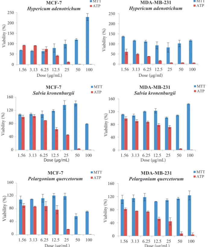
Figure 1 represents the viability curves of three different plant extracts. It was found that viability levels of the MTT assay were strikingly higher than those of the ATP assay. Depending on the doses, the viability levels were even higher than in the untreated control cells (100%). Surprisingly, although the cells were totally dead at especially higher concentrations, the MTT assay still produced extremely high viability values. These viability values were false and this finding was confirmed using both phase microscope evaluation and fluorescence imaging that confirmed cell death (Figures 2 and 3). In Figure 2, it is clearly shown that cell densities decreased when both cell lines were treated with 100 µg/mL plant extracts. However, at the same concentration, the MTT assay did not present any cytotoxic activity. In contrast, it even produced an abnormally high viability value. The results of microscopic imaging were more compatible with the ATP assay results. In fact, the ATP assay reliably showed these cell deaths (cytotoxic effects). According to these data, it should be thought that these plant extracts could interfere (reduce) with the MTT compound, leading to false-positive results for viability.
Decreased cell viability (cytotoxic effect) was also shown via fluorescent microscopy (Figure 3). Therefore,
the nuclei of living cells were examined after staining with Hoechst 33342 dye. Based on fluorescent imaging, all plant extracts caused a significant decrease in cell viability at 100 µg/mL concentration. In addition, the cell death by apoptosis was evident. The arrows in the figure show pyknotic nuclei, a well-known feature of apoptosis. These results are found to be compatible with the ATP viability assay. Taken together, the phase-contrast and fluorescent images confirm the ATP assay, not the MTT assay results.
Taking the false increments in viability into account, we suspected an interfering effect of the plants with the MTT compound. To determine interference of the plant extracts with the MTT compound, the plant extracts were tested in a cell-free culture system. For this purpose, the MTT compound was added to the extract solution and then incubated for 4 h. Table 1 shows the absorbance values of plant extracts without cells. According to the interference analysis results, the absorbance values of plant extracts were higher than the blank (with usual cell culture medium only) at the higher concentrations of extracts. This clearly demonstrates the existence of interference of plant extracts with the MTT compound.
To demonstrate the differences between the MTT and ATP assays, IC50 values (half-maximal inhibitory concentration) were also calculated (Table 2). Based on the MTT results, IC50 values of all plant extracts were found higher than 100 µg/mL in both cell lines. However, when IC50 values were calculated using the ATP assay, these values were found to be lower than the MTT assay results. These results also demonstrate that the IC50 values should be interpreted with caution if the MTT assay is employed when plants are concerned for cytotoxicity.
Taken together, the MTT assay can suffer from interference with the extracts, resulting in false-positive results for viability. Even though in the case of total cell death, it may still give rise to increases in viability due to the interfering (reducing) activity of the extracts with the MTT compound.
In conclusion, any data in the literature obtained from the MTT assay should be interpreted with great caution to avoid false-positive results for viability if plants are tested for their cytotoxic activity.
Acknowledgment
This study was supported by the Uludağ University Scientific Research Projects Unit (BUAP(F)-2014/3 coded project).
0 50 100 150 200 250 1.56 3.13 6.25 12.5 25 50 100 V ia bi lit y (% ) Dose (µg/mL) MCF-7 Hypericum adenotrichum MTT ATP 0 50 100 150 200 250 1.56 3.13 6.25 12.5 25 50 100 V ia bi lit y (% ) Dose (µg/ml) MDA-MB-231 Hypericum adenotrichum MTT ATP 0 40 80 120 160 1.56 3.13 6.25 12.5 25 50 100 V ia bi lit y (% ) Dose (µg/mL) MCF-7
Salvia kronenburgii MTTATP
0 40 80 120 160 1.56 3.13 6.25 12.5 25 50 100 V ia bi lit y (% ) Dose (µg/mL) MDA-MB-231 Salvia kronenburgii MTT ATP 0 40 80 120 160 1.56 3.13 6.25 12.5 25 50 100 Vi abi lit y (%) Dose (µg/mL) MCF-7
Pelargonium quercetorum MTTATP
0 40 80 120 160 1.56 3.13 6.25 12.5 25 50 100 Viability (%) Dose (µg/mL) MDA-MB-231 Pelargonium quercetorum MTT ATP
Figure 2. Phase-contrast imaging for demonstration of decrease in cell density. The cells were treated with 100 µg/mL plant extracts for 48 h, and then cells were imaged before the addition of the MTT salt. Magnification 100×.
Figure 3. Fluorescence imaging to confirm cell death/apoptosis. The cells were treated with 100 µg/mL plant extracts for 48 h and then stained as explained in Section 2, followed by visualization using fluorescence microscope. Pictures show Hoechst 33342 dye staining. Arrows show the pyknotic nuclei, a well-known feature of apoptosis. Magnification 100×
Table 1. Absorbance values of the extracts alone. H. adenotrichum, S. kronenburgii, and P. quercetorum extracts in usual culture medium were incubated with the MTT salt for 4 h in a cell-free culture system.
Plant extract Concentrations (µg/mL)
Blank 1.56 3.13 6.25 12.5 25 50 100
H. adenotrichum 0.053 0.017 0.034 0.068 0.135 0.271 0.542 1.084 S. kronenburgii 0.051 0.039 0.029 0.033 0.043 0.075 0.139 0.254 P. quercetorum 0.047 0.030 0.040 0.051 0.077 0.113 0.178 0.241 Table 2. IC50 values of the extracts determined by using the MTT and ATP assays. Note that great
variability occurs.
IC50 values
MCF-7 MDA-MB-231
MTT assay ATP assay MTT assay ATP assay H. adenotrichum >100 µg/mL 10.9 µg/mL >100 µg/mL 3.1 µg/mL S. kronenburgii >100 µg/mL 22.1 µg/mL >100 µg/mL 33.0 µg/mL P. quercetorum >100 µg/mL 18.1 µg/mL >100 µg/mL 16.3 µg/mL
References
Alley MC, Scudiero DA, Monks A, Hursey ML, Czerwinski MJ, Fine DL, Abbott BJ, Mayo JG, Shoemaker RH, Boyd MR (1988). Feasibility of drug screening with panels of human tumor cell lines using a microculture tetrazolium assay. Cancer Res 48: 589-601.
Altman FP (1976). Tetrazolium salts and formazans. Prog Histochem Cytochem 9: 1-56.
Andreotti PE, Cree IA, Kurbacher CM, Hartmann DM, Linder D, Harel G, Gleiberman I, Caruso PA, Ricks SH, Untch M et al. (1995) Chemosensitivity testing of human tumors using a microplate adenosine triphosphate luminescence assay: clinical correlation for cisplatin resistance of ovarian carcinoma. Cancer Res 55: 5276-5282.
Berridge MV, Tan AS, McCoy KD, Wang R (1996). The biochemical and cellular basis of cell proliferation assays that use tetrazolium salts. Biochemica 4: 14-19.
Burdon RH, Gill V, Rice-Evans C (1993). Reduction of a tetrazolium salt and superoxide generation in human tumor cells (HeLa). Free Radic Res Commun 18: 369-380.
Carmichael J, DeGraff WG, Gazdar AF, Minna JD, Mitchell JB (1987). Evaluation of a tetrazolium-based semi-automated colorimetric assay: assessment of chemosensitivity testing. Cancer Res 47: 936-942.
Crouch SP, Kozlowski R, Slater KJ, Fletcher J (1993). The use of ATP bioluminescence as a measure of cell proliferation and cytotoxicity. J Immunol Methods 160: 81-88.
Davis PH, Tan K, Mill RR (1988). Flora of Turkey and the East Aegean Islands (Supplement), Vol. 10. Edinburgh, UK: Edinburgh University Press.
Devika PT, Stanely Mainzen Prince P (2008). Epigallocatechin-gallate (EGCG) prevents mitochondrial damage in isoproterenol-induced cardiac toxicity in albino Wistar rats: a transmission electron microscopic and in vitro study. Pharmacol Res 57: 351-357.
Gabrielson J, Hart M, Jarelöv A, Kühn I, McKenzie D, Möllby R (2002). Evaluation of redox indicators and the use of digital scanners and spectrophotometer for quantification of microbial growth in microplates. J Microbiol Meth 50: 63-73. Hsu S, Bollag WB, Lewis J, Huang Q, Singh B, Sharawy M,
Yamamoto T, Schuster G (2003). Green tea polyphenols induce differentiation and proliferation in epidermal keratinocytes. J Pharmacol Exp Ther 306: 29-34.
Jaszczyszyn A, Gąsiorowski K (2008). Limitations of the MTT assay in cell viability testing. Adv Clin Exp Med 17: 525-529. Liu Y, Peterson DA, Kimura H, Schubert D (1997). Mechanism of
Lundin A, Hasenson M, Persson J, Pousette A (1986). Estimation of biomass in growing cell lines by adenosine triphosphate assay. Method Enzymol 133: 27-42.
Morgan DM (1998). Tetrazolium (MTT) assay for cellular viability and activity. Methods Mol Biol 79: 179-183.
Mosmann T (1983). Rapid colorimetric assay for cellular growth and survival: application to proliferation and cytotoxicity assays. J Immunol Methods 65: 55-63.
Mueller H, Kassack MU, Wiese M (2004). Comparison of the usefulness of the MTT, ATP and calcein assays to predict the potency of cytotoxic agents in various human cancer cell lines. J Biomol Screen 9: 506-515.
Page M, Bejaoui N, Cinq-Mars B, Lemieux P (1988). Optimization of the tetrazolium-based colorimetric assay for themeasurement of cell number and cytotoxicity. Int J Immunopharmacol 10: 785-793.
Peng L, Wang B, Ren P (2005). Reduction of MTT by flavonoids in the absence of cells. Colloid Surface B 45: 108-111.
Petty RD, Sutherland LA, Hunter EM, Cree IA (1995). Comparison of MTT and ATP-based assays for the measurement of viable cell number. J Biolumin Chemilumin 10: 29-34.
Plumb JA, Milroy R, Kaye SB (1989). Effects of the pH dependence of 3-(4,5- dimethylthiazol-2-yl)-2,5-diphenyl-tetrazolium bromide-formazan absorption on chemosensitivity determined by a novel tetrazolium-based assay. Cancer Res 49: 4435-4440.
Riss TL, Moravec RA, Niles AL, Benink HA, Worzella TJ, Minor L (2013). Cell Viability Assays. Assay Guidance Manual. Bethesda, MD, USA: Eli Lilly & Company and the National Center for Advancing Translational Sciences.
Sieuwerts AM, Klijn JG, Peters HA, Foekens JA (1995). The MTT tetrazolium salt assay scrutinized: how to use this assay reliably to measure metabolic activity of cell cultures in vitro for the assessment of growth characteristics, IC50-values and cell survival. Eur J Clin Chem Clin Biochem 33: 813-823.
Tunney MM, Ramage G, Field TR, Moriarty TF, Storey DG (2004). Rapid colorimetric assay for antimicrobial susceptibility testing of Pseudomonas aeruginosa. Antimicrob Agents Chemother 48: 1879-1881.
Ulukaya E, Colakogullari M, Wood EJ (2004). Interference by anti-cancer chemotherapeutic agents in the MTT-tumor chemosensitivity assay. Chemotherapy 50: 43-50.
Ulukaya E, Ozdikicioglu F, Oral AY, Demirci M (2008). The MTT assay yields a relatively lower results of growth inhibition than the ATP assay depending on the chemotherapeutic drugs tested. Toxicol In Vitro 22: 232-239.
Wang H, Cheng H, Wang F, Wei D, Wang X (2010). An improved 3-(4,5- dimethylthiazol-2-yl)-2,5-diphenyl tetrazolium bromide (MTT) reduction assay for evaluating the viability of