Summary
In this case report, the ultrasonographic appearance of the abomasum, changes in some biochemical and blood gases parameters in a cow with anterior abomasal displacement (ADA) were described. Hyperbasemia, hypokalemia, hypocalcemia and hyperlactatemia in the cow with anterior abomasal displacement was detected. Displaced abomasum was imaged approximately 10 cm cranial of the xyphoid process from the left and and right paramedian regions and from the ventral abdomen midline, immediately caudal to the reticulum. This aim of case report was to contribute to the litature data for anterior abomasal displacement that can be also rare in cows.
Keywords: Cow, Anterior abomasal displacement, Ultrasonography, Diagnose
Bir Sığırda Anterior Abomazum Deplasmanının
Ultrasonografik Bulgusu
Özet
Bu vaka raporunda anterior abomazum deplasmanlı (ADA) bir sığırda kan gazları ve bazı biyokimyasal parametrelerdeki değişiklikle birlikte abomazumun ultrasonografik görüntüsü tanımlandı. Materyali bir yaşında holştein ırkı sığır oluşturdu. Anterior abomazum deplasmanlı sığırda hiperbazemi, hipokalemi, hipokalsemi ve hiperlaktemia belirlendi. Deplase olan abomazum sol ve sağ paramedian bölgesinden ve ventral abdomenin ortasından ksifoid prossesin yaklaşık 10 cm kraniyalinde, hemen retikulumun kaudalinde görüntülendi. Bu olgu sunumunun amacı sığırlarda nadir görülen abomazumun anterior deplasmanına ilişkin literatüre katkı sağlamaktır.
Anahtar sözcükler: Sığır, Anterior abomazal deplasman, Ultrasonografi, Tanı
Ultrasonographic Finding in Anterior Displacement
of Abomasum in a Cow
Mahmut OK
1
Ramazan YILDIZ
2Amir NASERİ
11 2
Selcuk University, Faculty of Veterinary Medicine, Department of Internal Medicine, TR-42079 Konya - TURKEY Mehmet Akif Ersoy University, Faculty of Veterinary Medicine, Department of Internal Medicine, TR-15047 Burdur - TURKEY
Makale Kodu (Article Code): KVFD-2013-9846
Abomasal displacement are the most importance problem of dairy cows due to cause serious economic loss [1,2]. Abomasal displacement occurs most frequently
in high yielding cows during early lactation [3-5]. Left
displacement of the abomasum in dairy cattle occurs when the cow’s abomasum moves from its normal anatomic location and becomes entrapped between the rumen and left abdominal wall. Right displacement of the abomasum twists in 2 planes: on its longutidinal axis and on its mesenteric or omental axis when abomasum
moves from its normal anatomic location and becomes entrapped between liver/intestine and right abdominal wall [6-8]. Otherwise, anterior displacement of abomasum
in cattle very rare occurs and it’s diagnose is difficult. Van de Watering et al.[9] first described anterior displacement of
the abomasum in one cow. However, Radostits et al.[2] and
Zadnik [8] reported anterior dis-placement of abomasum
in cattle. It was reported that an ultrasonography a valuable techniques for evaluate of the size, position, and content of the abomasum[1,10].
INTRODUCTION
İletişim (Correspondence)
+90 332 2233584
[email protected]Journal Home-Page: http://vetdergi.kafkas.edu.tr
online SubmiSSion: http://vetdergikafkas.org
CASE REPORT
Kafkas Univ Vet Fak Derg20 (2): 317-319, 2014
318
Ultrasonographic Finding in ...
The ultrasonographic appearance of the abomasum, changes in some biochemical and blood gases para-meters in a cow with ADA was described in this case report.
CASE HISTORY
A 1- year old male Holstein cow was described anterior abomasal displacement. The cow with ADA had clinically appetite, depressive, abdominal pain, decreased rumen motility, no defecation, increased of hearth and respiration rate. In auscultation and percussion of the ventral abdomen, tympanic resonance (a ping sound) was not taken. Splashing sound was also not heard on ventral abdominal wall.
After cow received rutin clinical examination, heparinised and K-EDTA venous blood samples were taken from the jugular vein. RBC and WBC counts were measured by automatic haemocell counter (MS4, CFE 279, France). Blood gas analysis and sodium, potassium, ionised calcium (ICa) and lactate measurement were performed by GEM Premier Plus 3000 automatic analyzer (Model 5700, 74351, USA). Serum calcium and glucose was measured with an automatic analyzer (BT 3000 plus, Biotecnical Inc, SPA, Via lizenca, 18 00155, Rome, Italy).
Hyperbasemia, hypokalemia, hypocalcemia and hyperlactatemia, and metabolic alkalosis in the cow with ADA were detected (Table 1). Total WBC count was increased.
Ultrasonographic examination of the abomasum was performed to the ventral aspect of the thorax on both sides of the sternum and to the left and right lateral thorax up to the level of the elbow, and the area was examined from cranial to caudal (xyphoid process) using a real-time 3.5-5.0-MHz convex transducer [10,11].
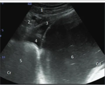
In ultrasonographic examination, displaced abomasum was imaged approximately 10 cm cranial of the xyphoid process from the left and and right paramedian regions and from the ventral midline, immediately caudal of reticulum
(Fig. 2). The abomasal content were visible heteroform due to fluid ingesta. The visible ingest in the abomasum was seen hypoechogenic. The walls of abomasum and reticulum were appeared thin echogenic line. The abomasal folds were seen as echogenic structures within the content of the abomasum. Reticulum content was not well imaged due to gaseous composition (Fig. 2). Abomasal content was taken from this area by ultrasound assisted paracentesis. pH of this content was 3.5. Also anterior abomasal displacement was confirmed by laparotomy. In a healthy cow, reticulum (3) and craniodorsal blind sac of the rumen (4) imaged from the left sternal region (Fig. 1).
DISCUSSION
The results of this case report indicate that the ADA could be easily diagnose by ultrasonograpic techniques. Displaced abomasum was imaged approximately 10 cm cranial of the xyphoid process from the left and and right paramedian regions and from the ventral midline, immediately caudal to the reticulum the displaced
Table 1. Blood gases, WBC, glucose, sodium, potassium, calcium,
inorganic calcium and lactate values in the cow with anterior displacement of abomasum
Tablo 1. Anterior abomazum deplasmanlı bir sığırda kan gazları, lökosit,
glikoz, sodyum, potasyum, kalsiyum, inorganik kalsiyum ve laktat değerleri
Parameters Values of Analysis Reference Range
WBC (x103/µl) 14.45 4.00-12.00 pH 7.54 7.35-7.50 pCO2 (mmHg) 41 35-45 HCO3 (mmol/L) 35.1 21-29 BE(W) (mmol/L) 12.6 4-12 pO2 (mmHg) 37 35-45 O2 saturation (%) 81 80-90 Sodium (mmol/L) 137 135-148 Potassium (mmol/L) 2.9 3.9-5.8 Glucose (mg/dl) 86 45-75 Lactate (mmol/L) 9.7 2-4 Calcium (mg/dl) 6.9 9.5-11.5
Inorganic calcium (mmol/L) 0.62 1.2-1.5
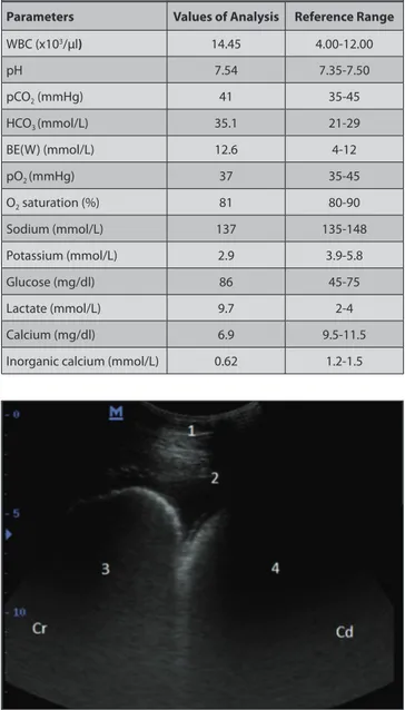
Fig1. Ultrasonogram of reticulum and craniodorsal blind sac of the
rumen in a healthy cow imaged from the left sternal region. Ventral abdominal wall (1), diaphragm (2), reticulum (3), craniodorsal blind sac of the rumen (4), Cr: cranial, Cd: caudal
Şekil 1. Sağlıklı bir sığırda sol sternal bölgeden rumenin kraniodorsal
kör kesesi ve retikulumun ultrasonogramı. Ventral abdominal duvar (1), diafram (2), reticulum (3), rumenin kraniodorsal kör kesesi (4), Cr: kranial, Cd: kaudal
319 OK, YILDIZ, NASERİ
abomasum could be easily distinguish from reticulum and craniodorsal blind sac of rumen by ultrasonography
(Fig. 1). Abomasal contents were seen as a hypoechogenic due to ingesta visible. However, the wall of the abomasum appeared as a narrow echogenic line. Parts of the abomasal folds were visible occasionally as echogenic structures within the abomasum (Fig. 2) [10,11].
In this case report, hyperbasemia, hypokalemia, hypo-calcemia and hyperlactatemia, and metabolic alkalosis in the cow with ADA were detected (Table 1). Decreased plasma concentration of K+ attributable primarily to
sequestration of gastric contents and anorexia and hyper- bicarbonatemia and increased concentrations attributable to obstruction of abomasal outflow and the resultant accumulation of HCO3- in the extracellular fluid space [4,12].
The plasma lactate value in the cow with ADA was high. Increase of lactate in this cow may be related to decreased abomasal tissue perfusion[13]. Because ischemic necrosis
was detected in abomasum by laparotomy. In additional there is fluid and fibrin deposits in abdominal cavity (Fig. 2). The serum calcium and plasma inorganic calcium values in the cow with ADA were low (Table 1). Hypocalcemia have
been described for cases of abomasal displacement [12].
Increased WBC in the cow with ADA may be related to abdominal cavity inflammation.
As a conclusion, this case reported is the first description of the diagnose of the anterior displacement of abomasum by ultrasonography. In ultrasonographic examination, displaced abomasum was imaged approximately 10 cm cranial of the xyphoid process from the left and right paramedian regions and from the ventral midline, immediately caudal to the reticulum. Ultrasonography is a valuable tool in the diagnose of the anterior displacement of abomasum.
REFERENCES
1. Braun U, Wild K, Guscetti F: Ultrasonograpic examination of the
abomasum of 50 cows. Vet Rec, 140, 93-98, 1997.
2. Radostits OM, Blood DC, Gay CC: Veterinary Medicine. A textbook
of the Diseases of Cattle, Sheep, Pigs, Goats and Horses. 9th ed. pp.324.
London, W.B. Saunders, 1999.
3. Güzelbektes H, Şen İ, Ok M, Costable PD, Boydak M, Çoskun A: The
levels of serum amiloid A and Haptoglobin concentrations and liver fat percentage in lactating cows with abomasal displacement. J Vet Intern
Med, 24, 213-219, 2010.
4. Şen İ, Ok M, Birdane FM, Güzelbektes H, Turgut K: The role of gastrin
in the aetiology of abomasal displecement in dairy cows. Vet Rec, 151, 26-27, 2002.
5. Sevinç M, Ok M, Başoğlu A: Liver function in dairy cows with
displacement of abomasum. Revue Vet Med, 153, 477-480, 2002.
6. Buckner R: Surgical correction of left displaced abomasum in cattle.
Vet Rec, 136, 265-267, 1995.
7. Huhn JC, Nelson DR: Right-sided abomasal problems in dairy cattle.
Vet Med, 90, 1169-1174, 1995.
8. Zadnik K: Review of anterior diplacement of the abomasum in cattle in
Slovenia. Vet Rec, 153, 24-25, 2003.
9. Van de Watering CC, Nemeth F, Breukink HJ: En geval van
lebmaagdilatatie dislokatie naar cranial. Tijdschrift Voor Diergeneeskunde, 21, 1478-1482, 1965.
10. Ok M, Arıcan, M, Turgut K: The Ultrasonographic finding in dairy
cows with left and right abomasal displacement. Revue Vet Med, 153 (1): 15-18, 2002.
11. Braun U: Ultrasonography of the gastrointestinal tract in cattle. Vet
Clin Food Anim, 25, 567-590, 2009.
12. Şen I, Ok M, Çoskun A: The level of serum ionised calcium, aspartate
aminotransferase, insulin, glucose, betahydroxybutyrate concentrations and blood gas parameters in cows with left displacement of abomasum.
Polish J Vet Sci, 9 (4): 227-232, 2006.
13. Grünberg W, Constable PD, Schröder U, Staufenbiel R, Morin D, Rohn M: Phoshorus homestasis indairy cow with abomasal displacement
or abomasal volvulus. J Vet Intern Med, 219, 894-898, 2005.
Fig 2. Ultrasonogram of reticulum and abomasum in a cow with
Anterior abomasal displacement imaged from the left sternal region. Ventral abdominal wall (1), deposits of fibrin (2) diaphragm (3), mild asites (4), reticulum (5), abomasum (6) Cr: cranial, Cd: caudal
Şekil 2. Anterior abamazum deplasmanlı bir sığırda sol sternal bölgeden
reticulum ve abomazumun ultrasonogramı. Ventral abdominal duvar (1), fibrin birikimi (2) diafram (3), sıvı birikimi (4), reticulum (5), abomazum (6), Cr: kranial, Cd: kaudal