Dicle Tıp Dergisi, 2007 Cilt: 34, Sayı: 2, (102-106)
* Dicle Üniv. Tıp Fak., Histoloji-Embriyoloji A.D. / Diyarbakır ** Dicle Üniv. Tıp Fak., Göz Hast. A.D. / Diyarbakır *** Dicle Üniv. Tıp Fak., Dermatoloji A.D. / Diyarbakır
102
Conjunctival Impression Cytology and Bulbar Surface Epithelium Changes
in Patients with Psoriasis
Sevda Söker*, Yusuf Nergiz*, Sevin Çakmak**, Sema Aytekin***
SUMMARY
In this study, we evaluated bulbar surface epithelium changes with conjunctival impression cytology (IC) in patients with psoriasis. Our study group consisted of 32 psoriatic patients (64 eyes), who were followed up at Dermatology Department of Dicle University Hospital. Control group comprised 32 healthy volunteers (64 eyes) who had no abnormality on routine ophthalmological examination and were in the same age and sex distribution. Specimens for conjunctival IC were obtained with a cellulose acetate filter paper from the upper bulbar conjunctiva and fixed with 70 % ethyl alcohol, 37 % formaldehyde and 20:1:1 glicial asetic acid solution. Specimens were stained with periodic acid Schiff’s and Hematoxylin-eosin. The grades of Nelson system were evaluated with light microscopy. Of the patients with psoriasis, 39 % had grade 0, 36 % grade I, and 25 % grade II conjunctival IC differentiation compared with 78, 22, and 0 %, respectively in the control group (p< 0.001). Snake-like appearance of nuclear chromatin in conjunctival epithelial cells was demonstrated in 3 % of eyes in group I but in no eyes in group II. In conclusion, we showed that there could be early conjunctival changes and squamose metaplasia as well as increased goblet cell density in patients with psoriasis when compared with control group.
Key Words: Psoriasis, Impression Cytology, Conjunctiva.
Psöriazisli Hastalarda Bulbar Yüzey Epitel Değişikliklerinin Konjunktival
İmpresyon Sitolojisi İle Değerlendirilmesi
ÖZET
Bu çalışmada psöriazisli hastalarda bulbar yüzey epitel değişiklikleri konjunktival impresyon sitolojisi ile değerlendirilmiştir. Çalışmamız Dicle Üniversitesi Tıp Fakültesi Dermatoloji AD’da Psöriazis tanısıyla takip edilen 32 hastanın 64 gözü (çalışma grubu) ile yapılan rutin göz muayenelerinde herhangi bir patoloji saptanmayan kontrol grubu 32 olgunun, 64 gözünü kapsamaktaydı. Üst bulbar konjuktivaya uygulanan impresyon sitolojisi yöntemi ile sellüloz asetat kağıdı yüzeyine alınan hücre grupları (%70’lik etil alkol, %37’lik formaldehit ve glisial asetik asitin 20:1:1 oranındaki) solüsyonunda fikse edildikten sonra Periodik Asid Schiff ve Hematoksilen-eosin ile boyandı. Gruplar, Nelson Evreleme sistemi ile ışık mikroskobunda değerlendirildi. Nelson Evreleme Sistemine göre Psöriazisli grubun %39’u Evre 0, %36 ‘sı Evre 1 ve %25 ‘i Evre 2 iken kontrol grubunda sırasıyla %78, %22 ve %0 bulundu (p< 0.001). Konjunktiva epitel hücrelerinde nükleer kromatinin yılan vari görünümü psöriazis grubunda %3 oranında saptanırken kontrol grubunda hiçbir olguda saptanmadı. Sonuç olarak, bu çalışmada psöriazisli hastalar kontrol grubu ile karşılaştırıldığında impresyon sitolojisi tekniği ile konjunktivalarında erken hücresel değişikliklerden skuamöz metaplaziye kadar varan değişiklikler ile artmış goblet hücre yoğunluğu olabileceği saptandı.
Anahtar Kelimeler: Psöriazis, İmpresyon Sitolojisi, Konjunktiva.
S. Söker ve ark. Dicle Tıp Dergisi 2007
103
INTRODUCTION
Psoriasis is a common, chronic and recurrent, inflammatory proliferative skin disease characterized by dry red patches covered with scales, occuring especially on the scalp and ears and genitalia and the skin over bony prominences. Although it does not usually involve the eye, ocular signs are
blepharitis, conjunctivitis, ectropion, madarosis,
and rarely, uveitis occur in 10 % of the psoriasis patients (1, 2).
Impression cytology (IC) is the technique of collection of the most superficial layers of the ocular surface by applying different collecting devices (usually filter papers) so that cells adherent to that surface can be subsequently removed from the tissue and further processed for a diversity of techniques (3). It is fast, non invasive, easy to perform, and economical (4). In this study, ocular-surface changes, and those progressing into squamose metaplasia, as well as increased goblet cell density were evaluated on the cell content of the surface conjunctival epithelium by conjunctival IC in patients with psoriasis.
METHODS
Our study group comprised 64 eyes of 32 patients with psoriasis (18 male, 14 female), whose diagnoses were confirmed with skin biopsy in Dermatology department of our University hospital. Control group consisted of 32 (15 male, 17 female) healthy volunteers (64 eyes) who had no abnormality on routine ophthalmological examination and were in the same age and sex distribution. The median age of patients was 32.94 ± 13.20 yr (range 11-63 yr) and control group was 32.53 ± 12.64 yr (range 17-61 yr) (p>0.05).
The specimens were collected from the upper bulbar conjunctiva, 5 mm from the limbus with 0.4% oxibupracaine hydrochlorid topical anesthesia. Specimens for conjunctival IC were obtained with a cellulose acetate filter paper of 0.020 µm pore size (Sartorious 11107-50-N) from the superior bulbar conjunctiva of both eyes of each subject by using the method described by Egbert et al (3,5). The cellulose acetate filters were firstly cut to obtain pieces of 3x4 mm size. The mat surface of these papers was gently pressed on the upper bulbar conjunctiva (at 12:00 o’clock direction for a period of 3-4 seconds), so
that the lower edge of the paper was kept 2 mm away from the limbus. The paper was then removed slowly from the conjunctiva, while keeping the conjunctiva epithelium samples obtained at cytological level on the upper surface of the paper. They were further fixed with 70 % ethyl alcohol, 37 % formaldehyde and 20:1:1 glicial asetic acid solution in an effort to dye, and then stored at a temperature of +4 oC to avoid the evaporation of alcoholic substances (5,6). Utmost care was taken to place the samples in their correct eye group during all these procedures. Specimens were stained with Periodic Acid Schiff (PAS), Hematoxylin-eosin (H&E) and conjunctival IC and goblet cell population were examined according to the classification of Nelson (4,7). The cytology was graded according to the scheme suggested by Nelson, the details of which are given in Table 1 (4, 7).
The Student –t test and x² test were used to compare the patients and control group.
RESULTS
We observed that 25 (39 %) eyes showed a grade 0 (figure 1); 23 (36 %) eyes, a grade I (figure 2); and 16 (25 %) eyes showed a grade II differentiation (figure 3) of the patients with psoriasis. None of the patients showed a grade III differentiation. In control group, 50 (78 %) eyes showed grade 0, 14 (22 %) eyes grade I differentiation. No one in the control group showed a grade II and III differentiation. There was a statistically significant difference between patients of psoriasis and control group in grading of the squamous metaplasia according to Nelson’s classification (p<0.001).
Figure 1. Grade 0 differentiation of
conjunctival IC in patients with psoriasis (original magnification, x 41, PAS and H&E stained).
Cilt: 34, Sayı: 2, (102-106)
104
Figure 2. Grade I differentiation of conjunctival IC in patients with psoriasis (original magnification, x 82, PAS and H&E stained).
Figure 3. Grade II differentiation of conjunctival IC in patients with psoriasis (original magnification, x 164, PAS and H&E stained).
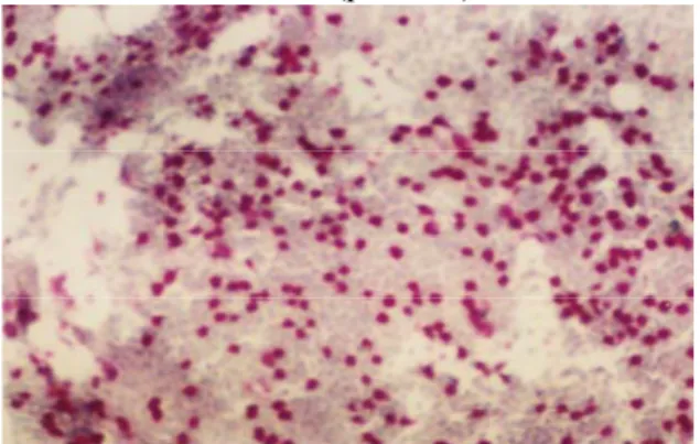
The impression cytologic examinations showed neutrophil clumping in 17 (27 %) eyes in patient group (figure 4).
Figure 4. Neutrophil clumping of conjunctival
IC (original magnification, x 82, PAS and H&E stained).
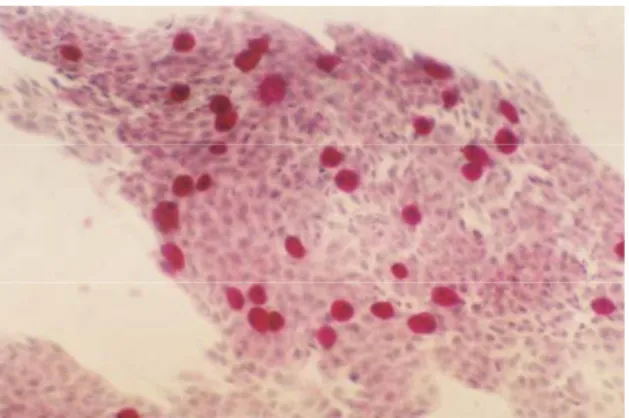
In patients with psoriasis, snake-like appearance of nuclear chromatin in conjunctival epithelial cells was demonstrated in 3 % of eyes in patients group with grade II (figure 5), but was not seen in the eyes of the control group. Also, in
terms of goblet cell population, there was an increasing trend in the number of goblet cells in psoriatic patients compared to controls, though this difference was not statistically significant (p=0.129) (Table 2).
Figure 5. This conjunctival IC from a patients
with psoriasis shows grade II differentiation and typical snake-like chromatin appearance in the cell nuclei (original magnification, x 164, PAS and H&E stained).
Table 1. Cytological gradation as per
Nelson&Wright (4, 7).
Grade 0: The epithelial cells are small and round
with eosinophilic staining cytoplasm. The nuclei are large, basophilic, with a nucleocytoplasmic ratio of 1:2. The goblet cells are abundant, plump and oval and have an intense PAS-positive cytoplasm.
Grade I: The epithelial cells are slightly larger
and more polygonal and have eosinophilic-staining cytoplasm. The nuclei are smaller, with nucleocytoplasmic ratio of 1:3. The goblet cells are decreased in number; however, they still maintain their plump, oval shape with an intensenly PAS-positive cytoplasm.
Grade II: The epithelial cells are larger and
polygonal, occasionally multinucleated, with variably staining cytoplasm. The nuclei are small, with a nucleocytoplasmic ratio of 1:4 to 1:5. The goblet cells are markedly decreased in number and are smaller, less intensely PAS- positive, with poorly defined cellular borders.
Grade III: The epithelial cells are large and
polygonal with basophilic-staining cytoplasm. The nuclei are small, pyknotic, and in many cells, completely absent. The nucleocytoplasmic ratio is greater than 1:6. Goblet cells are completely absent.
S. Söker ve ark. Dicle Tıp Dergisi 2007
105
Table 2. Goblet cell population in psoriasis
and control group.
DISCUSSION
Psoriasis is a chronic, inflammatory disease most commonly manifested by well-demarcated, erythematous, silvery-scaled plaques on the elbows, knees, scalp, and trunk. Ocular signs in patients with psoriasis have been investigated only in a small number of studies (2). In this study, all the psoriatic patients were evaluated bulbar conjunctiva surface epithelium changes with IC. Karabulut et al. reported 50 patients with psoriasis. Fifty percent of the patients had a grade 0, 30 % grade I, and 20 % grade II conjunctival IC differentiation (2).In our study, we observed that 25 (39 %) eyes showed grade 0, 23 (36 %) eyes grade I, and 16 (25%) eyes grade II differentiation. None of the patients showed grade III differentiation. The data of IC showed that there were important cell alterations in the psoriatic patients.
Tseng et al. theorized that the pathogenesis of squamous metaplasia of the ocular surface epithelia might be due to the loss of vascularization and scar formation (2,8). They further stated that circulating factors that maintain the normal epithelial differentiation were lacking, and intense inflammation might introduce different factors to facilitate the epithelial alterations (2). Squamous metaplasia is probably due to inflammatory nature of psoriasis. However no relationship was noted between the squamous metaplasia and the duration of the disease (2). This finding, hence, supports the view that squamous metaplasia is associated with primary psoriasis, whereas it might not be a side effect of the treatment or a complication of the disease.
The snake-like appearance of nuclear chromatin in conjunctival ephitelial cells was observed in 3 eyes (4 %) of psoriasis patients, and with grade II differentiation in accordance with Nelson’s classification. Karabulut et al.
…
previously reported the snake-like appearances of chromatin with 12 % of the psoriasis cases (2). Nuclear chromatin differences of this type, as observed in conjunctival epithelial cells, were found for the first time in patients with keratokonjunctivitis sicca (9). Though the snake-like appearance was initially attributed to an artifact related to the IC method, it was later understood to be free from the method through biopsies (2, 10). The snake-like appearance was defined to be only a squamous metaplasia case of cuboidal epithelial cells. The snake-like structure of central chromatin is a specific intense occurrence axially positioned at the center of the extended nucleus. The snake-like appearance was reported to develop from changes in fibrous lamina as a result of electron microscopy studies (2). This component of nuclear skeleton is formed by an internal thin layer covering the nucleus. Chronic irritation on epithelium surfaces can result in the effects such as extension of nucleus, dispersion of fibrous lamina and hence the displacement of peripheral heterochromatin (2). The snake- like appearance of chromatin in conjunctival epithelial cells with the psoriasis group was stated to be due to chronic mechanical irritation in connection with koebner phenomenon as observed with the patients (2, 11). The authors hope that further studies on snake-like chromatin will contribute greatly to better understanding of the etiology of psoriasis.
Also, in terms of goblet cell population, there was an increasing trend in the number of goblet cells in psoriatic patients compared to controls, though this difference was not statistically significant. We have not encountered any obvious data about this study until now in literature.
In summary, it may be concluded that the psoriasis may not only be related to the findings obtained from ophthalmological examination, it also develops from the changes in conjunctiva epithelium. Conjunctival IC may be a routine technique to evaluate squamous metaplasia and goblet cell changes in psoriatic patients.
n median SD
Psoriasis group 64 86.3 76.4