Synthesis of Novel Rhodium Complex of Biphenylic-Imidazole-Phenanthroline and
Photophysical Analysis of Metal Ions Selectively for Pb
2+1
Kiramat Shah,, 2Said Nadeem, 3Zahid Hussain* and 1Muhammad Raza Shah
1International Centre for Chemical and Biological Sciences, H.E.J. Research Institute of Chemistry, University of Karachi, Karachi-75270, Pakistan.
2Department of Chemistry, Mugla University, Turkey. 3
Department of Chemistry, University of Karachi, Karachi-75270, Pakistan.
(Received on 10th June 2015, accepted in revised form 1st December 2015)
Summary: A novel rhodium biphenylic-imidazole-phenanthroline metal-organic complex has been synthesized and the photophysical properties were studied against different metal ions. The rapid and sensitive detection of metal ions have an important impact on making the various environmental, biological and chemosensor devices. The new chemosensing properties have shown the affinity for Pb2+ toward Rhodium biphenyl-imidazole phenanthroline 6. These studies significantly conceded out that the UV/Vis and flourescent characteristic of the receptor has selectively responded at 344 nm with absorbance of 0.341 to the Pb2+, it is also obviously evident from the partial 1H-NMR results that the disappearance of NH at 7.129 and 10.081 ppm respectively. The remaining metal ions appeared small difference in their hypso- and hyperchromic shifts of UV/Vis results. Furthermore characterization and analytical studies of metal-organic complexes as chemosensors for different metal ions such as Ca2+, Cs+, Cu2+, Fe3+, K+, Mn2+, Na+, Pb2+, Sb+, Sn2+, Sn4+, and Zn2+ were carried using NMR, MS-ESI, Fluorescence and UV/Vis spectroscopic technique.
Keywords: Phenanthroline; Chemosensors; Metal-Organic Complex; UV/Vis, Flourescence, Photo-physical. Introduction
The synthetic development for molecular recognition of metal ions is the most attractive and growing research area of supramolecular chemistry [1-3]. These chemosensors that can selectively associated to specific metal ions has substantial attention, and having exciting development for scientific society, especially among chemists, biologists, physicists and material scientists. In biochemistry, clinical and medical sciences, and cell biology, freely mobile sensor molecules are employed generally in microscopy the use offers possibility of performing real-space measurements, for analytical and environmental sciences have studied on these sensors and elucidated various applications in clinical toxicology, bioremediation, waste management and cancer detection [4-6]. Lead among the heavy metal ions is the most abundant and have second position as a substance in the list of toxic elements, which is often encountered for its broad sharing in the environment as well as its presence in the earlier utilization of deferent substances like batteries, gasoline, and pigments [7]. Lead is a harmful metal ion causing large perturbation in environment and create health problems, the main symptoms are memory loss, irritability, anemia, muscle paralysis, and mental retardation have been attributed to lead poisoning [8-9]. The market demand and the increased resistance various toxin have attracted the attention toward the chemosensors which selective and specific for heavy and toxic metal ions is a constantly creating a worth
provoking demand day by day [10]. The development of fluorescent sensors for lead is helping in the advancement to make a clear cellular role of lead ions
in- vivo as well as in-vitro to observe lead
concentration in the contaminated sources [11]. There are various effective and efficient fluorescent sensors with certain limitation have been successfully developed for alkali, alkaline earth, and zinc ions, while only few have been synthesized for lead [12-14]. The heavy metals are well known as fluorescence quenchers via enhanced spin orbital coupling, energy, or electron transfer, development of selective as well as sensitive fluorescent sensors for lead presents a challenge [15]. There are methods available for the detection of lead including atomic absorption spectroscopy and inductively coupled plasma atomic emission spectroscopy have also been useful for the quantitative detection of lead ions all of these techniques are highly expensive, therefore researchers are continuously efforts to synthesis novel, new and cheap molecular entities for the developments of the smart chemosensors and fluorescence devices with efficient sensitivity [16-21].
The synthesis of a novel biphenylic imidazole phenanthroline 4 and its metal-organic complex of rhodium 6 were carried out, whereas various metal ions like Ca2+, Cs+, Cu2+, Fe3+, K+, Mn2+, Na+, Pb2+, Hg2+, Sb3+, Sn2+, Sn4+, and Zn2+ were screened. The effect on UV and fluorescence photophsical properties of the metal organic
Kiramat Shah et al., J.Chem.Soc.Pak., Vol. 38, No. 01, 2016
92
molecular building block was investigated. The promising results indicate that this receptor has selective and sensitive response for Pb2+ metal ion, the interaction of this complex with the rest of metal ions have not shown significance response in the intensity of absorbance at respective wavelengths as compared to the Pb2+.
Experimental
In these studies all chemicals were bought from Organics, Sigma-Aldrich, Acros Chemicals and Fisher Scientific Ltd. All chemicals used for the synthesis of starting materials without further purification. The deuterated solvents of Apolo were used for the NMR analysis. The characterization of the compounds was carried out on 1H Spectroscopic data using 300 MHz Bruker NMR Instrument, Perkin Elmer LS 55 flourescence Spectroscopic data, Mass Spectrometric (QSTAR XL MS/MS system) and UV/Vis spectrophotometer (Thermo Scientific Evolution 300)
1,10-Phen-5,6-dione (2)
A mixture of 1,10-phenenthroline 1 (5 g, 25.5 mmol) of and potassium bromide (5 g, 42 mmol) were taken in 100 mL round bottom flask kept under ice bath than the a mixture of 50 mL of concentrated sulfuric acidand 25 mL nitric acid was added slowly with stirring at 0oC and the reaction mixture was refluxed for 3 hrs. When the color of reaction mixture changed to yellow it was poured into 500 mL of ice cold distilled water and concentrated sodium hydroxide solution was added slowly to make the pH 4. The aqueous phase was extracted with dichloromethane (20 mL X 3) and dried with Na2SO4. The solvent was evaporated under
vacuum and characterized; EI-MS m/z: 212; 1H NMR (300 MHz, CDCl3), : 9.12 (dd, 2H, J = 1.8 Hz, J = 4.8 Hz, ArH), 8.51 (dd, 2H J = 1.5, J = 7.8 Hz), 7.69 (dd, 2H, J = 8.1, J =1.6 Hz). 4'-(1H-imidazo[4,5-f][1,10]phenanthrolin-2-yl)biphenyl-4-carbaldehyde (4) Mixture of 1,10-phenanthroline-5,6-dione 2 (0.212g,1mmol), 4,4-biphenyldicorboxyl adehyde 3 (0.210g, 1mmol) and (1g,15mmol) of ammonium ion were taken in a 50 mL round bottom flask, 5mL of glacial acetic acid was added as solvent and refluxed for 3 h. The reaction mixture was poured in to 300 mL water and ammonium hydroxide solution was added drop wise to neutralize the reaction mixture till pH 7. Yellow precipitates were were collected and washed with water, and cold acetone. The compound was purified by column chromatography by using ethanol as eluent. The yellow solid product obtained was further dried under vacuum. MS-ESI m/z: 400;
1 H-NMR (300 MHz, DMSO-D6), : 10.07 (s, 1H,), 9.05 (dd, 2H, 4J = 1.8 Hz, J = 4.2), 8.10 (m, 3H), 8.457 (dd, 2H,3J=5.1Hz, 2J=8.1), 8.109 (m, 6H), 7.869 (dd ,2H ,J=4.5 ,3J=8.1); UV/Vis (DMSO:water 1:1) nm: 247 nm (0.454), 288 nm (0.277), 345 nm (0.271). Rh-complex of 4'-(1H-imidazo[4,5-f][1,10] phenanthrolin-2-yl)biphenyl-4-carbaldehyde (6) Mixture of (0.020g,1mmol) of 4'-(1H- imidazo[4,5-f][1,10]phenanthrolin-2-yl)biphenyl-4-carbaldehyde (3) and (0.015g,1mmol) of Rh-complex (Bis[(pentamethylcyclopenta dienyl)dichloro-rhodium] CAS#12354-85-7) , (0.018g,1mmol) of ammonium hexafloro phpsphate and 2 ml methanol were placed in 100 ml round bottom flask. The reaction mixture was stirred at room temperature for 30 minutes and then reaction into 100 ml water, filtered and washed with acetone, dried in vacuum and characterized by ESI-MS: 673.113 (calculated for [C36H31ClN4ORh]2- = 673.13); 1H NMR (300
MHz, CDCl3), : 10.10 (s, 1H), 9.34 (dd, 2H, J =
4.8), 8.50 (d, 1H, J = 8.4), 8.29 (s, br, 1H), 8.12 (m, 3H), 7.01 (m, 4H), 1.73 (s, 15H), UV/Vis (DMSO:H2O 1:1) nm: 274 (0.297), 345 (0.101).
Preparation of sample solution for analysis
1 mmol/L stock solution was prepared from both compounds 4 and 6 in DMSO, wherein from the stock solution a 30 micro molar dilute solution was prepared for sample analysis. Meanwhile with the same concentration the following samples of salt solutions were prepared in water and photophysical studied of these samples were carried out. The samples of i.e CsOH, FeCl3, KBr, KSCN, MnSO4,
NaI, NaOAc, PbOAc, PhHgOAc, SbF, SnCl2, SnCl4,
and ZnOAc were mixed with both compounds 4 and 6 separately, hence with the 1:1 ratio of the respective samples for organic-metal complex using the DMSO and water (1:1v/v) at 25 oC were investigated.
Result and Discussion
Synthesis: The synthesis of dione phenenthroline 2 was carried out according to the literature procedure [22-23]. A yellow solid compound 2, one equivalent of 4,4`-biphenylcarboxaldehyde 3 and ammonium ion in acetic acid as solvent was mixed and the content of reaction mixture was refluxed for 3 hours with continous stirrering (scheme-1). The formation of imidazole ring resulted in the coupling of phenenthroline dione 2 with 4,4`-biphenylcarboxaldehyde 3 is using ammonium ion, where ammonium ion is the source of nitrogen in this especial type of condensation reaction.
Scheme-1: Synthesis of compound 4
Scheme-2: Synthesis of rhodium complex of 4'-(1H-imidazo[4,5-f][1,10] phenanthrolin-2-yl)-[1,1'-biphenyl]-4-carbaldehyde 6
The compound 4 was obtained as yellow precipitates after neutralization when the hot reaction mixture was poured into cold water. These precipitates were washed with acetone to remove traces of the reactant; furthermore the crude product 4 was purified on column chromatography using ethanol as eluent. The pure compound 4 was treated with Rhodium solution for complexation with rhodium dimer 5 in methanol and stirred for 30 minute at room temperature as shown in scheme 2.
After completion of the reaction time, a concentrated solution of ammonium hexaflorophosphate was added to the reaction mixture. Yellow precipitates of compound 6 were obtained which were washed with cold methanol and dried. Compound 4 was soluble in methanol and not in acetone, but this condition was reversed in case of compound 6 was soluble in methanol. The compound 2, 4 and 6 were characterized by NMR, ESI, UV/Vis and Fluorescence spectroscopic analysis.
Kiramat Shah et al., J.Chem.Soc.Pak., Vol. 38, No. 01, 2016
94
UV/Vis studies
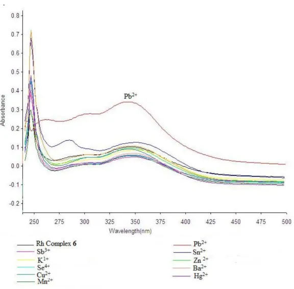
UV-spectra of compound 6 were obtained in DMSO and water mixture (1:1 v/v) as well as with solutions of the salts above were obtained and are shown in Figure 1.
Compound 6 showed a max of 247 nm in
UV spectrum (Figure 1). Lead showed red hyperchromic shift while the other metals showed hyper- or hypso-chromatic deviation in absorbance from the reference compound 4. It was found from the UV studies that both ligands 4 and 6 on addition of Pb2+ appeared at remarkable difference in absorbance value at 247 nm. Compound 4 showed absorbance at 0.345 while at the same concentration, compound 6 showed absorbance of 0.5 so Pb2+ also resulted in hyperchromic shift.
Compound 4 gives absorption of 0.454 and after complex formation with rhodium metal, compound 6 showed quenching of absorption band which was observed 0.297 and the prominent peak in compound 4 at 286 nm with absorption at 0.227 is almost disappeared in compound 6 due to the complex formation with rhodium metal.
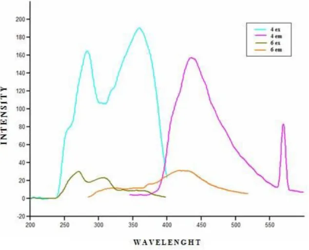
Fluorescence Studies
In the fluorescence studies two peaks were observed in the excitation spectrum of reference compound 4 i.e. 282 and 358 nm with excitation intensities of 165 and 192 respectively. The first excitation and emissions of compound 4 and 6 were compared as shown in figure 2.
.
Fig. 2: Comparison of excitation and emission behavior of compound 4 and 6.
Kiramat Shah et al., J.Chem.Soc.Pak., Vol. 38, No. 01, 2016
96
It was studied in earlier step that the two peaks which were observed in the excitation spectrum of reference compound 4 i.e. 282 and 358 nm with excitation intensities of 165 and 192 respectively and emission maxima was observed at 434 nm with intensity after making complex of 4 with rhodium compound 6, the maxima at 282 shifted to 271 nm with reduction in intensity to 30 while the maxima at 358 shifted to 307 nm with reduction in intensity 23. The similar changes were observed in the emission maxima from 434 to 420 nm with reduction in emission intensity from 158 to 31 (Table 1).
It is indicated in Table 1, where 81 % and 88 % reduction occurred in the peaks at 282 and 358 nm respectively, while in the excitation maxima and the emission maxima were showed 80 % decrease in the intensity on complex formation with rhodium.
Excitation studies
The fluorescence studies like compound 4 and compound 6 were also subjected to the same above mentioned different salts solutions. The
excitation spectrum of compound 6 obtained after the addition of metal solution is shown in figure 3. Table-1: Comparison of emission and excitation on fluorescent studies of compound 4 and 6.
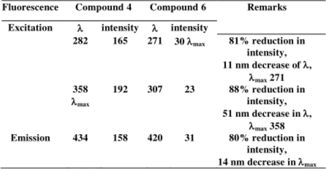
Fluorescence Compound 4 Compound 6 Remarks
Excitation intensity intensity
282 165 271 30 max 81% reduction in intensity, 11 nm decrease of , max 271 358 max 192 307 23 88% reduction in intensity, 51 nm decrease in , max 358 Emission 434 158 420 31 80% reduction in intensity, 14 nm decrease in max
The excitation of compound 6 showed excellent hyperchromic shifts as in previous case, no metal showed increase in intensity than the in the intensity of reference compound, after the detailed studies, 3 peaks were observed at 260, 310 and 360 nm respectively in the excitation spectra of 6. The increment of excitation intensity in response of different metal interaction is plotted in figure 4, it clearly shows an increase in almost all three peaks with few exceptions.
Fig. 5: Excitation intensities of compound 6 observed with different metals.
As there were three peaks observed, all of them were plotted in the above figure 5. The plot shows the highest increase in the intensity of lead (II) ion with 2000 folds. This increase in intensity is high enough to notice and also as was observed from the UV plot in figure 1, compound 6 also showed high absorbance than the reference compound 6 in case of lead (II) ion which can conclude that compound 6 can be used as a molecular sensor for lead metal ion. The deviation from the maxima of excitation was also studied in ascending order to understand the behavior of interaction of the metals and metalloid ions with compound 6.
The deviation was observed in case of sodium ion solution, 11 nm of decrease was observed from the reference compound and the rest of compounds did not show any noticeable deviation in excitation of compound 6.
Emission studies
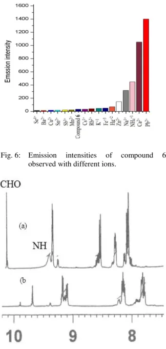
Like excitation studies of compound 6, emission studies have also shown a remarkable increase in the emission maxima with different amount in case of different metal solutions, it is shown in figure 6 that the increase of intensity of emission of compound 6 with different interactions.
The highest amount of increase in emission intensity was observed again in case of lead (II) ion solutions (Figure 6) followed by tellurium chloride and calcium chloride. The emission of reference compound 6 was found 434 nm, only tin chloride and sodium ion solutions showed red shift from the reference compound 6 up to 11 nm while the other metal solutions showed blue shift of around 27 nm as in case of zinc (II) ion.
Fig. 6: Emission intensities of compound 6 observed with different ions.
Fig. 7: Comparison Partial 1H-NMR of Compound 6 only (a) and Compound 6 (b) with Pb2+
Partial 1H-NMR Analysis
The partial 1H-NMR of the compound 6 (Figure 7a) and the interaction with Pb2+ ion (Figure 7b) were observed, hence a significant indication in the deviation of the chemical shift and the disappearance of the characteristic signals of aldehydic and NH protons at 10.2 and broad signal at 7.61 ppm of imidazole moiety respectively; it is evidently that lead has influenced over the receptor compound 6.
Kiramat Shah et al., J.Chem.Soc.Pak., Vol. 38, No. 01, 2016
98
Conclusion
A novel derivative of phenanthroline derivatives containing imidazole and biphenylic aldehyde 4 was synthesized for the first time. Aldehyde moiety is an important functional group which can be used for further reactions very easily. A rhodium complex 6 was also synthesized which can be useful in the studies of molecular sensors against different metals as was found in this study for Pb2+. The comparative UV studies of compound 4 showed complete disappearance of the main peak at 247 while other metals showed different amount of quenching behavior. The same study of compound 6 showed the enhancement of absorbance and the appearance of a shoulder peak at 261 nm. This type of behavior was also observed in the excitation and emission spectra of compound 6 with hyperchromic shift for Pb2+. It can be concluded from these lines that compound 6 can be used as a molecular sensor against lead.
Acknowledgment
We are indebted to the Higher Education Commission of Pakistan for financial support and HEJ Research Institute of Chemistry, International Center for Chemical and Biological Sciences, University of Karachi for the availability of instruments and chemicals.
References
1. Y. Zeng, G. Zhang, D. Zhang and D. Zhu, A Dual-Function Colorimetric Chemosensor for Thiols and Transition Metal Ions Based on ICT Mechanism, Tet. Lett., 49, 7391 (2008).
2. N. Singh, N. Kaur, R. C. Mulrooney and J. F. Callan, A Ratiometric Fluorescent Probe for Magnesium Employing Excited State Intramolecular Proton Transfer, Tet. Lett. 49, 6690 (2008).
3. F. Qian, C. Zhang, Y. Zhang, W. He, X. Gao, P. Hu and Z. Guo, Visible Light Excitable Zn2+ Fluorescent Sensor Derived from an Intramolecular Charge Transfer Fluorophore and Its in Vitro and in Vivo Application, J. Am,
Chem. Soc., 131, 1460 (2009).
4. J. Liu and Y. Lu, Accelerated Color Change of Gold Nanoparticles Assembled by DNAzymes for Simple and Fast Colorimetric Pb2+ Detection
J. Am. Chem. Soc. 126, 12298 (2004).
5. P. Chen, c. He, A General Strategy to Convert the MerR Family Proteins into Highly Sensitive and Selective Fluorescent Biosensors for Metal Ions, J. Am. Chem. Soc., 126, 728 (2003).
6. D. Prabhakaran, Y. H. Ma, H. Nanjo and H. Matsunaga, Naked-Eye Cadmium Sensor: Using Chromoionophore Arrays of Langmuir−Blodgett Molecular Assemblies,
Anal. Chem. 79, 4056 (2007).
7. F. Zapata, A. Caballero, A. Espinosa, A. Tarraga and P. Molina, Imidazole-Annelated Ferrocene Derivatives as Highly Selective and Sensitive Multichannel Chemical Probes for Pb(II) Cations J. Org. Chem., 74, 4787 (2009). 8. C. T. Chen and W. P. Huang, A Highly
Selective Fluorescent Chemosensor for Lead Ions J. Am. Chem. Soc., 124, 6246 (2002). 9. J. S. Lin-Fu, Lead Poisoning, a Century of
Discovery and Rediscovery, in Human Lead Exposure, Needleman, H. L. Ed., Lewis Publishing: Boca Raton, FL, (1992).
10. Y. K. Yang, K. J. Yook and J. A Tae, Rhodamine-Based Fluorescent and Colorimetric Chemodosimeter for the Rapid Detection of Hg2+ Ions in Aqueous Media, J. Am. Chem.
Soc., 127, 16760 (2005).
11. A. P. de Silva, D. B. Fox, A. J. M. Huxley and T. S. Moody, Combining Luminescence, Coordination and Electron Transfer for Signaling Purposes, Coord. Chem. Rev., 205, 41 (2000).
12. V. Amendola, L. Fabbrizzi, M. Licchelli, C. Mangano, P. Pallavicini, L. Parodi and A. Poggi, Correlations of the Distribution of Spin States in Spin Crossover Compounds, Coord.
Chem. Rev., 192, 649 (1999).
13. B. Valeur, and I. Leray, Design Principles of Fluorescent Molecular Sensors for Cation Recognition, Coord. Chem. Rev., 205, 3 (2000). 14. S. C. Burdette, G. K. Walkup, B. Spingler, R.
Y. Tsien and S. J. Lippard, Fluorescent Sensors for Zn2+Based on a Fluorescein Platform: Synthesis, Properties and Intracellular Distribution, J. Am. Chem. Soc., 123, 7831 (2001).
15. D. S. McClure, Spin-Orbit Interaction in Aromatic Molecules, J.Chem. Phy. 20, 682 (1952).
16. S. Goswami and R. Chakrabarty, Highly Selective Colorimetric Fluorescent Sensor for Pb2+, Eur. J. Org. Chem. 3791 (2010).
17. B. Valeur, Molecular Fluorescence:
Principles and Applications;
Wiley-VCH: Weinheim, Germany, (2002).
18. A.
W.
Czarnik,
Fluorescent
Chemosensors of Ion and Molecule
Recognition, Recent Applications to
Sensing,
Interfacial
Design
and
Chemical Sensing; Am. Chem. Soc.,
Chapter 27
p. 314 (1994).
19. L. Fabbrizzi and A. Poggi, Sensors and Switches from Supramolecular Chemistry,
Chem. Soc. Rev., 24, 197 (1995).
20. J. Y. Kwon, J. H. Soh, Y. J. Yoon and J. Yoon., Highly Effective Fluorescent Sensor for Hg 2+ in Aqueous Solution Supra. Chem., 16, 621 (2004).
21. S. Y. Moon, N. J. Youn, S. M. Park and S. K. Chang, Diametrically Disubstituted Cyclam Derivative Having Hg2+-Selective
Fluoroionophoric Behaviors, J. Org. Chem. 70, 2394 (2005).
22. S. M. Ji, H. M. Guo, X. L. Yuan, X. H. Li, H. D. Ding, P. Gao, C. X. Zhao, W. T. Wu, W. H. Wu and J. Z. Zhao, A Highly Selective OFF-ON Red-Emitting Phosphorescent Thiol Probe with Large Stokes Shift and Long Luminescent Lifetime, Org. Lett. 12, 2876 (2010).
23. M. R. Shah, M. Hassan, S. Nadeem and M. I. Bhanghar, Synthesis, Characterisation and Supramolecular Interaction of Rh-Biphenylic-Imidazole-Phenanthroline with Antibiotics,