Aksac et al. BMC Res Notes (2019) 12:82 https://doi.org/10.1186/s13104-019-4121-7
DATA NOTE
BreCaHAD: a dataset for breast cancer
histopathological annotation and diagnosis
Alper Aksac
1*, Douglas J. Demetrick
2, Tansel Ozyer
3and Reda Alhajj
1,4Abstract
Objectives: Histopathological tissue analysis by a pathologist determines the diagnosis and prognosis of most
tumors, such as breast cancer. To estimate the aggressiveness of cancer, a pathologist evaluates the microscopic appearance of a biopsied tissue sample based on morphological features which have been correlated with patient outcome.
Data description: This paper introduces a dataset of 162 breast cancer histopathology images, namely the breast
cancer histopathological annotation and diagnosis dataset (BreCaHAD) which allows researchers to optimize and evaluate the usefulness of their proposed methods. The dataset includes various malignant cases. The task associ-ated with this dataset is to automatically classify histological structures in these hematoxylin and eosin (H&E) stained images into six classes, namely mitosis, apoptosis, tumor nuclei, non-tumor nuclei, tubule, and non-tubule. By providing this dataset to the biomedical imaging community, we hope to encourage researchers in computer vision, machine learning and medical fields to contribute and develop methods/tools for automatic detection and diagnosis of cancerous regions in breast cancer histology images.
Keywords: Breast cancer, Histopathology, H&E staining, Annotation, Nottingham histologic score, Dataset
© The Author(s) 2019. This article is distributed under the terms of the Creative Commons Attribution 4.0 International License (http://creat iveco mmons .org/licen ses/by/4.0/), which permits unrestricted use, distribution, and reproduction in any medium, provided you give appropriate credit to the original author(s) and the source, provide a link to the Creative Commons license, and indicate if changes were made. The Creative Commons Public Domain Dedication waiver (http://creat iveco mmons .org/ publi cdoma in/zero/1.0/) applies to the data made available in this article, unless otherwise stated.
Objective
Histopathological tissue analysis by a pathologist plays an important role in the diagnosis and prognosis of many types of cancer, such as breast. Staging and grading sys-tems may vary for different types of cancer. Breast can-cer is one of the most common types of cancan-cer; it has its own grading systems. Nottingham grading system (also called the Elston-Ellis [1] modification of
Scarff-Bloom-Richardson [2] grading system) is widely used criteria
for the grade of breast tissues based on three main fea-tures, namely nuclear pleomorphism, tubular formation, and mitotic count, each of which is given 1 to 3 points. The scores of these three features are added together to determine an overall final score (in the range of 3–9) and the grade of the breast cancer. However, manually spot-ting and annotaspot-ting the affected area(s) on histopathol-ogy images with high accuracy is regarded as the gold
standard in cancer diagnosis and grading, but it is also a time-consuming and tedious task that requires consid-erable effort, expertise and experience of pathologists. These skills are mostly gained over time by analyzing more cases. Whereas this visual interpretation has strict guidelines, it brings a certain subjectivity to the histologi-cal analysis, and therefore leads to inter/intra-observer variability [3, 4] and some reproducibility issues. Besides, these issues may have a direct effect on patient prognosis and treatment planning. These problems can be allevi-ated by developing automallevi-ated image analysis tools in dig-itized histopathology. Thanks to the rapid development in the image capturing and analysis technology which could be employed to not only give more insight to but also guide pathologists in detecting and grading infected cases. These quantitative computational tools aim to improve the quality of pathology researchers concerning speed and accuracy.
Thus, it is imperative to develop an automatic assess-ment tool for the quantitative and qualitative analysis in order to help in removing this drawback. However, his-topathological examination of tissues is still a challenging
Open Access
BMC Research Notes
*Correspondence: [email protected]
1 Department of Computer Science, University of Calgary, Calgary, AB T2N
1N4, Canada
Page 2 of 3 Aksac et al. BMC Res Notes (2019) 12:82
problem since fixation, embedding, sectioning and stain-ing steps in tissue preparation produce large amounts of artifacts and differences [5]. Besides, the variability in size, shape, location, texture of nuclei turn automated detection into a tedious and more difficult task. We believe that our various annotations from different cases will help to provide good enough information about these challenging situations.
Data description
In this paper, we present a dataset of breast cancer
his-topathology images named BreCaHAD (Table 1, Data
set 1) which is publicly available to the biomedical
imag-ing community [6]. The images were obtained from
archived surgical pathology example cases which have been archived for teaching purposes. Nottingham Grad-ing System is an international gradGrad-ing system for breast cancer recommended by the World Health Organiza-tion, where the assessment of three morphological fea-tures (tubule formation, nuclear pleomorphism, and mitotic count) is used for scoring to decide on the final grade of the cancer case. To get these features, the H&E stained histological images are annotated or marked by a pathologist as either mitosis, apoptosis, tumor nuclei, non-tumor nuclei, tubule, and non-tubule. The sample cases are collected from various scenarios ranging from histological structures with clear boundaries to poorly differentiated structures with lack of typical features.
The BreCaHAD dataset contains microscopic biopsy images which are saved in uncompressed (.TIFF) image format, three-channel RGB with 8-bit depth in each channel, and the dimension is 1360 × 1024 pixels and each image is annotated (see Table 1, Data file 2–3). These annotations are mitosis, apoptosis, tumor nuclei, non-tumor nuclei, tubule, and non-tubule. They are used in the assessment of three morphological features, namely nuclear pleomorphism, tubular formation, and mitotic count. Besides, breast tissue biopsy slides are used to generate samples is stained with hematoxylin and eosin (H&E). The same acquisition conditions and settings are used to obtain digitized images from tissue sample slides with a 0.514 µm × 0.527 µm per pixel at 40×, the camera
at 40× objective captures 700 microns by 540 microns of microscopic image with a chip of 1360 × 1024 pixels. The images were captured under brightfield illumination with a Zeiss 40× oil objective on a Ziess Axiophot microscope through a 10× magnifier to a Spot Pursuit PR3440 cam-era controlled by Spot v5.2 software. While an automatic exposure mode is selected for the camera, the focusing is done manually for each slide.
All specimens were breast tissue fixed in 10% neutral buffered formalin (pH 7.4) for 12 h, processed in graded ethanol/xylene to Surgiplast paraffin. All sections were cut at 4 microns thickness, deparaffinized and stained with Harris’ hematoxylin and 1% eosin as per standard procedures. Specimens have been archived from 2 to 20 years, hence slight differences in staining and color characteristics reflect the procedures and reagents used over time. The dataset currently contains four malignant tumors (breast cancer): ductal carcinoma (DC), lobular carcinoma (LC), mucinous carcinoma (MC), and tubular carcinoma (TC). The distribution of annotations in the previously mentioned six classes and the format of the annotations for the BreCaHAD dataset can be found in Table 1, Data file 1.
The annotations for the BreCaHAD dataset are pro-vided in JSON (JavaScript Object Notation) format. In the given Table 1, Data file 4, the JSON file (ground truth) contains two mitosis and only one tumor nuclei annota-tions. Here, x and y are the coordinates of the centroid of the annotated object, and the values are between [0, 1] (divided by width and height of an image).
By providing this dataset for research purposes, we wish to promote research in computer-aided diagno-sis for breast cancer histopathology. Thus, researchers can optimize and prove the usefulness of their proposed methods while experimenting with this dataset.
Limitations
The limited pixel/image tonal range of the images due to the camera, slight differences in color due to differing batches of hematoxylin over time, and the optical resolu-tion of the 100× oil objective and immersion oil medium as these images were meant to reflect actual surgical
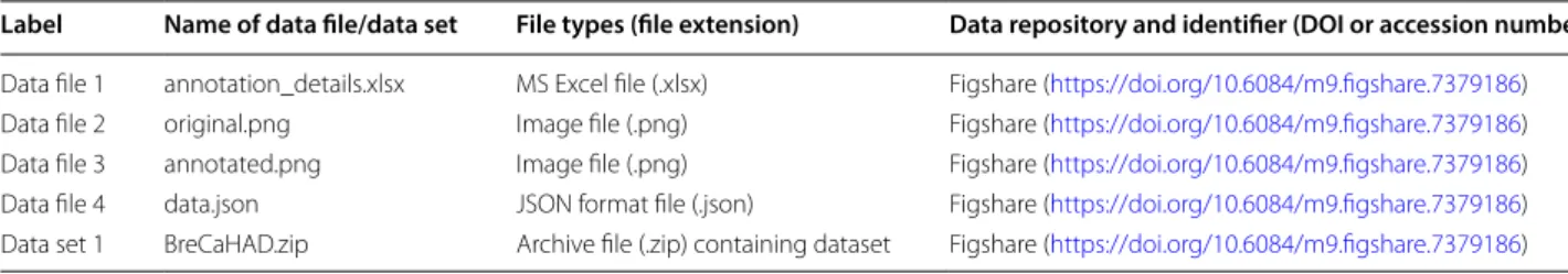
Table 1 Overview of data files/data sets
Label Name of data file/data set File types (file extension) Data repository and identifier (DOI or accession number) Data file 1 annotation_details.xlsx MS Excel file (.xlsx) Figshare (https ://doi.org/10.6084/m9.figsh are.73791 86) Data file 2 original.png Image file (.png) Figshare (https ://doi.org/10.6084/m9.figsh are.73791 86) Data file 3 annotated.png Image file (.png) Figshare (https ://doi.org/10.6084/m9.figsh are.73791 86) Data file 4 data.json JSON format file (.json) Figshare (https ://doi.org/10.6084/m9.figsh are.73791 86) Data set 1 BreCaHAD.zip Archive file (.zip) containing dataset Figshare (https ://doi.org/10.6084/m9.figsh are.73791 86)
Page 3 of 3 Aksac et al. BMC Res Notes (2019) 12:82
•fast, convenient online submission
•
thorough peer review by experienced researchers in your field
• rapid publication on acceptance
• support for research data, including large and complex data types
•
gold Open Access which fosters wider collaboration and increased citations maximum visibility for your research: over 100M website views per year
•
At BMC, research is always in progress. Learn more biomedcentral.com/submissions
Ready to submit your research? Choose BMC and benefit from:
pathology images typically used by diagnostic surgical pathologists to evaluate breast biopsies. In addition, the overall grading score for each case is not available and also the classification label is not included as either ductal carcinoma, lobular carcinoma, mucinous carcinoma or tubular carcinoma for each image.
Abbreviations
BreCaHAD: breast cancer histopathological annotation and diagnosis dataset; H&E: Hematoxylin and Eosin; DC: ductal Carcinoma; LC: lobular Carcinoma; MC: mucinous Carcinoma; TC: tubular Carcinoma; JSON: JavaScript Object Notation.
Authors’ contributions
AA, TO and RA initiated and designed the study. DJM prepared and organized the dataset. AA wrote the manuscript. TO, DJM and RA proofread the manu-script. All authors contributed to the revision. All authors read and approved the final manuscript.
Author details
1 Department of Computer Science, University of Calgary, Calgary, AB T2N
1N4, Canada. 2 Department of Pathology & Laboratory Medicine, University
of Calgary and Calgary Laboratory Services, Calgary, AB T2L 2K8, Canada.
3 Department of Computer Science, TOBB University of Economics and
Tech-nology, Ankara 06510, Turkey. 4 Department of Computer Engineering,
Istanbul Medipol University, Istanbul, Turkey.
Acknowledgements
Not applicable.
Competing interests
The authors declare that they have no competing interests.
Availability of data materials
The data described in this Data note can be freely and openly accessed on Figshare at https ://doi.org/10.6084/m9.figsh are.73791 86 [6]. Please see Table 1 and reference list for details and links to the data.
Consent for publication
Not applicable.
Ethics approval and consent to participate
This study involves anonymized information and images from which it is not possible to identify corresponding individuals. The necessary ethics approval has been granted by the Health Research Ethics Board of Alberta (HREBA. CC-17-0631). Data used in this study was collected for the routine diagnosis of patients. It was prepared and digitized at the University of Calgary. No intervention was made with patients for research purposes.
Funding
Not applicable.
Publisher’s Note
Springer Nature remains neutral with regard to jurisdictional claims in pub-lished maps and institutional affiliations.
Received: 24 November 2018 Accepted: 7 February 2019
References
1. Elston CW, Ellis IO. Pathological prognostic factors in breast cancer. I. The value of histological grade in breast cancer: experience from a large study with long-term follow-up. Histopathology. 1991;19(5):403–10.
2. Bloom HJG, Richardson WW. Histological grading and prognosis in breast cancer: a study of 1409 cases of which 359 have been followed for 15 years. Br J Cancer. 1957;11(3):359.
3. Robbins P, Pinder S, De Klerk N, Dawkins H, Harvey J, Sterrett G, et al. Histological grading of breast carcinomas: a study of interobserver agree-ment. Hum Pathol. 1995;26(8):873–9.
4. Frierson HF, Wolber RA, Berean KW, Franquemont DW, Gaffey MJ, Boyd JC, et al. Interobserver reproducibility of the Nottingham modification of the Bloom and Richardson histologic grading scheme for infiltrating ductal carcinoma. Am J Clin Pathol. 1995;103(2):195–8.
5. Wynnchuk M. Minimizing artifacts in tissue processing: part 2 Theory of tissue processing. Hanover Walk: Maney Publishing Suite; 2013. 6. Aksac A, Demetrick DJ, Özyer T, Alhajj R. BreCaHAD: A Dataset for Breast
Cancer Histopathological Annotation and Diagnosis. Figshare. 2018. https ://doi.org/10.6084/m9.figsh are.73791 86.