Ankara Üniv Vet Fak Derg, 61, 233-236, 2014
Short Communication / Kısa Bilimsel Çalışma
Pathology and molecular identification of Anisakis pegreffii
(Nematoda: Anisakidae) infection in the John Dory, Zeus faber
(Linnaeus, 1758) caught in Mediterranean Sea
Banu YARDIMCI, G. Zafer PEKMEZCİ, E. Emek ONUKDepartment of Diseases of Aquatic Animals, Faculty of Veterinary Medicine, University of Ondokuz Mayıs, Kurupelit, Samsun, TURKEY.
Summary: One specimen of John Dory, Zeus faber (Linnaeus, 1758), was caught from commercial sites at the costs of Gulf of Antalya at Mediterranean Sea, Turkey. Zeus faber was dissected carefully. After examination Anisakidae larvae were recovered from in the abdominal cavity, liver and muscles. A total of eight larvae were found in the abdominal cavity and seven larvae encapsulated on surface of liver at necropsy. In the macroscopic examination, liver was seen to pale and surface of the capsula was haemorhagic with subcapsular cysts including larvae in 0.3 cm diameter. In the histopathologic examination severe degeneration and hepatic necrosis was identified. Just below the liver capsula, Anisakis larvae were seen covered with fibrous capsula. The larvae morphologically identified as Anisakis larvae Type I were subjected to further molecular characterization by polymerase chain reaction-restriction fragment length polymorphism (PCR-RFLP) to identify the species. All Anisakis larvae were identified at species level by PCR-RFLP as belonging to A. pegreffii. Within the present study, for the first time, we report on the molecular identification of A. pegreffii larvae from John Dory, Z. faber, and we describe their pathological significance in a paratenic or intermediate hosts in Turkish waters.
Keywords: Anisakis pegreffii, pathology, PCR-RFLP analysis, Zeus faber.
Akdeniz’de yakalanan Dülger balığındaki Anisakis pegreffii (Nematoda: Anisakidae) enfeksiyonunun moleküler identifikasyonu ve patolojisi
Özet: Çalışmanın materyalini Akdeniz’de Antalya körfezi avlanma sahasında yakalanan bir dülger balığı Zeus faber (Linnaeus, 1758) oluşturdu. Zeus faber dikkatli bir şekilde diseke edildi ve karın boşluğu, karaciğer ve kaslar Anisakidae ailesindeki nematod larvaları yönünden incelendi. Makroskopik olarak karın boşluğunda sekiz larva ile karaciğer yüzeyinde kapsüle olmuş 7 larva bulundu. Makroskobik incelemede, solgun görünümde olan karaciğer yüzeyinde kanamalara ve kapsül altında 0,3 cm çapında kistlere rastlandı. Histopatolojik incelemede, hepatositlerde şiddetli dejenerasyon ve nekroz tanımlandı. Karaciğer kapsülü altında fibröz kapsülle çevrili Anisakis larvaları gözlendi. Morfolojik olarak tanımlanan Anisakis Tip I larvalar tür teşhisi için PZR-RFLP ile moleküler olarak incelendi. Tüm Anisakis larvalar PZR-RFLP ile Anisakis pegreffii olarak teşhis edildi. Bu çalışma ile Türkiye sularında avlanan dülger balığında Anisakis pegreffii’nin moleküler identifikasyonu ve paratenik ya da ara konakdaki patolojik bulguları ilk kez tanımlandı.
Anahtar sözcükler: Anisakis pegreffii, patoloji, PZR-RFLP, Zeus faber.
The John Dory, Zeus faber (Linnaeus, 1758) is widely distributed in the Aegean Sea, Mediterranean Sea, western coastal waters of Europe, the English Channel, North Sea, and the coastal waters of South Africa, Australia and New Zealand. It inhabits waters of southern Japan, the East and South China Seas, and the Pacific, Indian and Atlantic Oceans (7). Anisakis
pegreffii is the most frequently isolated in many fish
species from Mediterranean waters and appears to be responsible for many cases of human anisakiasis (9). In Turkey, A. simplex and A. pegreffii were morphologically
identified different fish species (1,16); while A. pegreffii was molecularly detected in Trachurus trachurus (17). Moreover, A. simplex was morphologically found in Z.
faber from the coastal waters of Gökçeada
(1).Nematodes of genus Anisakis parasitize a wide range of marine hosts with marine mammals, especially cetaceans, serving as definitive hosts, while fish, squid and other invertebrates serve as paratenic or intermediate hosts. In fish, third-stage larvae of the genus Anisakis encapsulates on the surfaces of the visceral organs, mainly in the mesenteries and liver. Recently, molecular
Banu Yardımcı - G. Zafer Pekmezci - E. Emek Onuk 234
techniques, sequencing and restriction fragment length polymorphism (RFLP) analyses of the ribosomal DNA (rDNA) internal transcribed spacer region (ITS region), have been proven to be particularly useful for the accurate identification of ascaridoid nematodes at the species level for eggs, larvae, and adults (4,18).
Most reports about the pathogenic effect of larval
Anisakis in fish are correlated with the effects of the
larvae to the liver of the host. The most probable larval damage is mainly caused by compression of the tissues. Sometimes larval damage is more deleteriuous due to more deeply penetration to the liver. Some authors reported reduction in liver size in several fish species after heavy infections of larval Anisakis (3,8,14). Liver atrophy of hake in superficial but heavy infections of
Anisakis was reported by Remotti (14) but according to
Guiart (6) this liver damage was caused by toxins secreted by the worm. In contrast, no evidence of degenerative liver changes or general inflammatory reaction was found in the same host by Brian (3) who
concluded that atrophy of the liver was due to mechanical compression.
In this paper, for the first time, we report on the molecular identification of A. pegreffii larvae from John Dory, Z. faber (Linnaeus, 1758), and we describe their pathological significance in a paratenic or intermediate hosts in Turkish waters.
Parasite detection and pathological examination:
One specimen of Z. faber was caught from commercial sites at the costs of Gulf of Antalya at Mediterranean Sea, Turkey. Z. faber was dissected carefully and examined for larvae of Anisakidae nematodes in the abdominal cavity, liver and muscles. Firstly, total length of larvae found in abdominal cavity was measured with a microscope. Then, a small portion of the mid-body of larvae was excised and stored in 70% ethanol at -20 °C for molecular study. The remainder of the larvae was cleared in lactophenol for morphological examination and measured with a microscope. Morphological identification was conducted according to Berland (2).
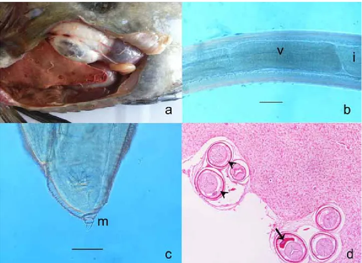
Figure 1. a. Macroscopical apperance of visceral organs and parasites. b. Anisakis Tip I larvae, ventriculus (v)-intestine (i) junction, scale 150 µ. c. Caudal end and mucron (m), scale 50 µ. d. Transverse sections of the Anisakis larvae below the liver capsula. Polymyarian muscle cells, separated into four quadrants by the chords were visible (arrow heads) and excretory cells were banana-shaped (arrow), HE x400.
Şekil.1. a. Parazitlerin ve iç organların makroskobik görünümü. b. Anisakis Tip I larva, ventriculus (v)-intestine (i) birleşim yeri, ölçü 150 µ. c. Arka uç ve mukron (m), ölçü 50 µ. d. Karaciğer kapsülü altındaki Anisakis larvalarının transversal kesiti. Polymyarian kas hücreleri (ok başları) ve muz şeklinde salgı hücreleri (ok), HE x400.
Ankara Üniv Vet Fak Derg, 61, 2014 235
The larvae morphologically identified as belonging to the genus Anisakis were subjected to further molecular characterization by RFLP to identify the species. Encapsulated nematodes were found on the liver tissue was fixed in 10% neutral formaldehyde solution for pathological examination. Tissue samples were routinely taken processed and embedded in paraffin. Tissue sections 4–6 µ in width were stained with haematoxyline-eosin (HE) and examined under light microscope (Nikon Eclipse 80i).
PCR-RFLP analysis: A total of 15 Anisakis larvae
Type I, at third-stage, recovered from the Z. faber were examined. Genomic DNA was extracted from larvae in abdominal cavity and paraffin-embedded encapsulated larvae on the liver, using the DNA purification kit (DNA DNeasy tissue kit, Qiagen) according to manufacturer's instructions. PCR amplification of internal transcribed spacer (ITS) of nuclear ribosomal DNA (rDNA) region of parasites was carried out according to Zhu et al (18) with NC5 (forward 5′-GTAGGTGAACCTGCGGAAGG ATCATT-3′) and NC2 (reverse 5′-TTAGTTTCTTCCT CCGCT-3′) primers. The restriction endonucleases, HhaI and HinfI, were used in RFLP analyses of ITS rDNA for the identification of the members of Anisakis genus according to the genetic markers defined by D'Amelio et al. (4).
Morphological and molecular identification of Anisakis larvae: A total of eight larvae were
macroscopically found in the abdominal cavity and seven larvae encapsulated on surface of liver at necropsy
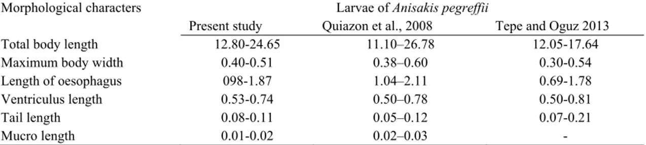
(Figure 1.a). Parasites were identified morphologically as Anisakis Type I larvae in abdominal cavity. The identification was based on the presence of a long ventriculus with an oblique ventricular-intestinal junction, without a ventricular appendage and intestinal caecum and a rounded tail possessing a mucron (Figure 1.b,c). Our morphological measurements were similar to literatures (11,16) except small differences (Table 1).
In the macroscopic examination, liver was seen to pale and surface of the capsula was haemorhagic with subcapsular cysts including larvae in 0.3 cm diameter. In the histopathologic examination severe degeneration and hepatic necrosis was identified. Just below the liver capsula, Anisakis larvae were seen covered with fibrous capsula. Larvae had a thin cuticle. Polymyarian muscle cells, separated into four quadrants by the chords were visible. Excretory cells were banana-shaped (Figure 1.d). The nematode was identified as a larva belonging to the genus Anisakis.
All Anisakis larvae were identified at species level by RFLP as belonging to A. pegreffii. All larvae amplifications of the ITS rDNA region produced a fragment of approximately 1000 bp. Three clear bands of 330, 280 and 240 bp for A. pegreffii were observed in RFLP patterns digested with HinfI (Figure 2), whilst digestion with HhaI produced two fragments of 550 and 430 bp in A. pegreffii (data not shown).
In the present study, for the first time, we report on the molecular identification of A. pegreffii larvae from John Dory, Z. faber, and we describe their pathological
Table 1. Comparative measurements (mm) of some morphological characters in third-stage larvae of Anisakis pegreffii. Tablo 1. Üçüncü dönem Anisakis pegreffii larvalarının bazı morfolojik karakterlerinin karşılaştırmalı ölçüleri (mm).
Morphological characters Larvae of Anisakis pegreffii
Present study Quiazon et al., 2008 Tepe and Oguz 2013
Total body length 12.80-24.65 11.10–26.78 12.05-17.64
Maximum body width 0.40-0.51 0.38–0.60 0.30-0.54
Length of oesophagus 098-1.87 1.04–2.11 0.69-1.78
Ventriculus length 0.53-0.74 0.50–0.78 0.50-0.81
Tail length 0.08-0.11 0.05–0.12 0.07-0.21
Mucro length 0.01-0.02 0.02–0.03 -
Figure 2. RFLP analysis with HinfI of the ITS PCR products amplified from the Anisakis type I larvae. Lane 1-11: A.
pegreffii. The 100-bp DNA ladder marker was used to estimate the size of the bands (lane M).
Şekil 2. Anisakis tip I larvalardan çoğaltılan ITS PCR ürününün HinfI ile RFLP analizi. 1-11: A. pegreffii. Bantların büyüklüğünü değerlendirmek için 100-bp DNA marker (M) kullanıldı.
Banu Yardımcı - G. Zafer Pekmezci - E. Emek Onuk 236
significance in a paratenic or intermediate hosts in Turkish waters.
Capsule formation around the Anisakis larvae is a feature common in the parasitic infection in the other species of fish (5,13). It was observed that, the visceral structure and the liver lesions of this larval form was found to be similar with Anisakiosis cases in the other species (10,15). Different studies have demonstrated that the ITS region (ITS1, the 5.8S and the ITS2) of nuclear rDNA provide useful genetic markers for the accurate identification of sibling species and morphospecies within Anisakid species (4,12,18). In this present study, the identification of the larval nematodes recovered from the examined John Dory, Z. faber (Linnaeus, 1758), as belonging to the species A. pegreffii was very well supported to PCR-RFLP technique by D’ Amelio et al. (4); Pontes et al. (12). A. pegreffii is the dominant species of Anisakis in the Mediterranean Sea, being widespread in all the fish species. Actually, this species is presently the most important anisakid larvae in different pelagic and demersal fish from Mediterranean waters (9). In the Mediterranean Sea, its definitive hosts include dolphins, such as Tursiops truncatus, Stenella coeruleoalba, and as intermediate/paratenic hosts, 18 fish species and 1 squid species have been reported by Mattiucci and Nascetti (9). On the other hand, the absence of other species of the genus Anisakis, for instance, A. simplex s.s., which is a species mainly distributed in the North Atlantic Sea, seems so far to suggest that these individuals of John Dory, Z. faber, were from a Mediterranean population. Indeed, Anisakis spp. have been proved to used as a good biological tags in suggesting the possible origin and migratory routes of their intermediate/paratenic hosts (9).
Acknowledgement
This study was presented in The 12th Symposium
Prospects for The 3rd Millennium Agriculture 26th-28th
of September 2013, Cluj-Napoca, Romania.
References
1. Akmırza A (2013): Gökçeada kıyı sularındaki balıkların
parazitik nematodları. T Parazitol Derg, 37, 199-202.
2. Berland B (1961): Nematodes from some Norwegian
marine fishes. Sarsia, 2, 1-50.
3. Brian L (1958): Ricerche sul ciclo biologico e l’ecologia
di Ascaris capsularia Rud. Atti dell‘Accademia ligure di
scienze e lettere, 14, 249-263.
4. D'Amelio S, Mathiopoulos KD, Santos CP, Pugachev ON, Webb SC, Picanco M, Paggi L (2000): Genetic
markers in ribosomal DNA for the identification of members of the genus Anisakis (Nematoda: Ascaridoidea) defined by polymerase chain reaction-based restriction fragment length polymorphism. Int J Parasitol, 30, 223–226.
5. Elarifi AE (1982): The histopathology of larval anisakid
nematode infections in the liver of whiting, Merlangius merlangus (L.), with some observations on blood leucocytes of the fish. J Fish Dis, 5, 411-419.
6. Guiart J (1938): Étude parasitologique et épidémiologique
de quelques poisons de mer. Bulletin de l’lnstitut
océanographique de Monaco, No. 755, 15 pp.
7. Janssen GM (1979): The occurrence of Zeus faber
(Linnaeus, 1758) in the coastal waters of the Netherlands (Pisces, Zeiformaes). Bull Zool Mus Univ Amsterdam, 6,
153–158.
8. Kahl W (1938): Nematoden in Seefischen. 11. Erhebungen
uber den Befall von Seefischen mit Larven von Anacanthocheilus rotundatus (Rudolphi) und die durch diese Larven hervorgerufenen Reaktionen des Wirtsgewebes.
Zeitschrift fur Parasitenkunde, 10, 513-534.
9. Mattiucci S, Nascetti G (2008): Advances and trends in
the molecular systematics of Anisakis nematodes, with implications for their evolutionary ecology and host-parasite co-evolutionary processes. Adv Parasitol, 66, 47–148.
10. Mattiucci S, Paoletti M, Borrini F, Palumbo M, Palmieri RM, Gomes V, Casati A, Nascetti G (2011):
First molecular identification of the zoonotic parasite Anisakis pegreffii (Nematoda: Anisakidae) in a paraffin-embedded granuloma taken from a case of human intestinal anisakiasis in Italy. BMC Infect Dis, 31, 11–82.
11. Quiazon KMA, Yoshinaga T, Ogava K, Yukami R (2008): Morphological differences between larvae and in
vitro cultured adults of Anisakis simplex (sensu stricto) and Anisakis pegreffii (Nematoda: Anisakidae). Parasitol
Int, 57, 483–489.
12. Pontes T, D’Amelio S, Costa G, Paggi L (2005): Molecular
characterization of larval anisakid nematodes from marine fishes of Madeira by a PCR-based approach, with evidence for a new species. J Parasitol, 91, 1430–1434.
13. Prusevich TO (1964): On the formation of capsules around
larvae of Anisakis sp. in the tissues of the shorthorn sculpin Myoxocephalus scorpius. Trudy Murmanskogo
Biologicheskogo Instituta, 5, 265-273. (In Russian) 14. Remotti E (1933): Ancora sull’ Ascaris capsularia Rud.
Bollettino dei Musei e laboratorii di zoologia e di anatomia comparata della R. Universit di Genova, 68, 1-15.
15. Smith,JW, Wootten R (1978): Anisakis and anisakiasis. Adv Parasitol, 16, 93–163.
16. Tepe Y, Oguz MC(2013): Nematode and acanthocephalan
parasites of marine fish of the eastern Black Sea coasts of Turkey. Turk J Zool, 37, 753–760.
17. Utuk AE, Pişkin FÇ, Balkaya İ (2012): Molecular
detection of Anisakis pegreffii in horse mackerels (Trachurus trachurus) sold for human consumption in Erzurum province of Turkey. Kafkas Univ Vet Fak Derg,
18, 303–307.
18. Zhu XQ, Gasser, RB, Podolska M, Chilton N (1998):
Characterization of anisakid nematodes with zoonotic potential by nuclear ribosomal DNA sequences. Int J
Parasitol, 28, 1911–1921.
Geliş tarihi: 22.11.2013 / Kabul tarihi: 20.03.2014
Address for correspondence:
Yrd. Doç. Dr. Banu Yardımcı Ondokuz Mayıs Üniversitesi Veteriner Fakültesi
Su Ürünleri Hastalıkları AD. Kurupelit, Samsun