Cukurova Medical Journal
Cukurova Med J 2018;43(4):1060-1061ÇUKUROVA ÜNİVERSİTESİ TIP FAKÜLTESİ DOI: 10.17826/cumj.384846
Yazışma Adresi/Address for Correspondence: Dr. Necmi Baykan, 1Erciyes Üniversitesi Tıp Fakültesi, Acil Tıp Anabilim Dalı, Kayseri, Turkey E-mail: [email protected]
Geliş tarihi/Received: 07.01.2018 Kabul tarihi/Accepted: 31.01.2018
EDİTÖRE MEKTUP / LETTER TO THE EDITOR
Emergency presentation of ophthalmic zona
Oftalmik zonanın acil sunumu
Polat Durukan
1, Necmi Baykan
2, Ömer Salt
3, Şule Yakar
4, İsmail Tekin
5, Cemil Kavalcı
6,
Seda Özkan
71Erciyes Üniversitesi Tıp Fakültesi, Acil Tıp Anabilim Dalı, Kayseri, Turkey 2Nevşehir Devlet Hastanesi, Acil Tıp Kliniği, Nevşehir, Turkey
3Trakya Üniversitesi Tıp Fakültesi, Acil Tıp Anabilim Dalı, Edirne, Turkey 4Ünye Devlet Hastanesi. Acil Tıp Kliniği, Ordu, Turkey
5Kayseri Eğitim ve Araştırma Hastanesi, Acil Tıp Anabilim Dalı, Kayseri, Turkey 6Başkent Üniversitesi Tıp Fakültesi. Acil Tıp Anabilim Dalı, Ankara, Turkey
7Dışkapı Yıldırım Beyazıt Eğitim Araştırma Hastanesi, Acil Tıp Kliniği, Ankara, Turkey
Cukurova Medical Journal 2018;43(4):1060-1061
To the Editor,
Herpes Zoster Ophthalmicus (HZO), known as shingles, is a viral disease characterized by a painful skin rash in one or more dermatome distributions of the fifth cranial nerve, shared by the eye and orbit. It is usually caused by reactivation of the chicken pox virus from its dormant status in the dorsal ganglion cells of the central nervous system4,3,2. Normal
aging, poor nutrition, and immunocompromised status correlate with outbreaks of herpes zoster, and
certain factors such as physical or emotional stress and fatigue may precipitate an episode. The rash of HZO begins as a reddening of the skin followed by the appearance of fluid-filled blisters that quickly rupture and crust over. These crusted lesions take days to weeks to resolve and may result in significant scarring3. In such cases, the diagnosis of a
zoster-related orbitopathy is readily suspected because typical skin lesions are already present. We report a patient in whom orbital symptoms and signs preceded the onset of the zoster rash2.
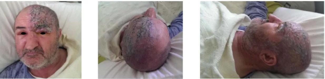
Figure 1. Diffuse periorbital edema of left eye, vesicular lesions and crusting on left periorbital area and forehead
66 year old male patient was admitted to emergency department with the complaints of edema over and around the left eye, fluid filled blisters and crust formation on the left forehead. He also had fever (37.1°C), pain in this area and malaise. From his
history, his complaints had started 4 days ago and increased progressively. His vital signs were as follows: TA: 120/80 mmHg, PR: 94/min, RR: 18 /min. Physical and biomicroscope examination revealed diffuse periorbital edema of left eye,
Baykan et al. Cukurova Medical Journal
1061
vesicular lesions and crusting on left periorbital area and forehead, hyperemia and chemosis of conjunctiva (Figure1).
His biochemistry and complete blood count examinations were normal. After consultation by ophthalmology and dermatology clinics, he was hospitalized and treated in dermatology clinics with the diagnosis of HZO. He was given 7 day treatment of intravenous Acyclovir (Zovirax®) and with the regression of lesions and sypmtoms. He was prescribed Acyclovir pomade and Lomefloxacin eye drop. After 1 month follow up, complete recovery was observed.
Ophthalmoplegia associated with HZO is not uncommon. The majority were due to involvement of the ocular motor cranial nerves, had onset of symptoms after appearance of the skin lesions2. Our
patient’s physical and biomicroscope examination revealed diffuse periorbital edema of left eye, vesicular lesions and crusting on left periorbital area and forehead, hyperemia and chemosis of conjunctiva. Cranial nerve palsies involving the third (most common), fourth, and sixth nerves may occur rarely. A majority of the cases will have spontaneous resolution within six months. In the pathogenesis directly or by hematogenous dissemination is suggested. The direct viral spread is thought to be able to pass through the perivascular plexus of the ophthalmic branch of the fifth cranial nerve of the virus, which is latent in the Gasser ganglion, to affect the ipsilateral middle cerebral arteries4. In
most people, the virus remains dormant forever and
never causes problems. In some people, however, the virus reactivates, or flares up. Most patients with HZO have a single attack and do not go on to get further attacks3.
Antiviral therapy provides rapid improvement in patients. Additionally, acyclovir administered within 72 hours of onset has been found to speed resolution of skin lesion4. Our patient’s history, his
complaints had started 4 days ago and increased progressively. A majority of the cases will have spontaneous resolution within six months5. HZO
should be kept in mind in the differential diagnosis of ophthalmic region lesions to treat on time and not to cause further complications.
REFERENCES
1. Incesu A. Orbital apeks sendromu: nadir bir oftalmik zoster şekli. Turkiye Klinikleri J Med Sci. 2012;32:218-21.
2. Kawasaki A, Borruat F. An unusual presentation of herpes zoster ophthalmicus: orbital myositis preceding vesicular eruption. Am J Ophthalmol. 2003;136:574-5.
3. Hall AJ. Herpes zoster ophthalmicus. Opthalmology. 2008;222-6.
4. Benbir G, Özekmekçi S, Ertan S, Albayram S. İnternal karotis arterde vaskülite yol açan oftalmik zona. Cerrahpaşa Tıp Dergisi. 2006;37:103-5.
5. Shaikh S, Ta CN. Evaluation and management of herpes zoster ophthalmicus. Am Fam Physician. 2002;66:1723-30.