1530
Scientific Abstracts
Methods: Patients with a diagnosis of BD who visited our outpatient clinic untilDecember 2019 were included in the study. Clinical data were recorded since diagnosis until the latest follow-up visit, analyzing clinical features, flares and therapeutic strategies adopted.
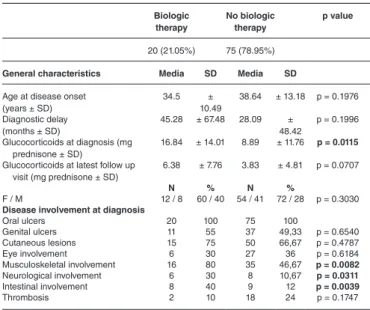
Results: A total of 95 patients were included in the study with a medium fol-low-up of 108.54 ± 169.59 months. 20 of them (21. 05%) were treated with biologic agents. Patients treated with biologic therapy compared to those on conventional non-biologic therapies had a higher proportion of musculoskeletal (80% vs 46.67%, p = 0.008), neurological (30% vs 10.67%, p = 0.031), intestinal involvement (40% vs 12%, p = 0.004), and they were treated with a higher dose of glucocorticoids at diagnosis (16.84 mg ±14.01 vs 8.89 mg ± 11.76, p = 0.012). The most frequent indications for biologic step-up therapy were musculoskele-tal involvement (40%), eye involvement (25%), neurological involvement (15%) and intestinal involvement (10%). Most patients initiated a biologic treatment within the first year of follow-up. TNF-inhibitor (TNFi) were more frequently pre-scribed (95%) and one patient was treated with 8 therapeutic cycles of Rituximab (500 mg/weekly for 4 infusions to be repeated after at least 6 months) because of recurrent pancytopenia. All patients experienced non-biologic therapy before starting a TNFi. The preferred first-line TNFi was infliximab (50%), followed by adalimumab (40%) and etanercept (5%). As second line treatment were also prescribed certolizumab (10%) and golimumab (5%). 10 patients switched to a second line treatment because of inefficacy of the first biologic agent, mainly because of refractory arthritis, intestinal and mucocutaneous involvement. One patient switched from infliximab to certolizumab during pregnancy with subse-quent worsening of arthritis.
85% of patients treated with biologic agents reached a clinical remission by the time of the latest follow up visit without any safety or tolerability issues. Conclusion: A relevant proportion of patients in our BD cohort were treated with biologic therapy, because of severe or refractory manifestations. The most frequent indications were musculoskeletal, neurological or intestinal involvement. Biologic agents were a generally effective and safe therapeutic approach. References:
[1] F. Alibaz-Oner, M. H. Sawalha, H. Direskeneli. Management of Behçet dis-ease, Curr. Opin. Rheumatol, 2018
Table 1. General characteristics and disease involvement at diagnosis Biologic therapy No biologic therapy p value 20 (21.05%) 75 (78.95%)
General characteristics Media SD Media SD
Age at disease onset (years ± SD) 34.5 ± 10.49 38.64 ± 13.18 p = 0.1976 Diagnostic delay (months ± SD) 45.28 ± 67.48 28.09 ± 48.42 p = 0.1996 Glucocorticoids at diagnosis (mg prednisone ± SD) 16.84 ± 14.01 8.89 ± 11.76 p = 0.0115
Glucocorticoids at latest follow up visit (mg prednisone ± SD)
6.38 ± 7.76 3.83 ± 4.81 p = 0.0707
N % N %
F / M 12 / 8 60 / 40 54 / 41 72 / 28 p = 0.3030
Disease involvement at diagnosis
Oral ulcers 20 100 75 100 Genital ulcers 11 55 37 49,33 p = 0.6540 Cutaneous lesions 15 75 50 66,67 p = 0.4787 Eye involvement 6 30 27 36 p = 0.6184 Musculoskeletal involvement 16 80 35 46,67 p = 0.0082 Neurological involvement 6 30 8 10,67 p = 0.0311 Intestinal involvement 8 40 9 12 p = 0.0039 Thrombosis 2 10 18 24 p = 0.1747
Disclosure of Interests: None declared DOI: 10.1136/annrheumdis-2020-eular.4582
AB0463 ETIOLOGY OF PALPABLE PURPURA; A SINGLE
CENTER EXPERIENCE
G. Kaynar1, O. C. İçaçan1, S. Çelik1, M. Yalçın Mutlu1, C. Bes1. 1University of Health Sciences, Bakırköy Dr. Sadi Konuk Training and Research Hospital, Department of Rheumatology, Istanbul, Turkey
Background: Purpura are lesions that occur after bleeding on the skin, mucous or serosal surfaces. Purpura can be classified into 2 subgroups; which are linked to thrombocytopenia and non-trombocytopenic purpura. While thrombocytopenic purpura often occurs due to a hematological disease; in non-trombocytopenic purpura etiological causes are very variable, and systemic vasculitis has an important place among them.
Objectives: The demographic features of the patients applying with purpura and the underlying causes and diseases were aimed to be revealed.
Methods: 44 consecutive patients (22 women, 22 men) who were admitted to the hospital in the last 6 months, due to purpura were evaluated. Average age of patients was 49.6 ± 19.6 years. Patients were questioned about a recent infec-tion, drug use, concomitant or underlying diseases. The serological tests and other laboratory tests for etiology were performed and biopsy was taken from the skin lesions which are appropriate.
Results: While 4 (9%) patients had widespread purpura on the trunk-upper-lower extremities, in 22 (50%) patients purpuric lesions were limited only to the lower limb. The period between the onset of the first symptom and the admission to the hospital was longer than 4 weeks in 35 (79.5%) patients, and shorter than 1 week in 9 (20.5%) patients. 24 (54%) patients had an anamnesis of infection 2-3 weeks before purpura, and 20 (45%) patients had an anamnesis of drug use. The most common accompanying symptom was abdominal pain and was present in 15 (34%) patients. Biopsy was performed from the skin lesion in 37 patients. Histopathological examination of all was compatible with leukocytoclas-tic vasculitis. In indirect immunofluorescence staining, 17 were found to be IgA positive. 2 (4.5%) patients were diagnosed PR3-ANCA positive granulomato-sis with polyangiitis. 1 patient had Hepatitis B virus infection was detected in 1 patient (2.2%), HIV infection was detected in 1 patient (2.2%) and malignancy was detected in 1 patient (2.2%).
Conclusion: In our study, the most common reason was found as IgA vasculitis in patients presenting with palpable purpura. Although vasculitic involvement was limited to the skin in most patients, organ-threatening systemic vasculitis was detected in a few patients. Patients applying with Purpura should be questioned for infection and drug use, should be examined for underlying diseases including systemic vasculitis, and closely monitored for organ involvement.
Disclosure of Interests: None declared DOI: 10.1136/annrheumdis-2020-eular.5471
AB0464 THE ROLE OF CYCLOPHOSPHAMIDE
CHEMOTHERAPY IN THE TREATMENT OF EOSINOPHILIC GRANULOMATOSIS WITH POLYANGIITIS
B. Bitik1, M. Aydın2, G. Sahin Dalgic3, D. Kaskari4, A. E. Yucel3. 1Baskent University, Ankara, Turkey; 2Baskent University, Adana, Turkey; 3Baskent University, Ankara, Turkey; 4Baskent University, Istanbul, Turkey
Background: Eosinophilic granulomatosis with polyangiitis (EGPA) is a rare type of ANCA associated Vasculitis (AAVs). Cyclophosphamide (CYC) is generally recommended for the induction of remission in life/organ threatening AAVs in combination with glucocorticoids. However, due to its rarity, randomized con-trolled trials regarding the efficacy of treatment modalities in EGPA are hard to perform. Therefore, the level of evidence for the use of CYC in the treatment of EGPA is lower when compared to other AAVs (1).
Objectives: The aim of this study is to investigate common therapeutic agents used for the treatment of patients with EGPA.
Methods: Medical records of patients who were followed-up with the diagnosis of EGPA between 2007-2020, in rheumatology clinics of Ankara and Adana Hos-pitals of Başkent University, were analyzed retrospectively. Treatment outcomes were assessed.
Results: Records of 11 patients (six females) were analyzed. The median age was 47 (19-77) years. The median follow-up time of the patients was 24 (9-156) months. Six patients were diagnosed with asthma. The median time between the diagnosis of asthma and EGPA was 4.5 (1-3) years. Five patients had tissue biopsies. Biopsy locations were terminal ileum, lung, myocardium and nerve. The most common forms of involvement were asthma, eosinophilic pneumonia and / or nodule, cardiovascular involvement, mononoritis multiplex, vasculitic skin rash, arthritis and bowel involvement, respectively. P-ANCA was positive in 8 patients. Three patients had myocarditis and cardiomyopathy, and two patients had isolated valve problems. The median BVAS value at the time of diagnosis and the third month of treatment was 17 (6-27) and 4 (2-7), respectively. Nine patients used oral 1mg/ kg methylprednisolone (MP) and 500mg CYC every two weeks as an induction therapy. The cumulative median CYC dose was 4.5 g (1.5-8). Neither of the patients developed CYC related side effects. MP was tapered to 2 mg in five patients, and was quited in two patients. Azathioprine (AZA) was used in remission treatment following CYC therapy. Rituximab (RTX) therapy 1 g twice, 2 weeks apart was initiated in two patients due to unrespon-siveness to CYC. While RTX was effective in one patient, newly developed renal involvement was detected after the third cycle of RTX therapy in other patient. Two patients had pregnancy plan therefore they used AZA plus MP as induction. A patient had mycophenolate mofetil plus MP due to AZA allergy. All patients are currently in remission except one patient.
Conclusion: In seven out of 11 EGPA patients, long-term remission was achieved with CYC treatment. CYC appears to be an effective and inexpensive method of first-line treatment for organ threatening EGPA.
FT Total. Protected by copyright.
on May 21, 2021 at Baskent Universitesi ANKOS Consortia
http://ard.bmj.com/
Scientific Abstracts
1531
References:[1] Yates M, Watts RA, Bajema IM, et al. EULAR/ERA-EDTA recommendations for the management of ANCA-associated vasculitis. Ann Rheum Dis. 2016 Sep;75(9):1583-94.
Disclosure of Interests: None declared DOI: 10.1136/annrheumdis-2020-eular.6481
AB0465 ANALYSIS OF VASCULITIS PATTERNS IN PATIENTS
WITH GIANT CELL ARTERITIS COMPARED TO PATIENTS WITH GIANT CELL ARTERITIS AND POLYMYALGIA RHEUMATICA
L. C. Burg1, P. Brossart2, C. Behning3, V. S. Schäfer1. 1Clinic for Internal Medicine III, University Hospital Bonn, Rheumatology, Bonn, Germany; 2Clinic for Internal Medicine III, University Hospital Bonn, Oncology, Haematology and Rheumatology, Bonn, Germany; 3University Hospital Bonn, Institute for Medical Biometry, Informatics and Epidemiology, Bonn, Germany
Background: Giant cell arteritis (GCA) and polymyalgia rheumatica (PMR) often coexist.1 The role of modern ultrasound in diagnosis of GCA as well as PMR is
well known.2 To date it is unknown, whether patients with GCA and PMR have
a different vasculitis pattern in ultrasound (US) examination than patients with GCA only.
Objectives: To prospectively identify differences in vasculitis patterns in con-secutive patients with newly diagnosed GCA and PMR compared to newly diag-nosed GCA patients without PMR.
Methods: US examination of the arteries typically affected in GCA, such as axil-lary arteries, vertebral arteries, superficial temporal arteries with both frontal and parietal branches and facial arteries was performed in patients with GCA and PMR (GCA-PMR-group) as well as in patients with GCA only (GCA-group) at time of first diagnosis. Arteries were defined as pathological, if measured inti-ma-media-thickness by US was above respective cut-off values.3
Results: The GCA-PMR-group consisted of 27 patients, the GCA-group of 18 patients. In the GCA-PMR-group, a total of 206 arteries were affected, while in the GCA-group 131 arteries were affected. Mean age and gender distribution was 74 years (SD± 9) with 10 (37%) females in the GCA-PMR-group and 76 years (SD± 9) with 10 (55%) females in the GCA-group. Median values of C-reactive
protein (CRP) were 57.2 (IQR 31.7-75.7) in the GCA-group and 48.3 (IQR 17.5- 79.9) in the GCA-PMR-group, no significance was observed (p= 0.3577). Mean number of affected arteries per patient was 7.63 and 7.28 in the GCA-PMR-group and GCA-group, respectively. Altogether, no significant difference in vascular pat-tern between the two groups was observed. Exact numbers, distribution and IMT-values for all measured arteries are depicted in table 1.
Conclusion: In our cohort, we did not observe a significant difference in vascular patterns between patients with GCA and PMR and GCA only patients. References:
[1]Salvarani C, Cantini F, Hunder GG. Polymyalgia rheumatica and giant-cell arteritis. The Lancet 2008;372:234–45.
[2]Dejaco C, Ramiro S, Duftner C, et al. EULAR recommendations for the use of imaging in large vessel vasculitis in clinical practice. Ann Rheum Dis 2018;77:636–43.
[3]Schäfer VS, Juche A, Ramiro S, Krause A, Schmidt WA. Ultrasound cut-off values for intima-media thickness of temporal, facial and axillary arteries in giant cell arteritis. Rheumatology (Oxford) 2017;56:1479–83.
Disclosure of Interests: None declared DOI: 10.1136/annrheumdis-2020-eular.5469
AB0466 EFFICACY AND SAFETY OF
INFLIXIMAB-BIOSIMILAR IN TAKAYASU ARTERITIS (TAKASIM): A MONOCENTRIC, OBSERVATIONAL, PROSPECTIVE, OPEN-LABEL STUDY
C. Campochiaro1, A. Tomelleri1, S. Sartorelli1, G. De Luca1, C. Sembenini1,
G. Cavalli1, P. Mapelli2, M. Picchio2, M. Papa3, E. Baldissera1, L. Dagna1. 1San Raffaele Scientific Institute - Vita-Salute San Raffaele, Unit of Immunology, Rheumatology, Allergy and Rare Diseases (UnIRAR), Milan, Italy; 2San Raffaele Scientific Institute - Vita-Salute San Raffaele, Unit of Nuclear Medicine, Milan, Italy; 3San Raffaele Scientific Institute - Vita-Salute San Raffaele, Unit of Radiology, Milan, Italy
Background: Treatment of Takayasu arteritis (TA) is mainly based on steroids, but, in approximately 50% of patients, disease-modifying antirheumatic drugs(D-MARDs) are required.
Objectives: To evaluate efficacy and safety of IFX-biosimilar in TA patients. Methods: Both bDMARD-naïve and IFX-O treated patients were eligible. Pri-mary endpoint was the number of patients with active disease as assessed by magnetic resonance angiography(MRA), 18FDG PET/CT, ITAS2010 and ITAS-ESR/CRP at month 6. Secondary endpoints were safety and tolerability, number of patients with active disease at month 12, quality of life. Non-parametric statis-tic tests were used.
Results: Twenty-three patients(21 female) were recruited. At baseline, mean age was 43.8±14.4 years and mean disease duration 95.5±61.3months. Two patients were IFX-O-naïve. Mean time on IFX-O was 51.5±37.9 months. Four patients had been previously treated with other biologics(tocilizumab, 3; adalimumab, 1). Twenty-one patients(91.3%) were on concomitant ster-oids(mean dose, 4.8±2.0 mg daily) and 82% on concomitant csDMARDs, kept unchanged throughout the study. At baseline, 4 patients(17%) were clas-sified as active according to ITAS2010, ITAS-ESR, and ITAS-CRP; mean HAQ was 3.48±5.26. Over the study period two patients dropped out the study because of poor disease control (1 at month 3 and 1 at month 6). PET/CT was not available for one patient who was on lactation during the study period and 1 patient refused to undergo imaging re-evaluation. At month 6, MRA was available for 21 patients: it was stable in 11(52%), improved in 5(24%), worsened in 5(24%). PET/CT was available for 20 patients: it was negative in 12(65%), improved although still positive in 3(16%), stable in 1(5%), wors-ened in 3(16%). At month 6, among 22 patients, 4(18%) were clinically active according to ITAS2010, ITAS-ESR and ITAS-CRP; mean steroid dose was significantly lower compared to baseline(4.2±2.0 mg daily, p=0.009); HAQ didn’t significantly change(mean, 3.35±6.59, p=0.357). At month 12, MRA was available for 20 patients. It was stable in 9 patients(45%), improved in 8 (40%), worsened in 3(15%). PET/CT was available for 19 patients: it was neg-ative in 14(74%), improved although still positive in 2(10%), stable in 3(10%), worsened in 1(5%). At month 12, 3 patients(14%) were active according to ITAS2010 and 2(9%) according to ITAS-ESR and ITAS-CRP; mean ster-oid dose was significantly lower compared to baseline(3.4±2.56 mg daily, p=0.034); HAQ didn’t significantly change(mean, 3.84±6.34, p=0.919). Nine patients(39%) experienced low-grade side effects related to TNFα-blockade (6, herpes reactivation; 3, urinary tract infection; 1 gastroenteritis). No IFX-B therapy modification was required.
Conclusion: Our study suggests that IFX-B is effective and safe both in IFX-O switch and IFX-O naïve TA patients.
Disclosure of Interests: : Corrado Campochiaro Speakers bureau: Novar-tis, Pfizer, Roche, GSK, SOBI, Alessandro Tomelleri: None declared, Silvia Sartorelli: None declared, Giacomo De Luca Speakers bureau: SOBI, Novar-tis, Celgene, Pfizer, MSD, Camilla Sembenini: None declared, Giulio Cavalli Speakers bureau: SOBI, Novartis, Pfizer, Paola Mapelli: None declared, Maria Picchio: None declared, Maurizio Papa: None declared, Elena Baldissera Speakers bureau: Novartis, Pfizer, Roche, Alpha Sigma, Sanofi, Lorenzo Dagna Grant/research support from: Abbvie, BMS, Celgene, Janssen, MSD, Mundipharma Pharmaceuticals, Novartis, Pfizer, Roche, SG, SOBI, Consult-ant of: Abbvie, Amgen, Biogen, BMS, Celltrion, Novartis, Pfizer, Roche, SG, and SOBI
DOI: 10.1136/annrheumdis-2020-eular.3250
AB0467 REMISSION AND LOW DISEASE ACTIVITY STATE
IN PATIENTS WITH GRANULOMATOSIS WITH POLYANGIITIS AND MICROSCOPIC POLYANGIITIS: PREVALENCE AND IMPACT ON DAMAGE ACCRUAL P. Delvino1, F. Sardanelli2, S. Monti1,3, P. Cohen4, X. Puéchal4, C. Montecucco1,
L. Mouthon4,5, L. Guillevin4,5, B. Terrier4,5on behalf of French Vasculitis Study
Group (FVSG). 1IRCCS Policlinico S. Matteo Foundation, University of Pavia,
Table. Prevalence of vasculitic affection of respective arteries in patients with giant cell arteritis and polymyalgia rheumatica and patients with giant cell arteritis only.
Affected artery Group
PMR-GCA-group (n=27)
GCA-group (n=18)
Unilateral Bilateral None Unilateral Bilateral none
Axillary artery 9 (33%) 12 (45%) 6 (22%) 5 (28%) 7 (39%) 6 (33%) Common superficial temporal artery 3 (11%) 21 (78%) 3 (11%) 5 (28%) 13 (72%) 0 (0%) Frontal branch 6 (22%) 17 (63%) 4 (15%) 3 (17%) 11 (61%) 4 (22%) Parietal branch 5 (18%) 21 (78%) 1 (4%) 3 (17%) 13 (72%) 2 (11%) Facial artery 7 (26%) 17 (63%) 3 (11%) 4 (22%) 11 (61%) 3 (17%) PMR-GCA-group: patients with diagnosis of giant cell arteritis and consecutive polymyalgia rheumatica
GCA-group: patients with diagnosis of giant cell arteritis only