A single-center experience of post-transplant
lymphoproliferative disorder (PTLD) cases after pediatric
liver transplantation: Incidence, outcomes, and association
with food allergy
Zeren Barış1 , Figen Özçay1 , Özlem Yılmaz Özbek2 , Nihan Haberal3 , Faik Sarıalioğlu4 , Mehmet Haberal5 1Department of Pediatric Gastroenterology, Başkent University Hospital, Ankara, Turkey
2Department of Pediatric Allergy, Başkent University Hospital, Ankara, Turkey 3Department of Pathology, Başkent University Hospital, Ankara, Turkey 4Department of Pediatric Oncology, Başkent University Hospital, Ankara, Turkey
5Department of General Surgery and Transplant Surgery, Başkent University Hospital, Ankara, Turkey
ABSTRACT
Background/Aims: We evaluated our 16-year single-center experience of pediatric post-transplant lymphoproliferative disorder (PTLD) cases who underwent liver transplantation between 2001 and 2017.
Materials and Methods: Of the 236 pediatric patients who underwent liver transplantation between 2001 and 2017, the clinical and laboratory data of eight patients diagnosed with PTLD were reviewed. The pre-transplant Epstein-Barr virus (EBV) status of 172 patients was also recorded.
Results: The total incidence of PTLD was 3.4%. The incidence of PTLD was 10% in pre-transplant EBV immunoglobulin G (IgG)-sero-negative patients and 0.8% in pre-transplant EBV IgG-seropositive patients. The mean age of the patients at liver transplantation was 2.71±3.21 years, and four patients were aged below 1 year at the time of transplantation. PTLD was diagnosed at 21.81±18.1 months after transplantation. The primary site of involvement was variable among patients: peripheral and mediastinal lymph nodes, stomach and intestine, transplanted graft, bone marrow, and nasopharynx. The eosinophil count varied greatly among patients, with a mean value of 524.62±679/mm3. Three patients had a food allergy and were administered an elimination diet at the time of PTLD diagnosis. Six patients had PTLD of B-cell origin. One patient died due to neutropenic sepsis during chemotherapy, whereas seven patients were followed up in full remission for 7.75±4 years.
Conclusion: PTLD is a life-threatening complication of solid-organ transplantation with a heterogeneous clinical spectrum. Food allergy had a close association with PTLD. A close follow-up of patients with risk factors and an early diagnosis with appropriate treatment may lead to a better outcome.
Keywords: Post-transplant lymphoproliferative disease, liver transplantation, EBV, food allergy
INTRODUCTION
Post-transplant lymphoproliferative disease (PTLD) is a sig-nificant complication of pediatric solid-organ transplanta-tion, with a reported incidence of 6%-20% and a mortality rate of 12%-60% (1-3). The development of PTLD is asso-ciated with discordance between immunosuppression and immune reaction. The risk of graft rejection has decreased with the use of potent immunosuppressants; however, the impairment of T-cell functions can increase the risk for PTLD (4). Most pediatric PTLD cases are associated with
Epstein-Barr virus (EBV) infection, and EBV-related PTLD tends to occur in the first 1 to 2 years after transplantation (5,6). The disease has a wide clinical spectrum changing from polyclonal PTLD to lymphoma (5). In this study, we present our 16-year single-center experience of pediatric PTLD cases after liver transplantation.
MATERIALS AND METHODS
Of the 236 pediatric patients who underwent liver trans-plantation between 2001 and 2017, the clinical and lab-Cite this article as: Barış Z, Özçay F, Yılmaz Özbek Ö, Haberal N, Sarıalioğlu F, Haberal M. A single-center experience of post-trans-plant lymphoproliferative disorder (PTLD) cases after pediatric liver transpost-trans-plantation: Incidence, outcomes, and association with food allergy. Turk J Gastroenterol 2018; 29: 354-60.
ORCID IDs of the authors: Z.B. 0000-0002-4976-9924; F.Ö. 0000-0002-5214-516X; Ö.Y.Ö. 0000-0002-7712-6504; N.H. 0000-0001-9852-9911; F.S. 0000-0001-5705-5078; M.H. 0000-0002-4976-9924.
Corresponding Author: Zeren Barış; [email protected] Received: November 9, 2017 Accepted: January 11, 2018
© Copyright 2018 by The Turkish Society of Gastroenterology • Available online at turkjgastroenterol.org DOI: 10.5152/tjg.2018.17731
oratory data of eight patients diagnosed with PTLD were reviewed. The patient age, gender, primary diagnosis before transplantation, age at transplantation, donor type, and time of PTLD emergence after transplantation were recorded. PTLD was diagnosed when there was a histopathologi-cal confirmation of the lymph node or the involved organ. PTLD was histopathologically classified according to the World Health Organization classification into four groups: early type, polymorphic, monomorphic PTLD, and classical Hodgkin lymphoma. The B- or T-cell origin of the lesions was noted. The EBV association was defined according to EBV-encoded RNA (EBER) in situ hybridization in tissue samples. The EBV viral loads, which were measured by real-time polymerase chain reaction (PCR) of EBV DNA from peripheral blood, were also recorded. The pre-trans-plant EBV serology was available for 172 of 236 patients. Pediatric patients with negative EBV IgM and IgG levels were recorded as EBV-naive. Patients were clinically clas-sified as early and late PTLD and as early PTLD occurring within 1 year after transplantation. The primary site of PTLD was also recorded.
Laboratory values at the time of PTLD diagnosis were in-vestigated: serum albumin, lactate dehydrogenase (LDH) and C-reactive protein (CRP) levels, and eosinophil count, and the immunosuppressive drug levels before PTLD di-agnosis were noted. Upper gastrointestinal endoscopy and colonoscopy were performed for patients with gas-trointestinal symptoms. Postoperative complications like rejection, infection, food allergy, or recurrent surgery after liver transplantation were evaluated. Patients diagnosed with a food allergy were recorded.
All patients, except one, were administered tacrolimus for immunosuppression. Our center’s protocol for im-munosuppression was as follows: 10 mg/kg prednisolone was administered in theater, then tapered to 1 mg/kg in 1 month, and gradually weaned off in 6 months. Myco-phenolate mofetil was administered to all patients at a dose of 20 mg/kg/day for 6 months. Tacrolimus was the main immunosuppressive agent, and our study protocol targeted trough levels of tacrolimus from 10 to 12 ng/mL in the immediate postoperative period, then 6 to 8 ng/mL at 1 month after liver transplantation, and 4 to 6 ng/mL at 1 year after liver transplantation. The treatment for PTLD and prognosis after PTLD were also recorded.
Informed consent was obtained from all patients and their parents; the study was approved by the Ethics Com-mittee of Başkent University.
Statistical analysis was performed using Statistical Pack-age for Social Sciences (SPSS) version 20 (IBM Corp.; Ar-monk, NY, USA).
RESULTS
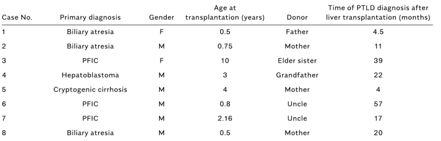
Of the 236 pediatric patients who underwent liver trans-plantation between 2001 and 2017, eight patients were diagnosed with PTLD, with an incidence of 3.4%, among which six were male. The mean age of the patients at liv-er transplantation was 2.71±3.21 years (min-max: 0.5-10 years), and four patients were aged below 1 year at the time of transplantation. All patients received a liver trans-plant from living related donors. Three patients were di-agnosed with biliary atresia, three patients had progres-sive familial intrahepatic cholestasis (PFIC), one patient had hepatoblastoma, and one patient had cryptogenic cirrhosis. PTLD was diagnosed at 21.81±18.1 months af-ter transplantation (min-max: 4-57 months). Since 2010, only two cases have been diagnosed with PTLD. The pri-mary diagnosis and demographic characteristics of the patients are shown in Table 1.
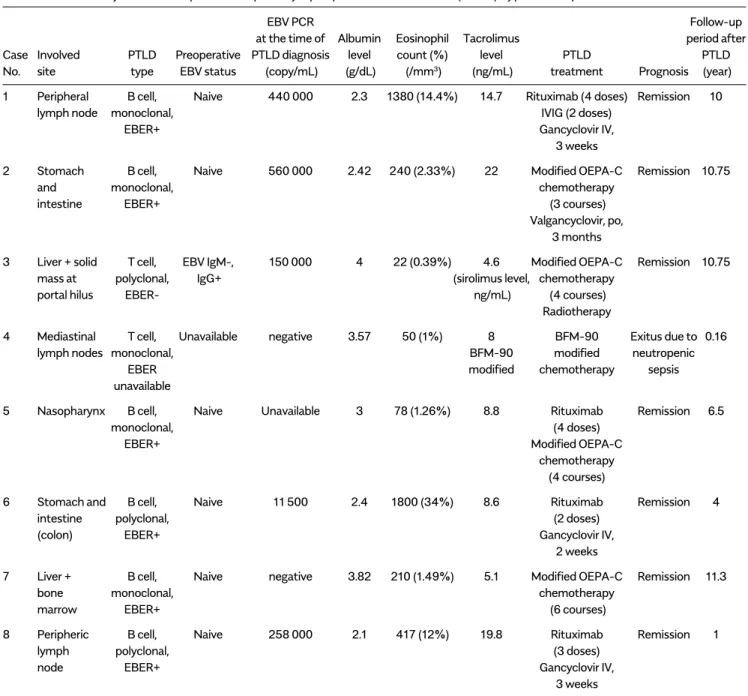
The presenting symptoms were cervical lymph node en-largement in three patients, abdominal pain and diarrhea in two patients, cough in one patient, and treatment-re-sistant nasal stuffiness in one patient. Two patients were diagnosed during routine abdominal ultrasonography screening. Some patients had multiple symptoms. The primary site of involvement was variable among patients: two patients had peripheral lymph node involvement; two patients had gastrointestinal PTLD; two patients had graft PTLD, one of whom also had bone marrow involve-ment; one patient had a nasopharyngeal tumor; and the remaining patient had a mediastinal mass.
The laboratory values of the patients at the time of presentation were as follows: The mean CRP level was 52.98±32.52 mg/dL (min-max: 0.8-86 mg/dL), and the mean albumin level was 2.95±0.75 g/dL (min-max: 2.1-4 g/dL). Patients with gastrointestinal and peripheral lymph node involvements had initial albumin levels <3 g/dL, whereas other patients had initial albumin levels >3 g/dL. The eosinophil count varied greatly among patients, with a mean value of 524.62±679/mm3 (min-max: 22-1800/ mm3), and the highest values occurred in two patients with peripheral lymph node involvement and one patient with gastrointestinal involvement. Three patients had a food allergy and were administered an elimination diet around the time of PTLD diagnosis. Patients were diag-nosed according to the positivity of specific IgEs in blood or skin prick test or eosinophilia in the gastrointestinal
epithelium and cessation of symptoms after receiving the elimination diet. Patient no.2 had a milk protein and wheat allergy; patient no.6 had an egg allergy; and patient no.8 had multiple food allergies including milk, egg, red meat, poultry, wheat, and lentil.
Tacrolimus levels vary among patients due to patient compliance and drug interactions. The highest tacrolim-us levels occurred in two patients who underwent trans-plantation in 2006 and one noncompliant patient who underwent transplantation in 2014. The mean trough level of tacrolimus in our patients was 12.42±6.48 ng/ mL (min-max: 5.1-22 ng/mL). One patient was on siroli-mus and mycophenolate mofetil treatment at the time of PTLD diagnosis, with a trough level of sirolimus as 4.6 ng/ mL. This patient had intractable epilepsy and was intoler-ant to calcineurin inhibitors.
The pre-transplant EBV status of 236 patients who un-derwent liver transplantation between 2001 and 2017 was evaluated; 172 patients had a pre-transplant EBV se-rology. Fifty-five patients (32%) were reported as EBV-naïve, whereas 117 (68%) patients were negative for EBV IgM and positive for EBV IgG. Seven patients with PTLD had a pre-transplant EBV serology. Six patients were EBV-naïve before liver transplantation and negative for EBV IgM and EBV IgG. One patient with PTLD occurring in the graft (liver) was negative for EBV IgM and positive for EBV IgG before transplantation. The incidence of PTLD among EBV-naive patients was 10% (6 of 55 patients), whereas the incidence of PTLD among patients who had positive pre-transplant EBV IgG was 0.8% (1 of 117 pa-tients) (p<0.001).
The EBV PCR status of seven patients at the time of PTLD diagnosis was evaluated. Two patients were EBV PCR-negative, whereas five patients were EBV PCR-pos-itive. The mean EBV PCR viral load values were 283, 900±219, 886 copy/mL (min-max: 11,500-560 000 copy/mL). The EBV PCR value was unavailable for one patient who was serologically EBV-naïve at the time of PTLD diagnosis. The laboratory values and PTLD type of our patients are given in Table 2.
Immunosuppressive drug doses were tapered in all pa-tients as a part of the PTLD treatment. Four papa-tients with B-cell PTLD were administered intravenous ganciclovir for 2 to 3 weeks or oral valganciclovir for 3 months. Six patients had lymphoproliferative disease of B-cell origin (B-cell PTLD), with a positive CD20 expression on lymphocytes. In all of these patients, the EBV viral load was high, and EBER was positive at histopathological ex-amination. Three patients with B-cell disease had been accepted in the polyclonal lymphoproliferation phase and treated with two to four doses of rituximab. The other three patients showed monoclonal proliferation and were in the lymphoma phase. In these patients, a modified OEPA-C chemotherapy regimen (prednisolone, etopo-side, vincristine, doxorubicin, and cyclophosphamide) for immunosuppressed patients was used in three to six courses. Patient no.3 and no. 4 had T-cell PTLD; one was in a polyclonal phase and the other was in a monoclonal phase. Modified BFM-90 (prednisone, vincristine, dauno-rubicin, l-asparaginase, cyclophosphamide, cytarabine,
6-mercaptopurine, and methotrexate) and modified OEPA-C regimens were administered to each patient
re-Age at Time of PTLD diagnosis after
Case No. Primary diagnosis Gender transplantation (years) Donor liver transplantation (months)
1 Biliary atresia F 0.5 Father 4.5
2 Biliary atresia M 0.75 Mother 11
3 PFIC F 10 Elder sister 39
4 Hepatoblastoma M 3 Grandfather 22
5 Cryptogenic cirrhosis M 4 Mother 4
6 PFIC M 0.8 Uncle 57
7 PFIC M 2.16 Uncle 17
8 Biliary atresia M 0.5 Mother 20
PFIC: progressive familial intrahepatic cholestasis
spectively. Conformal 3D radiotherapy was also admin-istered to the latter patient who partially responded to chemotherapy.
One patient with B-cell gastrointestinal PTLD devel-oped an inflammatory myofibroblastic tumor in the lung tissue during follow-up and underwent lung surgery.
The patient who underwent liver transplantation due to hepatoblastoma and had polyclonal T-cell disease in his mediastinum had died due to neutropenic sepsis during chemotherapy in the second month of the PTLD treat-ment. The other seven patients were followed up in full remission for 7.75±4 years (min-max: 1-11.3 years). There was no graft loss due to PTLD treatment. The PTLD type,
EBV PCR Follow-up
at the time of Albumin Eosinophil Tacrolimus period after
Case Involved PTLD Preoperative PTLD diagnosis level count (%) level PTLD PTLD
No. site type EBV status (copy/mL) (g/dL) (/mm3) (ng/mL) treatment Prognosis (year)
1 Peripheral B cell, Naive 440 000 2.3 1380 (14.4%) 14.7 Rituximab (4 doses) Remission 10
lymph node monoclonal, IVIG (2 doses)
EBER+ Gancyclovir IV,
3 weeks
2 Stomach B cell, Naive 560 000 2.42 240 (2.33%) 22 Modified OEPA-C Remission 10.75
and monoclonal, chemotherapy
intestine EBER+ (3 courses)
Valgancyclovir, po,
3 months
3 Liver + solid T cell, EBV IgM-, 150 000 4 22 (0.39%) 4.6 Modified OEPA-C Remission 10.75
mass at polyclonal, IgG+ (sirolimus level, chemotherapy
portal hilus EBER- ng/mL) (4 courses)
Radiotherapy
4 Mediastinal T cell, Unavailable negative 3.57 50 (1%) 8 BFM-90 Exitus due to 0.16
lymph nodes monoclonal, BFM-90 modified neutropenic
EBER modified chemotherapy sepsis
unavailable
5 Nasopharynx B cell, Naive Unavailable 3 78 (1.26%) 8.8 Rituximab Remission 6.5
monoclonal, (4 doses)
EBER+ Modified OEPA-C
chemotherapy
(4 courses)
6 Stomach and B cell, Naive 11 500 2.4 1800 (34%) 8.6 Rituximab Remission 4
intestine polyclonal, (2 doses)
(colon) EBER+ Gancyclovir IV,
2 weeks
7 Liver + B cell, Naive negative 3.82 210 (1.49%) 5.1 Modified OEPA-C Remission 11.3
bone monoclonal, chemotherapy
marrow EBER+ (6 courses)
8 Peripheric B cell, Naive 258 000 2.1 417 (12%) 19.8 Rituximab Remission 1
lymph polyclonal, (3 doses)
node EBER+ Gancyclovir IV,
3 weeks
EBV: Epstein-Barr virus; PTLD: post-transplant lymphoproliferative disease; EBV Naive: EBV IgM-, IgG-; EBER: EBV-encoded RNA; Modified OEPA-C: Prednis-olone, Etoposide, Vincristine, Doxorubicin, Cyclophosphamide; BFM-90 modified: Prednisone, Vincristine, Daunorubicin, L-Asparaginase, Cyclophosphamide, Cytarabine, 6-Mercaptopurine, Methotrexate; IV: intravenous; IVIG: intravenous immunoglobulin
treatment, and the prognosis of the patients are shown in Table 2.
Postoperative complications other than PTLD after liver transplantation in these seven patients were as follows: acute rejection (1), food allergy (3), hepatic artery and portal vein thrombosis with bile duct stenosis (1), tacro-limus-associated cardiac hypertrophy (1), and diaphrag-matic hernia (1).
DISCUSSION
In our study, the first pediatric PTLD case was experi-enced in 2005, with a total incidence of PTLD after liv-er transplantation as 3.4%, which is lowliv-er than the in-cidence of PTLD reported in the literature (1). Six of the eight patients with PTLD underwent transplantation be-fore 2010. We observed a further reduction in PTLD cases in subsequent years. A decrease in the incidence of PTLD in recent years was also reported by different transplant centers (1-3). A preference of lower trough levels of tac-rolimus in the recent years, full awareness of PTLD signs and symptoms, close monitoring of the EBV viral load, and adjustment of immunosuppressant blood levels ac-cordingly may lead to a decrease in the incidence of PTLD worldwide and also at our center (1,7).
In our PTLD case series, three patients had biliary atresia and three had PFIC, all of whom (except one) underwent liver transplantation at <2.5 years of age confirming that younger age at the time of solid-organ transplantation is a significant risk factor for PTLD. One of the reasons for young age being a risk factor is that most young pa-tients are EBV-naïve at the time of liver transplantation, and they usually receive a graft from an EBV seropositive donor (8). Six patients in our PTLD case series were EBV-naïve at the time of liver transplantation, and these pa-tients (except one) were aged below 2.5 years.
Older donor age increases the risk of PTLD (1). Because liver transplants and partial grafts from living donors were used in all of our patients, the donor age was older than that for cadaveric transplants, leading to a higher risk of EBV-positive donors for younger EBV-naive recipients. Approximately 70% to 80% of PTLD cases are EBV-relat-ed (4,5,8). PTLD relatEBV-relat-ed to EBV infection is most common within the first 2 years after liver transplantation, and non-EBV-related PTLD tends to occur later (6). Similarly, in our series, the incidence of PTLD was 10% in pre-trans-plant EBV-naïve patients and 0.8% in pre-transpre-trans-plant EBV seropositive patients. Additionally, most of our patients
were diagnosed with EBV-driven B-cell PTLD. PTLD of T-cell origin (T-cell PTLD) constitutes 10% of the cases in the literature (3,9). However, in our series, 25% of pa-tients had T-cell PTLD, which may be biased due to a low patient number.
Hypoalbuminemia and eosinophilia are important labo-ratory findings that may indicate PTLD (2). In our series particularly patients with lymph node and gastrointesti-nal involvement presented with these features.
Monitoring the EBV viral loads for early detection of PTLD is controversial (1,7,10,11). Some centers experience a decrease in the incidence of PTLD after monitoring the EBV viral load and reducing immunosuppression upon recognition of high-titer EBV of over 10,000 copies/mL (7). At our center, we did not perform routine EBV PCR screening in EBV-negative patients at the post-trans-plantation period. Instead of using this technique, we preferred observing patients at risk for PTLD such as recipient EBV-negative and donor EBV-positive cases, young infants, and patients who had clinical and labora-tory signs indicating the development of PTLD. We hardly attempted to avoid very high tacrolimus levels and also closely monitored the immunosuppressive drug levels in patients and families with drug adjustment and noncom-pliance problems.
Extranodal disease is commonly prevalent (75%-80%), but solid-organ grafts may also be involved in 15%-30% of PTLD cases (1,4). Consistent with the literature, our two patients had liver graft involvement at our center. Patients with organ involvement required more intense treatment compared with those with peripheral lymph node involvement. This may be related to early diagno-sis in nodal disease, as peripheral lymph nodes can be easily recognized with physical examination. Symptoms like chronic cough or nasal obstruction and difficulty in breathing necessitate prompt and early evaluation to exclude lymph node enlargement in the mediastinum or nasopharynx. Many studies have shown that early diag-nosis and early intervention with appropriate treatment leads to a better outcome (1,7,12). Similarly, Jeong et al. (7) have reported that rituximab therapy and reduction of immunosuppressive drug treatment were sufficient to induce remission in early recognized, localized, and poly-clonal benign lesions. Nevertheless, in our series, three monoclonal B-cell and two T-cell originated cases had re-ceived chemotherapy and experienced its complications. In fact, one patient was lost to follow-up due to death because of neutropenic sepsis during the chemotherapy
course. As a result, careful examination of patients, close follow-up of patients particularly with risk factors, and detection of eosinophilia or hypoalbuminemia and incon-sistent calcineurin drug levels may lead to early diagnosis of PTLD and enable us to take some precautions.
Giraldi et al. (9) applied a risk protocol in which FCD-R blocks (fludarabine, cyclophosphamide, doxorubicin, and rituximab) were used for polymorphic and non-Burkitt-like lymphomas, and a BFM-based program was used for Burkitt-like lymphomas; they also suggested to include rituximab as an initial therapy, decreasing immunosup-pression together with surgical resection in case of fully resectable masses in children. They also recommended discontinuation of immunosuppressive drugs together with rituximab and chemotherapy in high-risk patients. In this study, the disease-free survival was 94% and 75% in 1 and 5 years, respectively.
In our study, approximately 50% of patients with PTLD could be treated with immunosuppressive drug reduc-tion and rituximab therapy. In resistant cases, a low-dose cyclophosphamide with prednisolone and rituximab may result in high response rates. Radiotherapy and a more intensive chemotherapy were restricted to patients with refractory or recurrent cases. Although ganciclovir does not have any inhibitory effect in vivo, it has a theoretical advantage on EBV replication, and thus an oral or intra-venous ganciclovir/acyclovir was administered in more than half of our patients. Interestingly, three of eight PTLD cases (37.5%) had a food allergy. Food allergies are commonly observed in young patients, particularly those aged below 3 years after liver transplantation, possibly due to immature gastrointestinal and immune systems (13-15). Additionally, tacrolimus plays an important role in gastrointestinal immunotolerance by increasing the intestinal permeability and exposing the body to more allergens (16). The incidence of food allergy was 19.2% in pediatric patients who underwent liver transplantation at our center (unpublished data). Although the incidence of food allergy was higher in our patients who received liver transplants than in the general population, we ob-served a further increase in the frequency of food allergy in patients with EBV viremia in this study (17). We further observed that the incidence of food allergy was high as 37.5% in PTLD cases. This may be explained by several reasons. First, most of our patients with PTLD are aged below 3 years, which may lead to a relative increase in food allergy. Second, high tacrolimus levels might lead to the formation of both PTLD and disturbance in immuno-tolerance. Third, increased eosinophilia may further
sup-port the coexistence of immune dysregulation and food intolerance in patients with PTLD.
The limitations of our study are due to its retrospec-tive design and small patient cohort. Further large-scale multi-center studies are required for proper investigation of the association between PTLD, EBV, and food allergy. PTLD is a life-threatening complication of solid-organ transplantation with a heterogeneous clinical spectrum. Transplant clinicians should be aware of the subtle or overt symptoms of this disease and take precautions in early phases. A close follow-up of patients with risk fac-tors and an early diagnosis with appropriate treatment may lead to a good outcome. In summary, we emphasized the strong association between PTLD and food allergies after liver transplantation.
Ethics Committee Approval: Ethics committee approval was
received for this study from the Ethics Committee of Başkent University.
Informed Consent: Written informed consent was obtained
from all patients and their parents who participated in this study.
Peer-review: Externally peer-reviewed.
Author Contributions: Concept – Z.B., F.Ö.; Design - Z.B., F.Ö.;
Supervision - Z.B., F.Ö., Ö.Y.Ö., N.H., F.S., M.H.; Materials - Z.B., F.Ö., Ö.Y.Ö.; Data Collection and/or Processing - Z.B., F.Ö., Ö.Y.Ö.; Analysis and/or Interpretation - Z.B., F.Ö., Ö.Y.Ö., N.H., F.S., M.H.; Literature Search - Z.B., F.Ö.; Writing Manuscript - Z.B., F.Ö.; Crit-ical Review - Z.B., F.Ö., Ö.Y.Ö., N.H., F.S., M.H.
Conflict of Interest: The authors have no conflict of interest to
declare.
Financial Disclosure: The authors declared that this study has
received no financial support.
REFERENCES
1. Narkewicz MR, Green M, Dunn S, et al. Decreasing incidence of symptomatic Epstein-Barr virus disease and posttransplant lymph-oproliferative disorder in pediatric liver transplant recipients: report of the studies of pediatric liver transplantation experience. Liver Transpl 2013; 19: 730-40. [CrossRef]
2. Evens AM, Roy R, Sterrenberg D, et al. Post-transplantation lymphoproliferative disorders: diagnosis, prognosis, and current ap-proaches to therapy. Curr Oncol Rep 2010; 12: 383-94. [CrossRef] 3. Wistinghausen B, Gross TG, Bollard C. Post-transplant lymphopro-liferative disease in pediatric solid organ transplant recipients. Pedi-atr Hematol Oncol 2013; 30: 520-31. [CrossRef]
4. Özçay F. Lymphoproliferative Disease After Pediatric Liver Trans-plant. Exp Clin Transplant 2017; 15: 79-81.
5. Tsao L, Hsi ED. The clinicopathologic spectrum of posttransplan-tation lymphoproliferative disorders. Arch Pathol Lab Med 2007; 131: 1209-18.
6. Kremers WK, Devarbhavi HC, Wiesner RH, et al. Post-transplant lymphoproliferative disorders following liver transplantation: inci-dence, risk factors and survival. Am J Transplant 2006; 6: 1017-24. [CrossRef]
7. Jeong HJ, Ahn YH, Park E, et al. Posttransplantation lymphopro-liferative disorder after pediatric solid organ transplantation: ex-periences of 20 years in a single center. Korean J Pediatr 2017; 60: 86-93. [CrossRef]
8. Morscio J, Tousseyn T. Recent insights in the pathogenesis of post-transplantation lymphoproliferative disorders. World J Trans-plant 2016; 6: 505-16. [CrossRef]
9. Giraldi E, Provenzi M, Conter V, et al. Risk-adapted Treatment for Severe B-Lineage Posttransplant Lymphoproliferative Disease After Solid Organ Transplantation in Children. Transplantation 2016; 100: 437-45. [CrossRef]
10. Soriano-López DP, Alcántar-Fierros JM, Hernández-Plata JA, et al. A scheduled program of molecular screening for epstein-barr virus decreases the incidence of post-transplantation lymphopro-liferative disease in pediatric liver transplantation. Transplant Proc 2016; 48: 654-7. [CrossRef]
11. Jang JY, Kim KM, Lee YJ, Lee SG, Chi HS. Quantitative Epstein- Barr virus viral load monitoring in pediatric liver transplantation. Transplant Proc 2008; 40: 2546-8. [CrossRef]
12. Végso G, Hajdu M, Sebestyén A. Lymphoproliferative disorders after solid organ transplantation-classification, incidence, risk fac-tors, early detection and treatment options. Pathol Oncol Res 2011; 17: 443-54. [CrossRef]
13. Needham JM, Nicholas SK, Davis CM. Food allergies developing after solid organ transplant. Pediatr Transplant 2015; 19: 827-35. [CrossRef]
14. Shroff P, Mehta Rs, Chınen J, Karpen Sj, Davıs Cm. Presentation of atopic disease in a large cohort of pediatric liver transplant recip-ients. Pediatr Transplant 2012; 16: 379-84. [CrossRef]
15. Parashette Kr, Zeytinoğlu M, Kernek K, Molleston Jp, Subbarao G. Clinical, endoscopic, and histologic features of eosinophilic inflam-mation of the gastrointestinal tract in pediatric liver transplant pa-tients. Pediatr Transplant 2013; 17: 737-43. [CrossRef]
16. Gabe SM, Bjarnason I, Tolou-Ghamarı Z, et al. The effect of tacro-limus (FK506) on intestinal barrier function and cellular energy pro-duction in humans. Gastroenterology 1998; 115: 67-74. [CrossRef] 17. Ozbek OY, Ozcay F, Avcı Z, Haberal A, Haberal M. Food allergy after liver transplantation in children: A prospective study. Pediatr Allergy Immunol 2009; 20: 741-7. [CrossRef]