975
Araştırma Makalesi / Research Article
Fluorescent Copper Phosphate Nanoflowers: A Novel Toxicity Investigation
Study Based On Tenebrio molitor
Linnaeus, 1758 (Coleoptera:
Tenebrionidae) Larvae
Ata ESKİN
1, Cevahir ALTINKAYNAK
1*, Merve TÜRK
2, Nalan ÖZDEMİR
21Nevsehir Haci Bektas Veli University, Avanos Vocational School, Department of Plant and Animal Production, 50500, Nevsehir, Turkey
2Erciyes University, Faculty of Science, Department of Chemistry, 38039, Kayseri, Turkey (ORCID: 0000-0002-7953-654X) (ORCID0000-0003-0082-8521)
(ORCID:0000-0003-0833-3224) (ORCID: 0000-0002-8930-5198)
Abstract
Organic-inorganic hybrid nanoflowers with commercialization potential are the novel materials and are widely used in many experimental applications, recently. However their potential toxicity values of these nanoflowers are still unknown. In this study, fluorescent copper phosphate nanoflowers (FCPnfs) were first synthesized to constructed via organic-inorganic hybrid nanoflower synthesis method. The morphological features of FCPnfs (acridine orange amount, shape, size) were analyzed. The results showed that different amount of acridine orange caused different morphology and the organic component were homogeneously distributed inside the nanoflowers. These materials were characterized by SEM, EDX, XRD, and FTIR techniques. The toxic effects of the as-prepared FCPnfs were investigated in Tenebrio molitor Linnaeus, 1758 (Coleoptera: Tenebrionidae) larvae compared to pure copper phosphate nanoflowers (CPnfs). Nanoflowers were force feed to larvae at the different doses of 0.001, 0.005, 0.010, 0.025, 0.050, 0.100 mg/10 μl. According to probit assay, LC50 and LC99 values of
FCPnfs were found 0.490 and 0.145 mg/10 μl, respectively. On the other hand, LC50 and LC99 values of CPnfs
were detected 0.066 and 0.172 mg/10 μl, respectively. It was found that the insect exhibited slightly more resistance to CPnfs than FCPnfs when compared to both chemical toxic values. These findings will be offer a new insight into the researchers about knowing the toxic effect of the skeletal structure of new materials to be synthesized using the organic-inorganic hybrid nanoflower method.
Keywords: Copper phosphate, nanoflowers, toxic effects.
Tenebrio molitor Linnaeus, 1758 (Coleoptera: Tenebrionidae) Larvasında
Gerçekleştirilen Yeni Bir Toksik Araştırma: Floresan Bakır Fosfat Nano
Çiçekler
ÖzSon zamanlarda birçok deneysel uygulamada yaygın olarak kullanılan organik-inorganik hibrit nano çiçekler ticarileşme potansiyeline sahip yeni malzemelerdir. Ancak bu nano çiçeklerin toksik etkileri hala bilinmemektedir. Bu çalışmada, ilk defa organik-inorganik hibrit nano çiçek sentez metodu kullanılarak floresan bakır fosfat nano çiçekler (FCPnfs) sentezlendi, morfolojik özellikleri (akridin orange miktarı, şekli, boyutu) analiz edildi. Sonuçlar, farklı miktarda akridin orange’ın farklı morfolojiye neden olduğu ve organik bileşenin nano çiçeklerin içinde homojen bir şekilde dağıldığını gösterdi. Sentezlenen malzemelerin karakterizasyonları SEM, EDX, XRD ve FTIR teknikleri ile gerçekleştirildi. Model organizma olarak seçilen Tenebrio molitor Linnaeus, 1758 (Coleoptera: Tenebrionidae) larvalarında sentezlenen FCPnfs'lerin toksik etkileri, saf bakır fosfat nano çiçekleriyle (CPnfs) karşılaştırmalı olarak incelendi. Larvalara nano çiçekler, 0.001, 0.005, 0.010, 0.025, 0.050, 0.100 mg/10µl olacak şekilde farklı dozlarda zorla besleme yapıldı. Yapılan probit analizine göre, FCPnfs'in LC50 değeri 0.490 mg/10 µl
iken LC99 değeri 0.145 mg/10 µl bulundu. Öte yandan, CPnfs'nin LC50 değeri 0.066 mg/10 µl iken LC99 değeri
0.172 mg/10 µl bulundu. Elde edilen toksik veriler değerlendirildiğinde; Tenebrio molitor larvalarının CPnfs'ye
*Sorumlu yazar: [email protected]
976
FCPnfs'den biraz daha fazla direnç gösterdiği bulundu. Bu bulgular, organik-inorganik hibrit nano çiçek yöntemi kullanılarak sentezlenecek yeni malzemelerin iskelet yapısının toksik etkisinin bilinmesi hakkında araştırmacılara yön gösterecektir.
Anahtar kelimeler: Bakır fosfat, nano çiçekler, toksik etki. 1. Introduction
Organic-inorganic hybrid nanoflowers have received considerable attention with their high potential use in catalysis, adsorption, separation, sensing, drug delivery, energy storage, chemical and biological areas over the last six years [1-5]. The hybrid nanoflowers are synthesized via self-assembly method which included an organic and inorganic components [6-8]. In order to synthesize hybrid nanoflowers, proteins, enzymes, DNA, plant extracts and some organic molecules have been used as the organic components, while the copper (II) ion has commonly used as the inorganic component. [9-12]. Different metal ions such as Co2+[13], Mn2+ [14], Zn2+[15], and Ca2+[16] can also be used for the synthesis of the hybrid nanoflowers. The technique that had been mentioned previously included three following steps: (1) nucleation, (2) growth and (3) completion [1, 6, 10]. The applications of hybrid nanoflowers are increasing broadly in every field, and they are being tested many novel industrial applications for the benefit of mankind at laboratory conditions [4, 10, 14, 17]. Up to date; Ildiz et al. successfully synthesized copper hybrid nanoflowers containing the extract of Viburnum opulus Linnaeus, 1758 (Dispacales: Caprifoliaceae) and reported peroxidase-like activity [11]. Then they proved that their effective antimicrobial activity against bacterial (Escherichia coli (Migula, 1895) (Enterobacteriales: Enterobacteriaceae), Salmonella typhi (Schroeter, 1886) (Enterobacteriales: Enterobacteriaceae), Enterococcus faecium (Orla-Jensen, 1919), E. faecalis (Andrewes and Horder, 1906) (Lactobacillales: Enterococcaceae), Bacillus cereus Frankland and Frankland, 1887 (Bacillales: Bacillaceae), Staphylococcus aureus Rosenbach, 1884 (Bacillales: Staphylococcaceae), except Pseudomonas aeruginosa (Schroeter, 1872) (Pseudomonadales: Pseudomonadaceae) and Hemofilus influenza (Lehmann and Neumann, 1896) (Pasteurellales: Pasteurellaceae) and fungal pathogens (Candida albicans (Robin) Berkhout (1923), and C. glabrata (Anderson, 1917) (Saccharomycetales: Saccharomycetaceae). The good antimicrobial materials should have either little or no toxic effect on environment.
Nanomaterials have potent risks to the environment with their concentration, morphology, migration, and transformation processes [18]. The smaller nanoparticles having large surface area are more toxic than larger particles [19]. In addition, the toxicity of nanomaterials might be stemmed from the rigid particle frameworks [20], including metal ions [21] or organic component type. Previous studies have reported that copper and copper nanoparticles damaged the liver and kidney of Cyprinus carpio Linnaeus, 1758 (Cyrpniformes: Cyprinidae) [22] and proved that copper sulfate caused more severe damages compared to copper nanoparticle. Another study indicated that the toxicity of CuO nanoparticles was based on the dissolved Cu2+ [20].
When the organic-inorganic hybrid nanoflowers are used as an industrial material, inevitably, it will be released into the environment like other nanoparticles [23, 24]. However, the harmfull effects of organic-inorganic hybrid nanoflowers and rigid framework's (pure copper phosphate nanoflowers) on living organisms are still unknown. Therefore; the major source of nanoparticle toxicity should be investigated due to the possibility of their industrial limitation.
Tenebrio molitor Linnaeus, 1758(Coleoptera: Tenebrionidae) is a globally harmful insect that
causing a high loss commodity in stored grains [25] and is an excellent model organism in toxicology researches because of similarities between nervous system of humans and insects [25]. On the other hand, T. molitor can be easily cultured in the laboratory with easily supplied materials such as flour, wheat bran, carrot and potato. Nanomaterials can be given into the insects via force-feeding [26, 27]. In the present study; T. molitor was used as a model organism for the reasons stated above. To the best of our knowledge, there has been no report on the formation of fluorescent copper phosphate hybrid nanoflowers including acridine orange (FCPnfs) which is both a fluorescent and toxic substance [28]. The aim of this study were to: (a) observe the distribution of acridine orange molecules to overall the organic-inorganic hybrid nanoflowers, and (b) also evaluate the toxic part and levels of the nanoflowers using T. molitor larvae as a model organism.
977 2. Materials and Methods
This studies are conducted at Science Technology Application and Research Center Laboratory and Plant and Animal Production Department Laboratory of Nevsehir Haci Bektas Veli University, Avanos Vocational School in 2018.
2.1. Chemicals and Materials
Acridine Orange (AO), copper sulfate pentahydrate (CuSO4.5H2O), and other chemicals were supplied
from Sigma-Aldrich f,irm. For the preparationf of phosphate buffer solution, Na2HPO4, NaOH, NaCl,
KCl, KH2PO4, HCl were supplied and buffer soliton was prepared with using ultrapure water (18.2 MΩ;
Millipore Co., USA) and these chemicals. The following materials were used in studies with insects; bath type sonicator, wheat bran plastic boxes (20 mL) (51 mm x 34 mm x 19 mm), micro-fine insulin syringe (29 gauges), ethyl alcohol (70%), purified water, stereo microscope and cotton.
2.2. Preparation of Fluorescent Copper Phosphate Nanoflowers
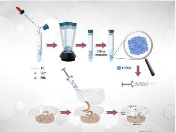
Fluorescent copper phosphate nanoflowers (FCPnfs) were synthesized according to the previous works with some modifications [1-7,9] At first, 120 mM copper sulfate pentahydrate solution was prepared. 333 µL of this solution (0.8 mM) was added to 50 mL of phosphate buffered saline (PBS) solution (10 mM, pH 7.4) containing different amounts of acridin orange (AO) (50 µL, 100 µL, 250 µL and 500 µL) in four different test tubes. Then, the mixture was vigorously vortexed for 30 sec. After incubation in the dark at +4°C for 3 days (Figure 1), the nanostructures were centrifuged 6.000 rpm for 15 min to obtain precipitates and washed with water for three times to remove unbounded molecules. The collected precipitates were dried 50°C under vacuum for using other analyses. For pure copper phosphate nanoflowers (CPnfs), copper (0.8 mM) were dissolved in PBS without any organic component. And similar process was followed.
Figure 1. Illustration of the experimental process for fluorescent copper phosphate nanoflowers and the
978
2.3. Characterization of Fluorescent Copper Phosphate Nanoflowers (FCPnfs)
The morphologies of the prepared fluorescent copper phosphate nanoflowers (FCPnfs) were inquired using field emission scanning electron microscope (SEM) (ZEISS). For this reason, dry FCPnfs were deposited on stub, after sputter coating device was coated with gold source. The energy-dispersive X-ray technique (EDX) performed for determining weight and atomic percentage of Cu element in FCPnfs. The crystal and chemical structure of FCPnfs were determined using the X-ray diffraction analysis (XRD) (BRUKER AXS D8) and the Fourier-Transform Infrared Spectroscopy (FTIR) (Perkin Elmer Spectrum 400), respectively. The phase contrast and fluorescent microscopy images were recorded on CNOEC, OPTO-EDU microscope.
2.4. Insects
The larvae of T. molitor (n=20) were maintained (and reared under 26 ± 2°C, 60 ± 5% relative humidity and dark conditions in our laboratory at Nevsehir Haci Bektas Veli University, Avanos Vocational School, Nevşehir, Turkey.
2.5. Experiments to Determine the Values of Lethal Concentration 50 (Lc50) of FCPnfs and CPnfs
on T. molitor Larvae
The toxicity studies were based on protocols from two different studies [25, 29]). In this context, FCPnfs and CPnfs were dissolved in purified water to prepare stock solution (0.001, 0.005, 0.01, 0.025, 0.05, 0.1 mg/10 μl/larva). FCPnfs and CPnfs doses were prepared to determine the acute toxicity on larvae, respectively. Both experiments were performed in separate arrangements and performed at different times. Larvae (85.50 ± 17 mg weight) for FCPnfs toxicity and larvae (93 ± 20 mg weight) for CPnfs toxicity were selected for the force feding treatment from the insect culture. Determinated larvae were starved for 1 day. The FCPnfs and CPnfs stock solutions were homogenized in eppendorf tube using a bath type sonicator with operating at 40 degrees for 10 minutes. After this, larvae were force fed with 10μl of these solutions with different doses (0.001, 0.005, 0.01, 0.025, 0.05, 0.1 mg/10 μl) (Figure 1). Also another control and experiment group larvae force fed with 10 μl of the homogenized CPnfs with a micro-fine insulin syringe (29 gauges) using the stereo microscope (Figure 2a). The micro-fine insulin syringe was sterilized with alcohol impregnated cotton (70%) at the end of each force fed application. After force feeding experiment, each larva was subjected to a 2g diet (only wheat bran) and was observed daily for 5 days until at any dose all larvae died (Figure 2b).
Figure 2. (a) Forcefeeding treatment to T. molitor larvae, (b) Dead T. molitor larvae (40X magnification) 20 larvae were used in the each different doses (a total of 4 replicate experiments were established with 5 larvae in one replicate). The lethal concentrations (LC50, LC60, LC70, LC80, LC90 and
LC99) of FCPnfs and CPnfs were established with probit analysis (SPSS software (IBM, NY, USA) 95%
979 3. Results and Discussion
3.1. Characterization of Fluorescent Copper Phosphate Nanoflowers (FCPnfs)
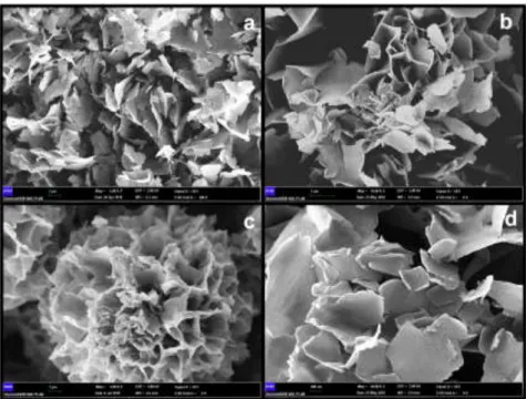
The synthesized FCPnfs were characterized using some techniques such as SEM, EDX, XRD, and FTIR. Firstly, FCPnfs SEM images including different amounts of AO were evaluated, to understand the effect of AO concentration on the morphology (Figure 3). The effect of AO amount on synthesis of nanostructures was evaluated ranging from 50 to 500 µL AO. At lower levels of organic component (50 µL AO), no flower morphology were formed due to lack of nucleus area formation (Figure 3a). When AO amount (100 µL AO) was doubled in synthesis solution, sufficient nucleus sites were observed and caused separate nanoscale petals to appear (Figure 3b). In growth process, Cu2+ binding sites on the
surfaces of the agglomerates cause copper phosphate crystals. When the organic component was presented in the optimum amount (250 µL AO) in the synthesis solution, nucleation sites are induced and result in anisotropic growth and cause more uniform and monodisperse flower-like morphology (Figure 3c). When using more number of AO (500 µL AO), the uniform and spherically shaped nanoflower morphology was scattered and creates large petals (Figure 3d). More number of organic molecules probably prevented the petals from binding together during the growth process. The concentrations of organic and inorganic components played a key role in the self-assembly process, hence the experimental condition must be optimized. The average particle size of optimized FCPnfs were determined to be 12 µm (Figure 3c).
To understand the distribution of organic components in FCPnfs, AO was added, and fluorescent microscopy images were analyzed as shown in Figure 4. Here, organic molecules (AO) were obtained that homogeneously distributed inside the nanoflowers like copper ions [17].
Figure 3. Scanning electron micrographs of fluorescent copper phosphate nanostructures formed with (a) 50 µL
AO; (b) 100 µL AO; (c) 250 µL AO; and (d) 500 µL AO
The EDX pattern of the FCPnfs was shown in Figure 5a. Figure 4a indicated to presence of C, O, Cu, P, and N atoms. The Cl peaks were stemmed from synthesis buffer. The trace element Ca ion was determined due to contamination of synthesis buffer. As shown in In Figure 5b; XRD analysis indicated the FCPnfs mainly matched with the crystal structure Cu3(PO4)2.3H2O (JCPDS card no 00-022-0548,
red line). This data explained about that the copper ion was incorporated into the crystalline structure of FCPnfs.
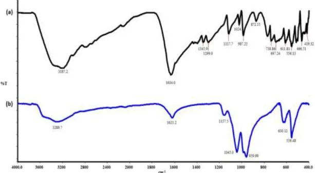
FTIR spectrums of pure AO and FCPnfs were shown in the range 4.000-400 cm-1 (Figure 6). The characteristic vibrations in the spectrum of AO was fit well with FCPnfs. The strong bands at around at 1.621 and 3.289 cm-1, can be assigned to N-H, H-O-H and C=C stretching vibrations [30]. In spectrum,
980
the peaks at 1.045, 959, 630, and 556 cm-1 demonstrated existence of phosphate groups [7]. These results illustrated that FCPnfs contains the AO molecules.
Figure 4. (a) The phase contrast and (b) Fluorescent microscopy images of Fnfs formed with 250 µl AO
(100x magnification)
Figure 5. (a) EDX analysis and (b) XRD diagram of synthesized FCPnfs (Cu3(PO4)2.3H2O, JCPDS card no
981
Figure 6. FTIR analysis of (a) acridin orange (AO) and (b) FCPnfs
3.2. Insect Toxicity Tests
To examine the different lethal concentrations of FCPnfs and CPnfs in T. molitor larvae, the larvae fed with FCPnfs and CPnfs at several concentrations (ranging between 0.001 and 0.100 mg/10 μl) (Table 1). Then larvae were placed into the experiment boxes (20 mL (51 mm x 34 mm x 19 mm) plastic boxes). The vitality of larvae were checked on a daily basis for 5 days. Different lethal concentrations of FCPnfs were determined as LC50: 0.49, LC60: 0.60, LC70: 0.71, LC80: 0.84, LC90: 0.102 and LC99: 0.145 mg/10
μl. On the other hand, lethal concentrations of CPnfs were also determined as LC50: 0.066, LC60: 0.078,
LC70: 0.090, LC80: 0.105, LC90: 0.125 and LC99: 0.172mg/10 μl as shown in Table 1.
The results demonstrated that FCPnfs at the concentration of 0.100 mg/10 μl were caused 100% mortality on T. molitor larvae in 5 days exposure time. However, CPnfs displays 85% mortality in the same nfs concentration. Probit analysis demonstrated that T. molitor was susceptible to FCPnfs. Table 1 also showed that at higher doses, such as >0.05 mg/10 μl were more toxic in both nanoflowers (with more than 10 dead larvae). This suggests that higher doses of nanoflowers were extremely disrupted the metabolisms of the larvae and caused the death of T. mollitor larvae within 5 days (Table 1). Based on these, FCPnfs including acridine orange is more toxic (LC50: 0.49 mg/10 μl) than CPnfs (LC50:0.066
mg/10 μl). Acridin orange may have made of increasing impact on the toxic effect of the FCPnfs. Because the cytocidal effect of free AO on bladder cancer cells was reported by Nishi and co-workers [30].
According to the previous results, the mechanisms of acute toxicity are defined as “necrosis, acetylcholinestrase inhibition, ion channel modulators, inhibitors of cellular” [31,32]. Carmona and co-workers indicated that CuONPs are genotoxic in Drosophila (Meigen, 1830) (Dpitera: Drosophilidae) and the oxidative stress may be mediated to these effects and most of the effects appear to be related to the presence of copper ions [33]. Another study, CuONP-induced toxicity was inquired using Drosophila as an in-vivo model. They observed that CuONPs increased in the body, and caused a dose dependent decrease in egg to adult survivorship and a delay in development [34]. Furthermore, the effects of CuO and copper sulphate nanoparticles on detoxification enzymes in the gut and fat body of Galleria mellonella (Lepidoptera: Pyralidae) as a model organism were also been determined by Tuncsoy and co-workers [35].
In conlusion, the reason of observed larval deaths seen in this study can be due to the mechanism of FCPnfs and CPnfs toxicity at the cellular, enzymatic, biochemical, and genetic levels in T. mollitor larvae mentioned above studies [30-33, 35].
982
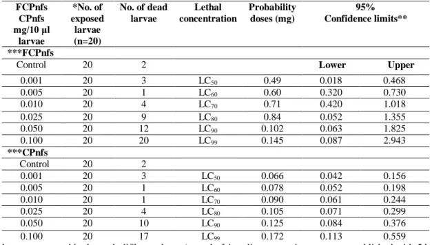
Table 1. Toxiciy of FCPnfs on T. molitor larvae (85.50 ± 17 mg weight) and toxicity of CPnfs on T. molitor
larvae (93 ± 20 mg weight) that exposed to nfs for 5d under laboratory conditions (26 ± 2ºC and 60 ± 5% RH). Purified water was used as a control
FCPnfs CPnfs mg/10 μl larvae *No. of exposed larvae (n=20) No. of dead larvae Lethal concentration Probability doses (mg) 95% Confidence limits** ***FCPnfs
Control 20 2 Lower Upper
0.001 20 3 LC50 0.49 0.018 0.468 0.005 20 1 LC60 0.60 0.320 0.730 0.010 20 4 LC70 0.71 0.420 1.018 0.025 20 9 LC80 0.84 0.052 1.355 0.050 20 12 LC90 0.102 0.063 1.825 0.100 20 20 LC99 0.145 0.087 2.943 ***CPnfs Control 20 2 0.001 20 3 LC50 0.066 0.042 0.156 0.005 20 1 LC60 0.078 0.052 0.198 0.010 20 1 LC70 0.090 0.061 0.244 0.025 20 4 LC80 0.105 0.071 0.299 0.050 20 10 LC90 0.125 0.084 0.376 0.100 20 17 LC99 0.172 0.113 0.559
*20 larvae were used in the each different doses (a total of 4 replicate experiments were established with 5 larvae in one replicate).
** Results are given with minimum and maximum confidence limits, FCPnfs-probit; Chi-Square=23.130, df=7, P=0.000, (Cu3(PO4)2) cyrstals probit; Chi-Square=13.343, df=7, P=0.020.
***Larvae (85.50 ± 17 mg weight) for FCPnfs toxicity and larvae (93 ± 20 mg weight) for CPnfs toxicity were selected for the force feeding treatment from the insect culture.
4. Conclusion
In summary, FCPnfs were prepared by simple and one-step via self-assembly method. The effects of acridine orange amount on hybrid nanoflower's morphology were investigated. The distribution of organic components in FCPnfs were imaged. Further, this study, for the first time, reported the toxic levels of copper phosphate nanoflowers with/without acridine orange on T. molitor larvae. The evaluating toxicological impact of FCPnfs and CPnfs are needed in the industrial production processes. Hereby, the knowledge of organic-inorganic hybrid nanoflowers chemical and toxicological behaviours, provide us to exhibit preventive behaviors on the materials to be produced and their effects on the environment. Importantly, FCPnfs toxicity was observed more toxic compared to CPnfs toxicity. The mechanisms of fluorescent copper phosphate nanoflowers toxicity will be detailed in the further studies. We continue to work on our studies on the effect of these two chemicals on the T. molitor and also another model organism insect species.
Acknowledgments
We also appreciate Elif Orgut ([email protected]) at the Cappadocia Technopark Corporation for assistance with schema drawings.
Authors’ Contributions
C.A. and A.E. conceived the original idea and build. N.Ö. supervised the project. The experiments were performed by A.E., C.A. and M.T. All authors contributed to the writing of the manuscript.
Statement of Conflicts of Interest
983 Statement of Research and Publication Ethics
The author declares that this study complies with Research and Publication Ethics. References
[1] Altinkaynak C., Tavlasoglu S., Özdemir N., Ocsoy I. 2016. A New Generation Approach In Enzyme Immobilization: Organic-İnorganic Hybrid Nanoflowers With Enhanced Catalytic Activity And Stability. Enzyme Microb. Technol., 93-94:105-12.
[2] Wang R., Zhang Y., Lu D., Ge J., Liu Z., Zare R.N. 2013. Functional Protein-Organic/Inorganic Hybrid Nanomaterials. Wiley Interdiscip Rev. Nanomed Nanobiotechnol, 5 (4): 320-328. [3] Lee S.W., Cheon S.A., Kim M.I., Park T.J. 2015. Organic-Inorganic Hybrid Nanoflowers:Types,
Characteristics, And Future Prospects. J. Nanobiotechnology, 13: 54.
[4] He X., Chen L., He Q., Xiao H., Zhou X., Ji H. 2017. Self-Assembled Metalloporphyrins– Inorganic Hybrid Flowers And Their Application To Efficient Epoxidation of Olefins. Journal of Chemical Technology & Biotechnology, 92 (10): 2594-2605.
[5] Gulmez C., Altinkaynak C., Özdemir N., Atakisi O. 2018. Proteinase K hybrid nanoflowers (P-hNFs) as a novel nanobiocatalytic detergent additive. International Journal of Biological Macromolecules 119: 803-810.
[6] Ge J., Lei J., Zare R.N. 2012. Protein-Inorganic Hybrid Nanoflowers. Nat. Nanotechnol, 7 (7): 428-432.
[7] Yu Y., Fei X., Tian J., Xu L., Wang X., Wang Y. 2015. Self-Assembled Enzyme-Inorganic Hybrid Nanoflowers and Their Application to Enzyme Purification. Colloids Surf B Biointerfaces, 130: 299-304.
[8] Jing M., Fei X., Ren W., Tian J., Zhi H., Xu L., Wang X., Wang Y. 2017. Self-Assembled Hybrid Nanomaterials With Alkaline Protease And A Variety of Metal Ions, RSC Advances, 7 (76): 48360-48367.
[9] Nadar S.S., Gawas S.D., Rathod V.K. 2016. Self-Assembled Organic-Inorganic Hybrid Glucoamylase Nanoflowers With Enhanced Activity and Stability. International Journal of Biological Macromolecules, 92: 660-669.
[10] Altinkaynak C., Tavlasoglu S., Kalin R., Sadeghian N., Ozdemir H., Ocsoy I., Özdemir N. 2017. A Hierarchical Assembly of Flower-Like Hybrid Turkish Black Radish Peroxidase-Cu2+ Nanobiocatalyst And Its Effective Use in Dye Decolorization. Chemosphere, 182: 122-128. [11] Ildiz N., Baldemir A., Altinkaynak C., Özdemir N., Yilmaz V., Ocsoy I. 2017. Self Assembled
Snowball-Like Hybrid Nanostructures Comprising Viburnum opulus L. Extract and Metal İons For Antimicrobial and Catalytic Applications. Enzyme and Microbial Technology, 102: 60-66. [12] Altinkaynak C., Kocazorbaz E., Özdemir N., Zihnioglu F. 2018. Egg White Hybrid Nanoflower
(EW-Hnf) With Biomimetic Polyphenol Oxidase Reactivity: Synthesis, Characterization And Potential Use in Decolorization of Synthetic Dyes. Int. J. Biol. Macromol, 109: 205-211.
[13] Kim K.H., Jeong J-M., Lee S.J., Choi B.G., Lee K.G. 2016. Protein-Directed Assembly Of Cobalt Phosphate Hybrid Nanoflowers. J. Colloid Interface Sci., 484: 44-50.
[14] Zhang Z., Zhang Y., He L., Yang Y., Liu S., Wang M., Fang S., Fu G. 2015. A Feasible Synthesis of Mn3(PO4)2@BSA Nanoflowers and Its Application as the Support Nanomaterial for Pt Catalyst.
Journal of Power Sources, 284: 170-177.
[15] Zhang B., Li P., Zhang H., Wang H., Li X., Tian L., Ali N., Ali Z., Zhang Q. 2016. Preparation of Lipase/Zn3(PO4)2 Hybrid Nanoflower and its Catalytic Performance as an Immobilized
Enzyme. Chemical Engineering Journal, 291: 287-297.
[16] Wang X., Shi J., Li Z., Zhang S., Wu H., Jiang Z., Yang C., Tian C. 2014. Facile One-Pot Preparation of Chitosan/Calcium Pyrophosphate Hybrid Microflowers. ACS Applied Materials & Interfaces (ACS Publications), 6(16):14522-14532.
[17] Jiao J., Xin X., Wang X., Xie Z., Xia C., Pan W. 2017. Self-Assembly Of Biosurfactant–Inorganic Hybrid Nanoflowers As Efficient Catalysts For Degradation Of Cationic Dyes, RSC Advances, 7 (69):43474-43482.
[18] Li J., Schiavo S., Xiangli D., Rametta G., Miglietta M.L., Oliviero M., Changwen W., Manzo S. 2018. Early Ecotoxic Effects of ZnO Nanoparticle Chronic Exposure in Mytilus Galloprovincialis
984
Revealed by Transcription of Apoptosisand Antioxidant-Related Genes. Ecotoxicology, 27 (3): 369-384.
[19] Gebel T. 2012. Small Difference in Carcinogenic Potency Between GBP Nanomaterials and GBP Micromaterials. Arch. Toxicol, 86 (7): 995-1007.
[20] Sun X., Chen B., Bin Xia N., Han Q., Zhu L., Qu K. 2017. Are CuO Nanoparticles Effects on Hemocytes of the Marine Scallop (Chlamys Farreri) Caused by Particles And/Or Corresponding Released Ions?. Ecotoxicol Environ. Saf., 139: 65-72.
[21] Adam N., Vakurov A., Knapen D., Blust R. 2015. The Chronic Toxicity of CuO Nanoparticles and Copper Salt to Daphnia Magna. J. Hazard Mater., 283: 416-422.
[22] Hoseini S.M., Hedayati A., Taheri Mirghaed A., Ghelichpour M. 2016. Toxic Effects of Copper Sulfate and Copper Nanoparticles on Minerals, Enzymes, Thyroid Hormones and Protein Fractions of Plasma and Histopathology in Common Carp Cyprinus carpio. Exp. Toxicol Pathol., 68 (9): 493-503.
[23] Rajput V., Minkina T., Fedorenko A., Sushkova S., Mandzhieva S., Lysenko V., Duplii N., Fedorenko G., Dvadnenko K., Ghazaryan K. 2018. Toxicity of Copper Oxide Nanoparticles on Spring Barley (Hordeum Sativum Distichum). Science of the Total Environment, 645: 1103-1113.
[24] Braz-Mota S., Campos D.F., MacCormack T.J., Duarte R.M., Val A.L., Almeida-Val V.M.F. 2018. Mechanisms of Toxic Action of Copper and Copper Nanoparticles in Two Amazon Fish Species: Dwarf Cichlid (Apistogramma agassizii) and Cardinal Tetra (Paracheirodon axelrodi). Sci. Total Environ., 630: 1168-1180.
[25] Collins L. 2012. Toxicity of Moist Snuff and Impact on Various Stages of Darkling Beetles. Biology and Biotechnology, 7.
[26] Ramarao N., Nielsen-Leroux C., Lereclus D. 2012. The Insect Galleria Mellonella As A Powerful İnfection Model to Investigate Bacterial Pathogenesis. J. Vis. Exp., 70: 4392.
[27] Dere B., Altuntaş H., Nurullahoğlu Z.U. 2015. Insecticidal and Oxidative Effects of Azadırachtın on the Model Organism Galleria mellonella L. (Lepidoptera: Pyralidae). Arch. Insect. Biochem Physiol, 89 (3): 138-152.
[28] Manente S., De Pieri S., Iero A., Rigo C., Bragadin M. 2008 A Comparison Between The Responses Of Neutral Red And Acridine Orange: Acridine Orange Should Be Preferential And Alternative To Neutral Red As A Dye For The Monitoring Of Contaminants By Means Of Biological Sensors. Anal. Biochem., 383 (2): 316-319.
[29] Altuntaş H., Duman E., Şanal Demirci S.N., Ergin E. 2016. Toxicological And Physiological Effects Of Ethephon On The Model Organism, Galleria mellonella L. 1758 (Lepidoptera: Pyralidae). Turkish Journal of Entomology, 40 (4).
[30] Nishi M., Hiruma H., Sasamoto H., Iwamura M., Isonaka R., Baba S. 2012. Cytocidal Effects Of Acridine Orange Evoked By Blue Light On Human Bladder Cancer Cells. Kitasato Med. J., 42: 128-137.
[31] Ali T.H., Abed A.A., Ellah A.A. 2016. Determination Of The Lethal Concentration 50% (Lc50) Of Cadmium Chloride in Mosquito Fish Gambusia Holbrooki. Tikrit Journal of Pure Science, 21: 1.
[32] Calabrese E.J., Baldwin L.A. 2001. U-Shaped Dose-Responses in Biology, Toxicology And Public Health. Annu. Rev. Public Health, 22: 15-33.
[33] Carmona E.R., Inostroza-Blancheteau C., Obando V., Rubio L., Marcos R. 2015. Genotoxicity Of Copper Oxide Nanoparticles in Drosophila melanogaster. Mutat. Res. Genet. Toxicol Environ. Mutagen, 791: 1-11.
[34] Baeg E., Sooklert K., Sereemaspun A. 2018. Copper Oxide Nanoparticles Cause a Dose-Dependent Toxicity via Inducing Reactive Oxygen Species in Drosophila. Nanomaterials (Basel), 8: 10.
[35] Sezer Tunçsoy B., Ozalp P. 2016. Toxic Effects Of Copper Oxide Nanoparticles In Midgut And Fat Body Of Galleria mellonella (Lepidoptera: Pyralidae). Toxicology Letters, 258: 270.