ISSN-L: 0034-8570
https://doi.org/10.3989/revmetalm.187
Microstructure analysis of welding fume of low and medium
carbon steels
Bekir Güney*
Karamanoğlu Mehmetbey University, Vocational School of Technical Sciences, Yunus Emre Yerleşkesi, 70100 Karaman, Turkey
(*Corresponding author: [email protected])
Submitted: 2 May 2020; Accepted: 20 January 2021; Available On-line: 7 April 2021
ABSTRACT: In this study, the sample of welding fume was obtained from low and medium carbon steels and the electrodes used in welding. The microstructures of the particles were analysed using scanning electron microscopy (SEM), energy dispersive spectrometer (EDS), X-ray diffractometer (XRD) and fourier transform infrared spectrometer (FTIR). In the experiments; Be, O, F, Fe, Si, Cl, K, Ca, Ti, V, Cr, Mn were found to be atomically more than 1%. Based on this finding, it is revealed that the structure is composed mainly of oxides such as Fe2O3, Fe3O4, MnO2, TiO2, SiO2, Fe3Mn3O8, FeMn2O4, BeO, CrO. It was also found with XRD analysis that the elements which were found to beatomically less 1% formed oxide phases. Because oxidized structures threaten the environment and human health, it has been experimentally found that the metals and heavy met-als emitted by welding fumes still keep polluting and threatening the environment.
KEYWORDS: Human healty; Microstructure; Pollution; Welding fume
Citation/Citar como: Güney, B. (2021). “Microstructure analysis of welding fume of low and medium carbon steels”.
Rev. Metal. 57(1): e187. https://doi.org/10.3989/revmetalm.187
RESUMEN: Análisis de la microestructura de humos de soldadura de aceros de bajo y medio contenido de carbon. En este estudio, la muestra de humo de soldadura se obtuvo a partir de aceros de bajo y medio carbono y los electrodos utilizados en la soldadura. Las microestructuras de las partículas se analizaron mediante microscopía electrónica de barrido (SEM), espectrómetro de dispersión de energía (EDS), difractómetro de rayos X (XRD) y espectrómetro de infrarrojos por transformada de Fourier (FTIR). En los experimentos se encontró que los elementos Be, O, F, Fe, Si, Cl, K, Ca, Ti, V, Cr, Mn tenían contenidos atómicos superiores al 1%. Con base en este hallazgo, se revela que la estructura está constituida principalmente por óxidos tipos Fe2O3, Fe3O4, MnO2, TiO2, SiO2, Fe3Mn3O8, FeMn2O4, BeO, CrO. También se encontró mediante análisis XRD que los elementos con contenidos inferiores al 1% atómico se encontraban también asociados a fases en forma de óxidos. Debido a que las estructuras oxidadas amenazan el medio ambiente y la salud humana, se ha descubierto experimentalmente que los metales emitidos por los humos de soldadura siguen contaminando y amenazando el medio ambiente. PALABRAS CLAVE: Contaminación; Humos de soldadura; Microestructura; Salud humana
ORCID ID: Bekir Güney (https://orcid.org/0000-0001-9764-9313)
Copyright: © 2021 CSIC. This is an open-access article distributed under the terms of the Creative Commons
1. INTRODUCTION
Metals have excellent mechanical properties
compared to other materials in terms of hardness,
toughness and strength (Shackelford et al., 2016).
In industrial applications, the bonding of these
metals is usually obtained by a welding technique.
During welding, it is necessary to have a base
met-al, additional metal and a heat source (Turan et al.,
2011). The welding arc forming is a process of
accu-mulating an electric arc between welding electrode
and base material, melting the metals at the joining
(Cary and Helzer, 2005; Erden et al., 2018).
According to the science of physics, welding arc
occurs when electrons emitted from the cathode
portion bombard the anode with a high speed as the
electric current passes from one conductive metal
to another. This bombardment causes a strong rise
in temperature since it causes the ionization of the
neutral molecules at the end of the impact (Anık,
2001). The temperature above 4000 ºC in the arc
(Palmer and Eaton, 2001) allows the metals to melt
and thus to bond (Howden, et al., 1988). Each
ma-terial is a potential source of fume when heated to
high temperatures. Welding fumes are produced as
a result of metallurgical reactions at high
temper-atures. Some of the metal components which are
heated well above the electrode boiling degree are
released as gases by burning or evaporating into the
atmosphere. The vaporous components are
con-densed again to become ultra-fine fume particles
smaller than 100 nm, which are light enough to fly
in the air and small enough to breathe.
Chemical composition of welding fumes depends
on the welding technique used, welding parameters,
melting, welding metal and welding electrode which
has a composition of metal
(Berlinger
et al.,
2019).
When welding, welding fumes, gases and
electro-magnetic energy (radiation) are usually released in
indoor areas.
Welding fumes are caused by melting and
evapo-ration of metal wire electrodes or dust during
join-ing or coatjoin-ing of metals. A variety of metallic and
non-metallic elements and compounds are present
in the fume composition (Sowards et al., 2010),
in-cluding, metallic oxides, silicates and fluorides, as
well as complex mixtures of heavy metal
contam-inants such as cadmium, aluminium, chromium,
copper and lead (Rana et al., 2019).
Some of these
particles vent into the atmosphere and some of them
hang in the air for a while and then accumulate on
the ground as a result of condensation, air
move-ment, gravity or atomic interactions.
Inhalation of toxic metals and metalloids poses
a risk to workers’ health in many industries. Today,
among these health-damaging factors, great
impor-tance is given to the toxic effects caused by
weld-ing fumes (Flechsig, 1988). It is estimated that more
than one hundred million workers worldwide work
as welders and more than three million employees
weld at certain intervals as part of their work
(Mc-Neilly et al., 2004).
Arc welding procedures emit solid particles and
gases that may have adverse health-related effects
following inhalation, including cardiovascular
(Sjogren et al., 2006), neurological (Fored et al.,
2006) respiratory signs and symptoms. Therefore,
it may cause environmental and health problems
(Lighty et al., 2000; Antonini, 2003; Donaldson et
al., 2005; Jenkins and Eagar 2005a; Oberdörster et
al., 2005). Welding fume has toxicity which may be
hazardous to human health if inhaled or swallowed
in pure form. Metal oxides exhibiting toxic
charac-teristics contain alloying elements which can be
dan-gerous in this sense (Jenkins and Eagar, 2005a)
.
Previous works have reported some specific
chemical composition of welding (Ehrman et al.,
1999; Jenkins and Eagar, 2005a; Jenkins and
Ea-gar, 2005b; Sowards et al., 2010; Golbabaei and
Khadem, 2015; Stebounova et al., 2018). However,
there is still need of a study involving comprehensive
analysis of chemical composition of welding fume
in order to have better understanding on possible
adverse health effects, and have create better
preven-tive and safety strategies. Accordingly, in this study,
the microstructure of low and medium carbon steels
and welding particles obtained from electrodes used
in their welding were characterized by using SEM,
EDS, XRD and FTIR techniques.
2. MATERIALS AND METHODS
2.1. Welding fume collection
The welding fume sample was obtained from the
fume of electrodes used in low and medium carbon
steels and their electric arc welding since carbon
steels are the most commonly used materials in the
world (Golbabaei and Khadem, 2015). These
parti-cles were deposited by vacuuming to a ceramic filter
at the room temperature. The studies were carried
out in the Material Characterization Laboratory at
Karamanoğlu Mehmetbey University, Scientific and
Technological Researches Application and Research
Center.
2.2. Micro structure analysis
The fume particles were aspirated at room
tem-perature and collected in a ceramic filter.
Micro-structure analyses were performed with a field
emis-sion SEM (HITACHI SU5000) equipped with EDS
operating at 10 kV. IR spectroscopy (Bruker Vertex
70 ATR) was used to measure the FTIR spectrum
of the sample. The data were collected by vibration
frequencies at 4000-400 cm
-1scanning range at 4 cm
-1spectral resolution. X-ray diffraction phase analysis
was performed with a Bruker D8 ADVACE with
DAVINCI XRD (Cu-K
α radiation, λ = 1,5406 Å
in the range 10° ≤ 2θ ≤ 90° operated at 40 kV and
40 mA) with secondary beam graphite
monochro-mator. The phase analyses were characterized by
the data obtained from the Diffract EVA software
and the International Centre for Diffraction Data
(ICCD).
3. RESULTS AND DISCUSSION
3.1.
Characterization by
XRD
The composition of the welding fume particles
comprises different structures due to the cooling
mechanism and the agglomerated method. X-ray
diffraction studies revealed that approximately 90%
of the fume is crystal structure (Fig. 1). Since the
source fume particles are composed of many
el-ements and molecules according to the results of
the EDS and FTIR analyses, many peaks of XRD
phase analysis were obtained (Fig. 1). According to
EDS analysis, there were many elements in the
struc-ture. X-ray diffraction analysis revealed that
differ-ent compounds had strong peaks at the same point.
The peak in the same range indicated the presence
of more than one compound. The peaks of the
com-pounds given in Table 1, were the strongest matches.
table 1. XRD diffraction spectra of welding fume with strong peaks
Name Formula Crystal System Peak Number
Zinc Manganese Iron Oxide ZnMnFeO4 Cubic 2, 3, 5, 6, 7
Copper Iron Nickel Zinc Oxide Cu0.1Fe1.9Ni0.65Zn0.35O4 Cubic 2, 3, 4, 6, 7, 8
Iron Manganese Oxide Fe3Mn3O8 Cubic 2, 3, 4, 6, 7, 8
Manganese Iron Titanium Oxide (FeMn)2TiO3 Rhombohedral 3, 6
Iron Manganese Oxide FeMn2O4 Cubic 2, 3, 4, 6, 7, 8, 9
Manganese Iron Zinc Oxide Mn0.09Fe0.08Zn1.83O4 Cubic 2, 3, 4, 6, 7, 8, 9 Zinc Manganese Iron Oxide Zn2Mn8Fe2O4 Cubic 2, 3, 4, 6, 7, 8, 9 Zinc Manganese Iron Oxide Zn4Mn6Fe2O4 Cubic 2, 3, 4, 6, 7, 8, 9 Zinc Manganese Iron Oxide Zn6Mn4Fe2O4 Cubic 2, 3, 4, 6, 7, 8, 9
Zinc Manganese Iron Oxide Zn9MnFe2O4 Cubic 2, 3, 4, 6, 7, 8, 9
Zinc Manganese Iron Oxide ZnMnFe3O8 Tetragonal 2, 3, 4, 6, 7, 8, 9
Magnetite Fe3O4 Orthorhombic 1, 2, 3, 4, 6, 7, 8, 9
Fayalite, Manganoan (FeMn)2SiO4 Orthorhombic 3, 9
Hematite Fe2O3 Tetragonal 1, 2, 3, 4, 6, 7, 8
Iron Oxide FeO Orthorhombic 2, 3
Aluminum Oxide Al2O3 Orthorhombic 2, 3, 4
Berylium Oxide BeO Hexagonal 5
Chromium Oxide Cr2O3 Rhombohedral 5
Copper Magnesium Mg2Cu Orthorhombic 3, 4, 5
Periclase MgO Cubic 4, 8, 9
Manganese Oxide MnO2 Hexagonal 4, 7, 9
Sodium Oxide Na2O2 Hexagonal 1, 3, 4
Nickel Titanium Oxide Ni2Ti4O Cubic 3, 7, 8
Silicon Oxide SiO2 Monoclinic 1, 3
Titanium Oxide TiO2 Cubic 3, 7
Zinc Titanium Oxide Zn2TiO4 Cubic 2, 3, 4, 6, 7, 8, 9
Zirconium Oxide ZrO2 Rhombohedral 2, 3
Since welding fumes consist of ultra-fine
par-ticles, these structures were essentially shapeless.
The structures of the phases obtained from the
XRD analysis given in Table 1 were composed of
different crystal lattice structures as reported in
previous studies
(Ehrman et al., 1999;
Stebouno-va et al., 2018).
During the condensation of these
particles, separate molecules may get together
to form different phases in a single structure. In
some structures, other oxide shells could be found
around the iron oxide core. Therefore, particle
structures are generally heterogeneous (Jenkins
and Eagar, 2005b)
.
Welding fume is a product of high temperature. It
is possible that a large number of elements or
mol-ecules present in the body can form very different
compounds at these elevated temperatures. Based
on this, information on the compounds giving peaks
in the XRD analysis of the fume material was given
in Table 1. When Table 2 was examined, it is very
difficult to analyze the structure in detail due to the
elements which can be included in the structure
un-controlled from the atmosphere depending on the
chemical content of the materials used in forming
the welding arc or due to the effect of high
tem-perature. However, it is possible to say that Fe and
Mn-based structures are predominant. It is
under-stood from the XRD analyses that intermetallics
such as NiAl, TiNi are formed in the structure due
to high temperature. When the peaks obtained by
XRD were evaluated together with EDS and FTIR
analyses, Be element BeO, K element K
2O, Ca
ele-ment CaO, V eleele-ment V1
6O
3, Ti element Zn
2TiO
4,
Ni
2Ti
4O, W element W
3O
8, and Cr element CrO are
available in the structure forming Si element SiO
2.
X-Ray diffraction analysis showed that the
domi-nant phase in the whole fume was highly
correlat-ed with Fe
3O
4in the magnetite structure and Fe
2O
3in the hematite structure which gives strong peaks
(Jenkins and Eagar, 2005b). Other possible
struc-tures were MgO, K
2CO
3, Na
2CO
3and MnFe
2O
4.
The results of the analysis reveal that welding fume
contains various oxides in very complex structures
and different combinations.
3.2. Characterization by scanning
electron microscopy
The images of such structures were difficult to
analyses with SEM. The small welding fume
par-ticles formed larger spherical agglomerated
parti-cles by the cooling mechanism from vapour state.
These agglomerates appear on the micrographs
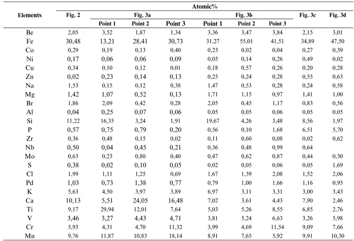
table 2. Atomic quantities of elements detected in welding fume according to EDS analysisElements
Atomic%
Fig. 2 Fig. 3a Fig. 3b Fig. 3c Fig. 3d
Point 1 Point 2 Point 3 Point 1 Point 2 Point 3
Be 2,05 3,52 1,87 1,34 3,36 3,47 3,84 2,15 3,01 Fe 30,48 13,21 28,41 30,73 31,27 55,01 41,51 34,89 47,50 Co 0,29 0,19 0,13 0,40 0,23 0,02 0,04 0,27 0,39 Ni 0,17 0,06 0,06 0,09 0,05 0,14 0,26 0,49 0,02 Cu 0,34 0,10 0,12 0,01 0,18 0,57 0,26 0,20 0,28 Zn 0,02 0,23 0,14 0,13 0,25 0,24 0,28 0,55 0,63 Na 1,53 0,15 0,12 0,38 1,47 0,53 0,28 0,24 0,58 Mg 1,42 1,07 0,52 0,13 1,71 1,15 0,97 1,41 1,00 Br 1,86 2,09 0,42 0,28 2,05 0,45 1,17 0,83 0,56 Al 0,04 0,25 0,07 0,06 0,05 0,05 0,06 0,05 0,05 Si 11,22 16,35 3,24 1,91 19,67 4,26 3,48 8,56 1,97 P 0,57 0,75 0,79 0,20 0,56 0,10 1,68 6,51 5,70 Zr 0,36 0,48 0,15 0,02 0,11 0,60 0,08 0,02 0,62 Nb 0,50 0,04 0,45 0,21 0,36 0,48 0,99 0,64 Mo 0,63 0,23 0,80 0,40 0,47 0,62 0,87 0,44 0,30 S 0,38 0,02 0,10 0,05 0,02 0,05 0,06 0,05 1,69 Cl 1,99 1,11 1,25 0,69 1,67 1,39 2,08 1,52 2,06 Pd 1,03 0,73 1,38 0,77 0,79 1,00 1,66 1,16 0,95 K 5,63 4,50 3,97 3,89 6,97 3,11 3,31 3,00 3,43 Ca 10,13 5,51 24,05 16,48 7,02 3,61 4,43 7,90 2,46 Ti 9,17 29,94 12,01 7,64 5,03 5,26 8,55 6,85 2,76 V 3,46 3,27 4,43 4,71 3,81 5,24 6,63 3,26 5,98 Cr 5,93 4,31 4,70 11,32 3,99 4,69 11,54 9,09 7,66 Mn 9,76 11,87 10,83 18,14 8,91 7,65 5,92 9,91 10,30
as foam or finely mixed hair (Fig. 2) (Jenkins and
Eagar, 2005b). However, larger particles can also
be produced by spattering from the welding arc.
Large particles were composed of Al, Si, K, Na, F
and water-soluble compounds, while small
parti-cles were predominantly composed of heavy
met-als such as Fe, Ni, Mo, Mn, Cr and their oxides.
These were the particles flaking during
condensa-tion and are the most commonly observed particles
at each stage. The other was common, although a
much lower amount of isolated spherical particles is
present. The third was the irregularly shaped
parti-cles with the lowest density. When the EDS analysis
conducted in point 1of Fig. 3a was examined in
Ta-ble 2, it is seen that Mn, Br, Ti, Ca, Si, V, Cr, K and
Be elements were atomically high. When the EDS
analysis of point 2 in Fig. 3a was examined in Table
2, it is seen that the elements Ti, Mn, Si, Cl, Cr, Ca,
K, V, Be and Pd were also atomically high.
When the EDS analysis of point 3 in Fig. 3 is
ex-amined in Table 2, it is seen that the elements Ti,
Mn, Si, Cr, Ca, K, V and Be were atomically high.
The micrograph of spherical particles constituting
the majority of the fume morphology was given
in Fig. 3b in 5.000x magnification. When the EDS
analysis of point 1 of Fig. 3b is examined in Table
2, it is seen that the elements Mn, Fe, Ti, Ca, Si, V,
Cr, K and Be were atomically high. When the EDS
analysis of point 2 of Fig. 3b is examined in Table
2, it is seen that the elements Mn, Fe, Ti, Ca, Si, V,
Cr, K and Be were atomically high. When the EDS
analysis of point 3 of Fig. 3b is examined in Table 2,
it is seen that the elements Mn, Fe, Ti, Ca, Si, V, Cr,
Cl, K and Be were atomically high. When the EDS
analysis of Fig. 3b in Table 2 is examined, it was
found that the elements Mn, Fe, Ti, Ca, Si, V, Cr, K
were atomically high. The micrograph of spherical
particles of different sizes and particles which tend
to agglomerate is given in Fig. 3c at 10.000x
magnifi-cation. The spherical particle size is shown in Fig. 3d
in 20.000x magnification. When the EDS analysis of
the surface of the micrograph was analysed in Table
2, it was found that the elements Fe, Si, P, Cl, K,
Ca, Ti, V, Cr, and Mn were atomically high. Particle
morphology needs to be considered as it determines
the surface area of a part and the aerodynamic
di-ameter of the particles. A pellet has a much larger
surface area than the individual spherical particle
having the same cross-section. These agglomerates
also have different aerodynamic properties that can
affect the degree to which they can be inhaled
(So-wards et al., 2008). When EDS analysis was
evaluat-ed in general, it was found that Be, Fe, Si, Cl, K, Ca,
Ti, V, Cr, and Mn elements were found to be high in
each point examined. When evaluated together with
FTIR and XRD analyses, it reveals that the
struc-ture is composed of the molecules and compounds
belonging to the elements that are detected more
atomically.
Smaller particles are subjected to higher degrees
of overcooling in the first fume vapor. This
caus-es the formation of primary particlcaus-es in the fumcaus-es
produced during welding. Thus, metallic particles
in the chemical elements found in the welding are
condensed and nucleated. The elements that are
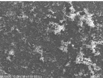
FiguRe 2. SEM micrograph of 110x magnification takenfrom welding fume surface.
The welding fume was shown in Table 1, where
the metallic elements which were present in the
composition are represented by different %
atom-ic ratios in the form of compounds. The elements
in the structure such as Be, Ca, Cl, Cr, Fe, Mn, K,
Si, Ti and V were found to be atomically higher
than 1% due to the welded material and the
struc-ture of the electrode. In addition, when the results
of XRD and EDS analyses were examined
togeth-er, the elements such as Al, Br, Co, Mg, Mo, Na,
Nb, Ni, P, Pd, S, Zn, and Zr were found to be less
than 1% or in trace amount. Since the
tempera-ture reached at the source is about 4000 ºC
(Palm-er and Eaton, 2001)
all elements in the structure
are almost gaseous. During the gas condensation
of welding fumes, the amount of O added from
the atmosphere to the composition is high in
con-centration. Metallic nanostructure particles tend
to compound rapidly with O. This tendency leads
to the formation of high amounts of metal oxides,
the main element of the fume concentration.
When the SEM micrograph in 110x magnification
was examined in Fig. 2, regarding fume
morphol-ogy, the structure consisting of oxide deposits and
predominantly spherical particles on the surface is
striking. When the EDS analysis of Fig. 2 was
ex-amined in Table 2, it is seen that the elements Mn,
Fe, Ti, Ca, Si, V, Cr, Cl, K and Be were atomically
high in the structure.
Figure 3a shows three different types of particles
in the 2.000x magnification micrograph. Spherical
particles are predominantly seen in the micrograph.
lighter in the fume may not be involved in
nuclea-tion and may be vented into the atmosphere. This
resulted in the formation of higher amounts of
el-ements such as Be, Fe, Si, Cl, K, Ca, Ti, V, Cr and
Mn in the source fume composition (Sowards et
al., 2010). Other structures consist of nanoparticles
having multiple oxidation states, which are formed
as amorphous or single nanoparticles or
agglomer-ates which have been able to achieve compounding
capacity during condensation.
3.3.
Characterization by FTIR
FTIR measurements were performed to
investi-gate the bonds of functional free and complex
mol-ecules in the source fume (Fig. 4). According to the
EDS analysis of the welding fume sample, the
bond-ing structures of the metallic-based elements which
are more than 1% by weight are examined.
Accord-ing to the peak values shown in Fig. 4; 3296, peaks
in the band range of 2921 cm
-1(Ehrman et al., 1999;
Jenkins et al., 2005a; Jenkins and Eagar, 2005b;
Wang et al., 2006; Chen and He, 2008; Sowards
et al., 2008; Sevilla and Fuertes, 2009; Gibot and
Vidal, 2010; Saikia and Parthasarathy, 2010; Zheng
et al., 2010; Vaculikova et al., 2011; Basu et al., 2011;
Farzaneh and Najafi, 2011; Lin et al., 2012;
Abdul-lah et al., 2014; Jamal et al., 2014; Golbabaei and
Khadem, 2015; Naushad et al., 2015; Diko et al.,
FiguRe 3. SEM micrograph of welding fume surface with a magnification of: a) 2.000x, b) 5.000x, c) 10.000x, and d) 20.000x.FiguRe 4. Characterization of welding fume particles FTIR spectrum.
2016; Sahai et al., 2016; Benykhlef et al., 2016; Yi et
al., 2018; Ge et al., 2019; Habtemariam et al., 2019;
Bahah et al., 2019; Abinaya et al., 2019; Alias et al.,
2019; Altunal et al., 2019; Boro
ń et al., 2019;
Red-dy et al., 2019; Karthik et al., 2019; Karunathilaka
et al., 2019; Kono et al., 2019; Mohammadi et al.,
2019; Ponmudi et al.2019; Scaccia et al., 2019; Wang
et al., 2019a; Wang et al., 2019b; Yang et al., 2019),
indicate O-H bonding in the structure. This
indi-cates the presence of an H
2O molecule. Peaks in the
band range of 1012 and 420 cm
-1were obtained due
to the tension of the metal oxide bonds. The FTIR
peaks of the elements in the source fume obtained
according to EDS analysis were consistent with the
literature Al-O (Jamal et al., 2014; Benykhlef et al.,
2016; Yi et al., 2018), Be-O (Altunal et al., 2019),
Br-O (Naushad et al., 2015), C-O, CaO and CH
(Scaccia et al., 2019), Cd-O (Karthik et al., 2019),
Cl-O (Wang et al., 2019a), Co-O (Gibot and Vidal,
2010), Cr-O (Basu et al., 2011; Farzaneh and
Na-jafi, 2011; Abdullah et al., 2014), Cu-O (Zheng et al.,
2010; Sahai et al., 2016; Ponmudi et al., 2019),
Fe-O
(Abdullah
et al.,
2014; Golbabaei and Khadem,
2015)
,
Mn-O (Chen and He, 2008; Sevilla and
Fuertes, 2009; Lin
et al.,
2012)
, Mo-O (Abinaya
et al., 2019), N-O (Boro
ń et al., 2019; Yang et al.,
2019), P-O (Kono et al., 2019), Pd-O (Reddy et al.,
2019), S-O (Yang et al., 2019),
Si-O (Saikia and
Parthasarathy, 2010; Vaculikova
et al.,
2011; Diko
et al.,
2016; Bahah
et al.,
2019; Mohammadi
et al.,
2019)
, Ti-O (Jamal et al., 2014), V-O (Wang et al.,
2006; Habtemariam et al., 2019), Zr-O (Wang et al.,
2019b) metal oxides and oxide structures in
differ-ent structures because metal oxides generally exhibit
peaks below 1000 cm
-1, which may be caused by
in-ter-atomic vibrations (Lagashetty et al., 2007)
.
Welded metal and additional metal have a rich
chemical composition. During joining, a certain
amount of this rich structure burns or evaporates
and thus forms welding fumes. Welding fumes
contain very different structures by its nature.
1139, 1257, 1407, 2848 and 2921 cm
-1peaks
ob-tained from FTIR analysis were
Al-O (Jamal et al.,
2014; Benykhlef et al., 2016; Yi et al., 2018),
Cl-O
(Wang et al., 2019a), Co-O (Gibot and Vidal, 2010),
C-H and CC (Scaccia et al., 2019)
, C-F
(Karuna-thilaka et al., 2019)
, C-Br (Nicasio-Collazo
et al.,
2019), N-H (Oswald
et al.,
2019), C-N (Panja
and Ghosh, 2019), Fe-O and Fe
2O
4(Abdullah
et
al.,
2014; Golbabaei and Khadem, 2015)
,
F
2O
3(Oberdörster et al., 2005),
Mn-O (Jamal
et al.,
2014;
Alias
et al.,
2019)
,
N-O (Boro
ń et al., 2019; Yang et
al., 2019), P-O (Kono et al., 2019), Pd-O (Reddy et
al., 2019),
Si-O (Saikia and Parthasarathy, 2010;
Vaculikova
et al.,
2011; Diko
et al.,
2016; Bahah
et
al.,
2019; Mohammadi
et al.,
2019) functional due
to the fact that stretching of bonds of functional
groups has increased.
4. CONCLUSIONS
• In this study, the molecular structure,
com-pound structure and crystal structure of the
elements which are formed after melting,
evapo-ration and combustion were investigated. With
this study; Be, Fe, Si, Cl, K, Ca, Ti, V, Cr and
Mn were found to be more than 1% of the
to-tal composition in the welding fume. Based on
this finding, it is concluded that the structure
is mainly composed of oxides such as Fe
2O
3,
Fe
3O
4, MnO
2, TiO
2, SiO
2, Fe
3Mn
3O
8, FeMn
2O
4,BeO and CrO. Welding fume is released into
the atmosphere as a high-temperature product.
Therefore, it has been experimentally explained
that combinations of oxidized structures
cha-racterizing welding fume have complex
morpho-logy and chemical properties. In addition, it was
determined by SEM micrographs that other
na-no-sized particles were found to be amorphous.
• These properties have potential effects on
toxici-ty mechanisms. However, previous studies have
experimentally showed that metals and heavy
metals emitted by welding fumes still pollute the
environment.
• Therefore, it can be clearly stated that these
ma-terials are vented into the atmosphere and
threa-ten the environment and human health because
the fume produced during the welding process
contains many different oxides and elements
(Stockmann-Juvala et al., 2013; Stebounova et
al., 2018; McCarrick, et al., 2019).
• The data obtained in this study provide
impor-tant information for understanding the effects
of welding fumes on health and environment.
More efforts should be made to reduce the
emis-sion values emitted by welding fumes to the
en-vironment.
ACKNOWLEDGMENTS
I would like to thank Mehmetbey University,
Sci-entific and Technological Research Application and
Research Center, Material Characterization
Labo-ratory staff, to whom I have received assistance in
conducting this study.
REFERENCES
Abdullah, M.M., Rajab, F.M., Al-Abbas, S.M. (2014). Structur-al and opticStructur-al characterization of Cr2O3 nanostructures: Evaluation of its dielectric properties. AIP Advances 4 (2), 027121. https://doi.org/10.1063/1.4867012.
Abinaya, M., Saravanakumar, K., Jeyabharathi, E., Muthuraj, V. (2019). Synthesis and Characterization of 1D-MoO3 Nanorods Using Abutilon indicum Extract for the Pho-toreduction of Hexavalent Chromium. J. Inorg.
Organom-et. Polym. Mater. 29 (1), 101-110. https://doi.org/10.1007/
Alias, S.S., Harun, Z., Azhar, F.H., Yusof, K.N., Jamalludin, M.R., Hubadillah, S.K., Basri, S.N., Al-Harthi, M.A. (2019). Enhancing the performance of a hybrid porous polysulfone membrane impregnated with green Ag/AgO additives derived from the Parkia speciosa. Vacuum 163, 301-311. https://doi.org/10.1016/j.vacuum.2019.02.034. Anık, S. (1991). Methods and Equipment, Welding Technique
Handbook. Gedik Foundation Publications, pp. 1-50.
Antonini, J.M. (2003). Health effects of welding.
Criti-cal reviews in toxicology 33 (1), 61-103. https://doi.
org/10.1080/713611032.
Altunal, V., Guckan, V., Ozdemir, A., Can, N., Yegingil, Z. (2019). Luminescence characteristics of Al-and Ca-doped BeO obtained via a sol-gel method. J. Phys. Chem. Solids 131, 230-242. https://doi.org/10.1016/j.jpcs.2019.04.003. Bahah, S., Nacef, S., Chebli, D., Bouguettoucha, A., Djellouli,
B. (2019). A New Highly Efficient Algerian Clay for the Removal of Heavy Metals of Cu (II) and Pb (II) from Aqueous Solutions: Characterization, Fractal, Kinetics, and Isotherm Analysis. Arab. J. Sci. Eng. 45 (1), 205-218. https://doi.org/10.1007/s13369-019-03985-6.
Basu, M., Sinha, A.K., Pradhan, M., Sarkar, S., Negishi, Y., Pal, T. (2011). Fabrication and functionalization of CuO for tuning superhydrophobic thin film and cotton wool.
J. Phys. Chem. C 115 (43), 20953-20963. https://doi.
org/10.1021/jp206178x.
Benykhlef, S., Bekhoukh, A., Berenguer, R., Benyoucef, A., Morallon, E. (2016). PANI-derived polymer/Al2O3 nano-composites: synthesis, characterization, and electrochemi-cal studies. Colloid Polym. Sci. 294 (12), 1877-1885. https:// doi.org/10.1007/s00396-016-3955-y.
Berlinger, B., Weinbruch, S., Ellingsen, D.G., Zibarev, E., Chas-hchin, V., ChasChas-hchin, M., Thomassen, Y. (2019). On the bio-accessibility of 14 elements in welding fumes. Environ.
Sci.- Proc. Imp. 21 (3), 497-505. https://doi.org/10.1039/
c8em00425k.
Boroń, P., Rutkowska, M., Gil, B., Marszałek, B., Chmielarz, L., Dzwigaj, S. (2019). Experimental Evidence of the Me-chanism of Selective Catalytic Reduction of NO with NH3 over Fe-Containing BEA Zeolites. ChemSusChem 12 (3), 692-705. https://doi.org/10.1002/cssc.201801883.
Cary, H.B., Helzer, S.C. (2005). Welding, Modern Welding
Tech-nology. Pearson Education, pp. 1-169.
Chen, H., He, J. (2008). Facile synthesis of monodisperse man-ganese oxide nanostructures and their application in water treatment. J. Phys. Chem. C 112 (45), 17540-17545. https:// doi.org/10.1021/jp806160g.
Diko, M., Ekosse, G., Ogola, J. (2016). Fourier transform infra-red spectroscopy and thermal analyses of kaolinitic clays from South Africa and Cameroon. Acta Geodyn. Geomater. 13 (2), 149-158. https://doi.org/10.13168/AGG.2015.0052. Donaldson, K., Tran, L., Jimenez, L.A., Duffin, R., Newby,
D.E., Mills, N., Stone, V. (2005). Combustion-derived na-noparticles: A review of their toxicology following inhala-tion exposure. Part. Fibre Toxicol. 2 (1), 1-14. https://doi. org/10.1186/1743-8977-2-10.
Erden, M.A., Gündüz, S., Çalıgülü, U., Boz, M. (2018). Tozal-tı kaynak yöntemi ile birleştirilen alaşımsız ve hardoks çeliklerin mikroyapı ve sertlik özelliklerinin araştırılması.
Journal of the Faculty of Engineering and Architecture of Gazi University 33 (1), 221-226.
Ehrman, S.H., Friedlander, S.K., Zachariah, M.R. (1999). Phase segregation in binary SiO2/TiO2 and SiO2/Fe2O3 nanoparti-cle aerosols formed in a premixed flame. J. Mater. Res. 14 (12), 4551-4561. https://doi.org/10.1557/JMR.1999.0617. Farzaneh, F., Najafi, M. (2011). Synthesis and characterization
of Cr2O3 nanoparticles with triethanolamine in water un-der microwave irradiation. J. Sci. I. R. Iran 22 (4), 329-333. Flechsig, R. (1988). What do we know today about
welding-fu-me effects on the respiratory system?. Ind. Health 26 (2), 93-100. https://doi.org/10.2486/indhealth.26.93.
Fored, C.M., Fryzek, J.P., Brandt, L., Nise, G., Sjögren, B., McL-aughlin, J.K., Ekbom, A. (2006). Parkinson’s disease and other basal ganglia or movement disorders in a large na-tionwide cohort of Swedish welders. Occup. Environ. Med. 63 (2), 135-140. https://doi.org/10.1136/oem.2005.022921.
Ge, Y., Shen, W., Wang, X., Feng, H., Feng, L. (2019). Synthesis and bactericidal action of Fe3O4/AgO bifunctional mag-netic-bactericidal nanocomposite. Colloid Surface A 563, 160-169. https://doi.org/10.1016/j.colsurfa.2018.11.063. Gibot, P., Vidal, L. (2010). Original synthesis of chromium (III)
oxide nanoparticles. J. Eur. Ceram. Soc. 30 (4), 911-915. https://doi.org/10.1016/j.jeurceramsoc.2009.09.019. Golbabaei, F., Khadem, M. (2015). Air pollution in welding
pro-cesses - Assessment and control methods. In Current Air Quality Issues. Chapter 2, In Tech, pp. 33-63.
Habtemariam, A.B., Kabtamu, D.M., Maaza, M. (2019). One-step hydrothermal synthesis and characterization of Mg/ Mo co-doped VO2 nanorods. SN Appl. Sci. 1 (5), 413. ht-tps://doi.org/10.1007/s42452-019-0448-x.
Howden, D.G., Desmeules, M.J.A., Saracci, R., Sprince, N.L., Herber, P.I. (1988). Respiratory hazards of welding: occu-pational exposure characterization. Am. Rev. Respir. Dis 138, 1047-1048.
Jamal, R., Osman, Y., Rahman, A., Ali, A., Zhang, Y., Abdir-yim, T. (2014). Solid-state synthesis and photocatalytic activity of polyterthiophene derivatives/TiO2 nanocompo-sites. Materials 7 (5), 3786-3801. https://doi.org/10.3390/ ma7053786.
Jenkins, N.T., Eagar, T.W. (2005a). Fume formation from spatter oxidation during arc welding. Sci. Technol. Weld. Joi. 10 (5), 537-543. https://doi.org/10.1179/174329305X48310. Jenkins, N.T., Eagar, T.W. (2005b). Chemical analysis of welding
fume particles. Weld. J. 84 (6), 87-93.
Karthik, K., Dhanuskodi, S., Gobinath, C., Prabukumar, S., Si-varamakrishnan, S. (2019). Ultrasonic-assisted CdO-MgO nanocomposite for multifunctional applications. Mater.
Technol. 34 (7), 403-414. https://doi.org/10.1080/10667857
.2019.1574963.
Karunathilaka, S.R., Choi, S.H., Mossoba, M.M., Yakes, B.J., Brückner, L., Ellsworth, Z., Srigley, C.T. (2019). Rapid classification and quantification of marine oil omega-3 su-pplements using ATR-FTIR, FT-NIR and chemometrics.
J. Food Compos. Anal. 77, 9-19. https://doi.org/10.1016/j.
jfca.2018.12.009.
Kono, T., Watanabe, A., Kanno, T., Ootani, Y., Tamamura, R., Sakae, T., Okada, H. (2019). Second Order Differentiation Analysis of Micro FTIR Method Revealed the Variable Erosion Characteristics of Carbonated Soft Drink for the Individual Human Teeth Enamel. J. Hard Tissue Biol. 28 (1), 7-12. https://doi.org/10.2485/jhtb.28.7.
Lagashetty, A., Havanoor, V., Basavaraja, S., Balaji, S.D., Venka-taraman, A. (2007). Microwave-assisted route for synthesis of nanosized metal oxides. Sci. Technol. Adv. Mater. 8 (6), 484-493. https://doi.org/10.1016/j.stam.2007.07.001. Lighty, J.S., Veranth, J.M., Sarofim, A.F. (2000). Combustion
aerosols: Factors governing their size and composition and implications to human health. J. Air Waste Manage. 50 (9), 1565-1618. https://doi.org/10.1080/10473289.2000.104641 97.
Lin, C.C., Chen, C.J., Chiang, R.K. (2012). Facile synthesis of monodisperse MnO nanoparticles from bulk MnO. J.
Cryst. Growth 338 (1), 152-156. https://doi.org/10.1016/j.
jcrysgro.2011.10.022.
McCarrick, S., Wei, Z., Moelijker, N., Derr, R., Persson, K.A., Hendriks, G., Odnevall Wallinder, I., Hedberg, Y., Karl-sson, H.L. (2019). High variability in toxicity of welding fume nanoparticles from stainless steel in lung cells and reporter cell lines: the role of particle reactivity and solubi-lity. Nanotoxicology 13 (10), 1293-1309. https://doi.org/10. 1080/17435390.2019.1650972.
McNeilly, J.D., Heal, M.R., Beverland, I.J., Howe, A., Gibson, M.D., Hibbs, L.R., MacNee, W., Donaldson, K., (2004). Soluble transition metals cause the pro-inflammatory effects of welding fumes in vitro. Toxicol. Appl. Pharm. 196 (1), 95-107. https://doi.org/10.1016/j.taap.2003.11.021. Mohammadi, M., Khorrami, M.K., Ghasemzadeh, H. (2019).
ATR-FTIR spectroscopy and chemometric techniques for determination of polymer solution viscosity in the presence of SiO2 nanoparticle and salinity. Spectrochim.
Acta A Mol. Biomol. Spectrosc. 220, 117049. https://doi.
Naushad, M., Khan, M.R., ALOthman, Z.A., AlSohaimi, I., Rodriguez-Reinoso, F., Turki, T.M., Ali, R. (2015). Remo-val of BrO3-from drinking water samples using newly de-veloped agricultural waste-based activated carbon and its determination by ultra-performance liquid chromatogra-phy-mass spectrometry. Environ. Sci. Pollut. Res. 22 (20), 15853-15865. https://doi.org/10.1007/s11356-015-4786-y. Nicasio-Collazo, J., Ramírez-García, G., Flores-Álamo, M.,
Gu-tiérrez-Granados, S., Peralta-Hernández, J.M., Maldona-do, J.L., Oscar, J., Jimenez-Halla, C., Serrano, O. (2019). A novel coordination mode of κ1
-N-Br-pyridylbenz-(imi-da, oxa or othia)-zole to Pt(II): synthesis, characterization, electrochemical and structural analysis. RSC Adv. 9 (25), 14033-14039. https://doi.org/10.1039/c9ra01856e.
Oberdörster, G., Maynard, A., Donaldson, K., Castranova, V., Fitzpatrick, J., Ausman, K., Karn, B., Kreyling, W., Lai, D., Olin, S., Warheti, D., Yang, H. (2005). Princi-ples for characterizing the potential human health effects from exposure to nanomaterials: elements of a scree-ning strategy. Part. Fibre Toxicol. 2 (1), 1-35. https://doi. org/10.1186/1743-8977-2-8.
Oswald, S., Suhm, M.A., Coussan, S. (2019). Incremental NH stretching downshift through stepwise nitrogen complexa-tion of pyrrole: a combined jet expansion and matrix iso-lation study. Phys. Chem. Chem. Phys. 21 (3), 1277-1284. https://doi.org/10.1039/C8CP07053A.
Palmer, W.G., Eaton, J.C. (2001). Effects of Welding on Health,
XIV. American Welding Society, pp. 1-66.
Panja, A., Ghosh, K. (2019). Cholesterol-based simple supramo-lecular gelators: an approach to selective sensing of CN-ion with applicatCN-ion in dye adsorptCN-ion. Supramol. Chem. 31 (4), 239-250. https://doi.org/10.1080/10610278.2018.15 62190.
Ponmudi, S., Sivakumar, R., Sanjeeviraja, C., Gopalakrishnan, C., Jeyadheepan, K. (2019). Tuning the morphology of Cr2O3: CuO (50:50) thin films by RF magnetron sputtering for room temperature sensing application. Appl. Surf. Sci. 466, 703-714. https://doi.org/10.1016/j.apsusc.2018.10.096. Rana, H.K., Akhtar, M.R., Islam, M.B., Ahmed, M.B., Lio,
P., Quinn, J.M., Moni, M.A. (2019). Genetic effects of welding fumes on the development of respiratory system diseases. Comput. Biol. Med. 108, 142-149. https://doi.or-g/10.1016/j.compbiomed.2019.04.004.
Reddy, G.K., Peck, T.C., Roberts, C.A. (2019). “PdO vs. PtO”-The Influence of PGM Oxide Promotion of Co3O4 Spinel on Direct NO Decomposition Activity. Catalysts 9 (1). 1-18. https://doi.org/10.3390/catal9010062.
Sahai, A., Goswami, N., Kaushik, S.D., Tripathi, S. (2016). Cu/Cu2O/CuO nanoparticles: Novel synthesis by ex-ploding wire technique and extensive characterization.
Appl. Surf. Sci. 390, 974-983.
https://doi.org/10.1016/j.ap-susc.2016.09.005.
Saikia, B.J., Parthasarathy, G. (2010). Fourier transform infrared spectroscopic characterization of kaolinite from Assam and Meghalaya, Northeastern India. J. Mod. Phys. 1 (4), 206-210. https://doi.org/10.4236/jmp.2010.14031.
Scaccia, S., Vanga, G., Gattia, D.M., Stendardo, S. (2019). Pre-paration of CaO-based sorbent from coal fly ash cenos-pheres for calcium looping process. J. Alloy. Compd. 801, 123-129. https://doi.org/10.1016/j.jallcom.2019.06.064. Sevilla, M., Fuertes, A.B. (2009). Chemical and structural
pro-perties of carbonaceous products obtained by
hydrother-mal carbonization of saccharides. Chem. Eur. J. 15 (16), 4195-4203. https://doi.org/10.1002/chem.200802097. Shackelford, J.F., Han, Y.H., Kim, S., Kwon, S.H. (2016).
Me-tals. In CRC Materials Science and Engineering Handbook.
CRC press, pp. 25-40.
Sjogren, B., Gyntelberg, F., Hilt, B. (2006). Ischemic heart di-sease and welding in Scandinavian studies. Scand. J. Work
Env. Hea. 2 (2), 50-53.
Sowards, J.W., Ramirez, A.J., Lippold, J.C., Dickinson, D.W. (2008). Characterization procedure for the analysis of arc welding fume. Weld. J. 87 (3), 76-83.
Sowards, J. W., Ramirez, A. J., Dickinson, D. W., Lippold, J.C. (2010). Characterization of welding fume from SMAW electrodes-Part II. Weld. J. 89, 82-90.
Stebounova, L.V., Gonzalez-Pech, N.I., Peters, T.M., Grassian, V.H. (2018). Physicochemical properties of air dischar-ge-generated manganese oxide nanoparticles: comparison to welding fumes. Environ. Sci.: Nano 5 (3), 696-707. ht-tps://doi.org/10.1039/c7en01046j.
Stockmann-Juvala, H., Hedberg, Y., Dhinsa, N.K., Gri-ffiths, D.R., Brooks, P.N., Zitting, A., Odnevall Wa-llinder, I., Santonen, T. (2013). Inhalation toxicity of 316L stainless steel powder in relation to bioaccessibi-lity. Hum. Exp. Toxicol. 32 (11), 1137-1154. https://doi. org/10.1177/0960327112472354.
Turan, E., Koçal, T., Ünlügençoğlu, K. (2011). Welding techno-logies in shipbuilding industry. TOJSAT 1 (4), 24-31. Vaculikova, L., Plevová, E., Vallová, S., Koutnik, I. (2011).
Cha-racterization and differentiation of kaolinites from selected Czech deposits using infrared spectroscopy and differential thermal analysis. Acta Geodyn. Geomater. 8 (1), 59-67. Wang, H., Yu, M., Lin, C.K., Lin, J. (2006). Core–shell
struc-tured SiO2@ YVO4: Dy3+/Sm3+ phosphor particles: sol–gel
preparation and characterization. J. Colloid Interf. Sci. 300 (1), 176-182. https://doi.org/10.1016/j.jcis.2006.03.052. Wang, S., Zhou, S., Huang, J., Zhao, G., Liu, Y. (2019a).
At-taching ZrO2 nanoparticles onto the surface of graphene oxide via electrostatic self-assembly for enhanced me-chanical and tribological performance of phenolic resin composites. J. Mater. Sci. 54 (11), 8247-8261. https://doi. org/10.1007/s10853-019-03512-w.
Wang, S., Wu, S.H., Fang, W.L., Guo, X.F., Wang, H. (2019b). Synthesis of non-doped and non-modified carbon dots with high quantum yield and crystallinity by one-pot hy-drothermal method using a single carbon source and used for ClO- detection. Dyes Pigm. 164, 7-13.
https://doi.or-g/10.1016/j.dyepig.2019.01.004.
Yang, K., Yi, H., Tang, X., Zhao, S., Gao, F., Huang, Y., Yang, Z., Wang, J., Shi, Y., Xie, X. (2019). Reducing the com-petitive adsorption between SO2 and NO by Al2O3@TiO2 core-shell structure adsorbent. Chem. Eng. J. 364, 420-427. https://doi.org/10.1016/j.cej.2019.02.009.
Yi, H., Yang, K., Tang, X., Zhao, S., Gao, F., Huang, Y., Yang, Z., Wang, Y., Xie, X. (2018). Simultaneous Desulfurization and Denitrification on the SAPO-34@Al2O3 Core–Shell Structure Adsorbent. Energy Fuels 32 (11), 11694-11700. https://doi.org/10.1021/acs.energyfuels.8b02847.
Zheng, M., Liu, Y., Jiang, K., Xiao, Y., Yuan, D. (2010). Al-cohol-assisted hydrothermal carbonization to fabricate spheroidal carbons with a tunable shape and aspect ratio.
Carbon 48 (4), 1224-1233.