Lung Segmentation Algorithm for CAD System in
CTA Images
H. Özkan, O. Osman, S. ùahin, M. M. Atasoy, H. Barutca, A. F. Boz and A. Olsun
Abstract—In this study, we present a new and fast algorithm
for lung segmentation using CTA images. This process is quite important especially at lung vessel segmentation, detection of pulmonary emboly, finding nodules or segmentation of airways. Applied method has been carried out at four steps. At first step, images have been applied optimal threshold. At the second one, the subsegment vessels, which have a place in lung region and which are in small dimension, have been removed. At the third one, identifying and segmentation of lungs and airway edges have been carried out. Lastly, by throwing away the airway, lung segmentation has been presented.
Keywords—Lung segmentation, computed tomography
angiography, computer-aided diagnostic system I.INTRODUCTION
OMPUTER-Aided Diagnosis (CAD) schemes for thoracic computed tomography (CT) are widely used to characterize, quantify, and detect numerous lung abnormalities, such as pulmonary emboli [1-3], emphysema [4,5], lung nodules [6,7], and interstitial lung disease (ILD) [8–10]. Therefore, segmentation of the lung regions in pulmonary CT images is a challinghing research area for several research groups. Sluimer’s et al. summarizes the methods of analyzing thoracic scans using computer in their survey paper [11], including segmentation of various pulmonary structures, registration of chest scans and applications of CAD.In [12], Haider et al. developed a region growing-based segmentation method followed by a manual revision for lungs with ILD. In [13], Sluimer et al. employed a fully automated atlas-driven method for lung segmentation, in which a normal lung scan was elastically registered to, and utilized to guide the segmentation of a scan with severe ILD. They achieved a mean overlap rate of 82% with their approach which was evaluated on 10 CT scans with severe ILD.
H. Özkan is with the University of Istanbul Kültür, Vocational Schools of Technical Sciences, in Istanbul, Turkey ([email protected])
O. Osman is with the University of Istanbul AREL University, Faculty of Electrical and Electronics Engineering, in Istanbul, Turkey
S.ùahin, M.M. Atasoy, H. Barutca and A. Olsun is with the Dr. Siyami Ersek Thoracic and Cardiovascular Surgery Training and Research Hospital, department of Radiology and cardiothoracic Surgery, in Istanbul, Turkey ([email protected])
A.F.Boz, is with the University of Sakarya, Faculty of Technical Education, in Sakarya, Turkey ([email protected])
In [14], Prasad et al. use an adaptive thresholding technique guided by the curvature of ribs to segment lungs. Their segmentation method obtained a mean overlap rate of 87% for 44 CT scans with different disease patterns of scleroderma, asthma, and emphysema. In [9], Uchiyama et al. use combination of the gray-level thresholding technique and morphological operation in segmentation of lungs with ILD. They applied manual correction to the cases with consolidation patterns to obtain accurate lung segmentation results.
II. MATERIALS ANDMETHODS
A. Data Retrieval
In this study, data was collected from Dr. Siyami Ersek thoracic and cardiovascular surgery training and research hospital. All pulmonary computed tomographic angiography exams performed with 16 detectors CT (Somatom Sensation 16, Siemens, AG, Erlanger, Germany) equipment. Patients were informed about the examination and also for breath holding. Imaging performed with Bolus tracking program. After scenogram, single slice is taken at the level of pulmonary truncus. A bolus tracking is placed at pulmonary truncus and trigger is adjusted to 100 HU (Hounsfield Unit). 70ml nonionic contrast agent at the rate of 4mL/sec with an automated syringe (Optistat Contrast Delivery System, Liebel-Flarsheim, USA) is used. When opacification is reached at the pre-adjusted level exam performed from the supraclavicular region to the diaphragms. Contrast injection performed via 18-20G intra venous cannula that was placed at antecubital vein. Scaning parameters were 120 kV, 80- 120 mA, slice thickness 1 mm, pitch 1.0-1.2. Images reconstructed with 1mm and 5mm thickness, and evaluated at mediastinal window (WW 300, WL 50) with advanced workstation (Wizard, Siemens, AG, Erlanger, Germany) in coronal sagittal and axial planes. Oblique plans used if needed. Each exam consists of 400-500 images with 512x512 resolution.
B. Method
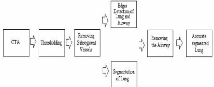
The stages, which have been followed while doing lung segmentation from CTA images at this work, are shown in figure 1.
Fig. 1 The diagram of lung segmentation method
C
World Academy of Science, Engineering and Technology International Journal of Biomedical and Biological Engineering
Vol:5, No:5, 2011
190
International Scholarly and Scientific Research & Innovation 5(5) 2011 scholar.waset.org/1307-6892/15507
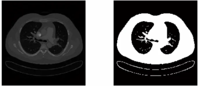
CTA images, which are in hands, are 250 as being 2D. The first step is thresholding the image. A thoracic CT contains two main groups of pixels: 1) high–intensity pixels located in the body (body pixels), and 2) low–intensity pixels that are in the lung and the surrounding air (non–body pixels). Due to the large difference in intensity between these two groups, thresholding leads to a good separation. In this study, thresholding has been tried out for the first time in a way that contains bigger parts than 700 HU. At the end of thresholding the new images are going to be in logical value.
Thresh=image>700;
Fig. 2 Thresholded image
In each of these new images, subsegment vessels exist in lung region. At the second step, this method has been used to get rid of these vessels: firstly each of 2D images has been considered one by one and each of components in the image have labeled with “connected component labeling algorithm”. Then, looking at the size of each labeled piece, items, whose pixel numbers are under 1000, were removed from the image. Figure 3.
Fig.3 Image that subsegment vessels are removed Next, the image in Figure 3 has been labeled with “connected component labeling algorithm”. The biggest size which is logical 1 is the patient’s body. This biggest size has been taken and the other parts have been removed from the image. And then the opposite of it has been gotten. So all of “0” turn into “1” and all of “1” turn into “0” Figure 4.
Fig. 4 Identifying lungs as logically
As the 1. or 512. pixels of the parts out of the body in the image which shown in Figure 4 is going to be logical 1, the
parts that achieve this condition have been removed and lung and airway are appeared like in Figure 5
Fig.5 segmentation of lung and airway
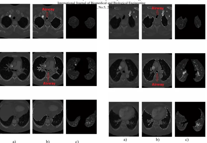
Due to the fact that airway in Figure 5 is going to be very small compared to the lung size, each of images have been labeled with “connected component labeling algorithm” and the component whose number of pixels are below 1000 have been determined as airways and then removed from the image. The last image in hand is the segmented form of target lung. Before airways removed, finding the edges of the image with sobel algoritm, it has been gathered to original image and the edges of lung and airway region have been shown in the original image Figure 6 (b). Also, by multiplying defined lung region with the original CTA lung image, original segmented lung image has been carried out. Figure 6 (c).
a) b) c) Fig. 6 a) Original CTA image b) Identified edges of airway and
lung c) Accurate segmented lung IV. CONCLUSION ANDDISCUSSION
The method, which has been tried at this work, applied to 30 different patients. Because of the fact that the segmentation in all CTA images (approximately 250) belonging to one patient is solved in a short time as 1-2 minutes, it has a fast working property. And it has been seen that the results at the end of applying adjust to the lung contours determined with hand by radiologist Dr. Sinan ùahin who is a doctor of Dr. Siyami Ersek Thoracic and Cardiovascular Surgery Training and Research Hospital, Istanbul,Turkey. With the help of this work, which performs lung segmentation in a fast way, lung vessel segmentation, detection of pulmonary emboly, finding nodules or segmentation of airways can be done in a faster way.
III. EXPERIMENTAL RESULTS
Different 2D images that belong to patient who is told at this work, the image results which have been taken from 2 different patients, and 3D image all of them are given at the following lines:
World Academy of Science, Engineering and Technology International Journal of Biomedical and Biological Engineering
Vol:5, No:5, 2011
191
International Scholarly and Scientific Research & Innovation 5(5) 2011 scholar.waset.org/1307-6892/15507
a) b) c)
Fig. 7 a) Original CTA image b) Identified edges of airway and lung c) Accurate segmented lung
a) b) c)
Fig. 8. a) Original CTA image b) Identified edges of airway and lung c) Accurate segmented lung
a) b) c)
Fig. 9 a) Original CTA image b) Identified edges of airway and lung c) Accurate segmented lung
a) b) c) Fig. 10 a) 3D images that belong to 1.patient b) 3D images that belong to 2.patient c) 3D images that belong to 3.patient
REFERENCES
[1] Kiraly A. P., Pichon E., Naidich D. P., and Novak C. L., “Analysis of arterial subtrees affected by pulmonary emboli,” in Medical Imaging 2004: Image Processing. Procs of the SPIE, 5370, pp. 1720–1729, May 2004.
[2] Y. Masutani, H. MacMahon, K. Doi, Computerized detection of pulmonary embolism in spiral CT angiography based on volumetric image analysis, IEEE Transactions on Medical Imaging 21 (12) (2002) 1517–1523.
[3] E. Pichon, C.L. Novak, A.P. Kiraly, D.P. Naidich, A novel method for pulmonary emboli visualization from high-resolution CT images, SPIE Medical Imaging 5367 (2004) 2004.
[4] R. Uppaluri, T. Mitsa, M. Sonka, E. Hoffman, and G. McLennan, “Quantification of pulmonary emphysema from lung computed tomography images,” Am. J. Respir. Crit. Care Med. 156, 248–254 _1997_.
[5] Y. Xu, M. Sonka, G. McLennan, J. Guo, and E. Hoffman, “MDCT-based 3-D texture classification of emphysema and early smoking related lung pathologies,” IEEE Trans. Med. Imaging 25, 464–475 _2006_.
World Academy of Science, Engineering and Technology International Journal of Biomedical and Biological Engineering
Vol:5, No:5, 2011
192
International Scholarly and Scientific Research & Innovation 5(5) 2011 scholar.waset.org/1307-6892/15507
[6] S. Armato III, M. Giger, and H. MacMahon, “Automated detection of lung nodules in CT scans: Preliminary result,” Med. Phys. 28, 1552– 1561 _2001_.
[7] Q. Li, F. Li, and K. Doi, “Computerized detection of lung nodules in thin-section CT images by use of selective enhancement filters and an automated rule-based classifier,” Acad. Radiol. 15, 165–175 _2008_. [8] R. Uppaluri, E. Hoffman, M. Sonka, P. Hartley, G. Hunninghake, and
G. Mclennan, “Computer recognition of regional lung disease patterns,” Am. J. Respir. Crit. Care Med. 160, 648–654 _1999_. [9] Y. Uchiyama, S. Katsuragawa, H. Abe, J. Shiraishi, F. Li, Q. Li, C.
Zhang, K. Suzuki, and K. Doi, “Quantitative computerized analysis of diffuse lung disease in high-resolution computed tomography,” Med. Phys. 30, 2440–2454 _2003_.
[10] I. Sluimer, P. Waes, M. Viergever, and B. Ginneken, “Computer-aided diagnosis in high resolution CT of the lungs,” Med. Phys. 30, 3081–3090 _2003_.
[11] I. C. Sluimer, A. M. R. Schilham, M. Prokop, and B. van Ginneken, Computer analysis of computed tomography scans of the lung: a survey, IEEE Transactions on Medical Imaging 25(4), pp. 385–405, (2006).
[12] C. Haider, B. Bartholmai, D. Holmes, J. Camp, and R. Robb, “Quantitative characterization of lung disease,” Comput. Med. Imaging Graph. 29, 555–563 _2005_.
[13] I. Sluimer, M. Prokop, and B. Ginneken, “Toward automated segmentation of the pathological lung in CT,” IEEE Trans. Med. Imaging 24, 1025–1038 _2005_.
[14] M. Prasad, M. Brown, S. Ahmad, F. Abtin, J. Allen, I. Costa, H. Kim, M. Mcnitt-Gray, and J. Goldin, “Automatic segmentation of lung parenchyma in the presence of disease based on curvature of ribs,” Acad. Radiol. 15, 1173–1180 _2008_.
World Academy of Science, Engineering and Technology International Journal of Biomedical and Biological Engineering
Vol:5, No:5, 2011
193
International Scholarly and Scientific Research & Innovation 5(5) 2011 scholar.waset.org/1307-6892/15507