TURCICA Acta Orthop Traumatol Turc 2008;42(3):178-183
Early results of autologous mononuclear bone marrow cell transplantation
in nontraumatic avascular necrosis of the femoral head
Travmaya bağlı olmayan femur başı osteonekrozunda
otolog konsantre mononükleer kemik iliği hücre naklinin erken dönem sonuçları
Omer KARATOPRAK,1 Mehmet Fatih KORKMAZ, Ayhan Nedim KARA,Abdullah GOGUS, Zekeriya Ugur ISIKLAR
Amaç: Erken dönem femur başı osteonekrozunda kor de-kompresyon ve otolog konsantre mononükleer kemik iliği hücre naklinin erken dönem klinik ve radyolojik sonuçları değerlendirildi.
Çalışma planı: Çalışmaya, Steinberg sınıflamasına göre evre I- II travmaya bağlı olmayan femur başı osteonekrozu olan dokuz hasta (1 kadın, 8 erkek; ort. yaş 46.5; dağılım 33-59) alındı. Kemik iliğinden elde edilen CD34 hücre konsantre-si, kor dekompresyon tüneli içerisinden femur başına enjekte edildi. Tüm olgular klinik olarak görsel ağrı skalası (GAS), Harris kalça skoru ve WOMAC osteoartrit indeksine göre değerlendirildi. Radyolojik kontrollerde, femur başında çök-me, koksofemoral eklem mesafesinde daralma, osteonekrotik bölgede artış olup olmadığı araştırıldı. Ortalama izlem süresi 27.2 ay (dağılım 24-38 ay) idi.
Sonuçlar: Ameliyat öncesi ile sonrası (24. ay) değerler karşı-laştırıldığında, GAS skoru 3.4±0.4’ten 1.2±0.6’ya, WOMAC osteoartrit indeksi 33±3’ten 11±6’ya gerilerken, Harrris kalça skoru 54’ten 92’ye yükseldi. Ameliyat öncesinde Steinberg sınıflamasına göre olguların ikisi evre I-B, dördü I-C, üçü evre II-A idi. Son kontrollerde ise bir olgu evre I-A’ya, diğer olgular ise evre 0’a geriledi. Radyografik değerlendirmede hiçbir olguda femur başında çökme, koksofemoral eklemde daralma görülmedi.
Çıkarımlar: Femur başı osteonekrozunda konsantre mono-nükleer kemik iliği hücre nakli eklem ağrılarını ve hastalığın ilerleyişini önleyerek, subkondral kırık oluşmasını engelle-mektedir; bu nedenle, özellikle evre I ve evre II olgularda seçilebilecek bir tedavi yöntemi olarak düşünülmelidir.
Anahtar sözcükler: Kemik iliği transplantasyonu; dekompres-yon, cerrahi; femur başı nekrozu/cerrahi; kalça eklemi/patoloji; osteonekroz/cerrahi.
Objectives: We evaluated early clinical and radiologic results of core decompression combined with autologous mononu-clear bone marrow cell implantation for early stage nontrau-matic avascular necrosis of the femoral head.
Methods: The study included nine patients (1 female, 8 males, mean age 46.5 years; range 33 to 59 years) with stage I-II nontraumatic avascular necrosis of the femoral head, ac-cording to the Steinberg classification. Bone marrow-derived CD34 cells were injected through a core decompression channel into the femoral head. Clinical assessment included a visual analog scale (VAS), Harris hip score, and the WOMAC Osteoarthritis Index. Radiologically, femoral head collapse, narrowing of the coxofemoral joint space, and the size of the osteonecrotic area were assessed. The mean follow-up was 27.2 months (range 24 to 38 months).
Results: Pre-and postoperative (24th month) evaluations showed that the mean VAS score and the WOMAC Osteoar-thritis Index decreased from 3.4±0.4 to 1.2±0.6, and from 33±3 to 11±6, respectively, with an increase in the Harris hip score (from 54 to 92). Preoperatively, two patients were Stein-berg I-B, four were I-C, and three were II-A. Finally, all the patients were stage 0 except for one patient who regressed to I-A. None of the patients exhibited femoral head collapse or narrowing of the coxofemoral joint space.
Conclusion: Autologous mononuclear bone marrow cell im-plantation relieves articular pain, prevents the progression of osteonecrosis, and hence subchondral fractures. Therefore, it may be treatment of choice particularly in stage I-II avascular necrosis of the femoral head.
Key words: Bone marrow transplantation; decompression, sur-gical; femur head necrosis/surgery; hip joint/pathology; osteone-crosis/surgery.
Correspondence / Yazışma adresi: Dr. Ömer Karatoprak. Kadikoy Florence Nightingale Hospital, Department of Orthopaedics and Traumatology,
34724 Kızıltoprak, Kadıköy, İstanbul. Phone: +90216 - 450 03 03 Fax: +90216 - 450 19 61 e-mail: [email protected]
Submitted / Başvuru tarihi: 23.11.2007 Accepted / Kabul tarihi: 06.05.2008
©2007 Türk Ortopedi ve Travmatoloji Derneği / ©2007 Turkish Association of Orthopaedics and Traumatology
1Kadikoy Florence Nightingale Hospital, Department of Orthopaedics and Traumatology;
Osteonecrosis of the femoral head (FH) is usually
seen by the second and fourth decade of the life.[1] The
aim of treatment is to prevent of the FH deformation
and to delay its degenerative changes.[2] The treatment
methods of osteonecrosis of FH can divide into two groups as invasive and non-invasive methods. Non-invasive methods are including drugs, electric stimu-lation, extracorporeal shock-wave therapy and
elect-romagnetic field application.[2] Invasive methods are
osteotomy and total hip replacement for the advanced stages. Core decompression can be used as a single procedure for the cases diagnosed in early stages or combined with vascular-non vascular bone grafts, physical agents like electromagnetic field and electric stimulation, bone marrow injection and biologic agents
like bone morphogenetic protein (BMP).[2]
Core decompression was used for the histological
diagnosis in 1964 by the Ficat and Arlet.[2,3] Later on,
it has performed as a surgical method which improves the venous circulation with decreasing the intraosse-os pressure in the intraosse-osteonecrintraosse-osis of FH. It has become the treatment of choice for the cases diagnosed in early
stage of disease.[2-5] The outcome of the core
decomp-ression is usually good in the cases without collapse of
FH and low disseminated disease.[2-6] While the results
have been reported as 23 % satisfactory with conserva-tive treatment, the results of only core decompression
have been reported between 62-72 % goo [3-6]
Autologous bone marrow transplantation has been performed for the treatment of osteonecrosis since
1990 and reported satisfactory results. [7] The effect of
the bone marrow transplantation in the osteonecrosis depends on the osteogenic effect of the transplanted mononuclear cells in the FH. This effect occurs with the angiogenic cytokines secreted from the injected bone marrow stromal cells to the FH and subsequent
angiogenesis.[7]
This prospective study evaluates the early results of the core decompression combined with bone marrow mononuclear cell transplantation in the treatment of early stage FH osteonecrosis.
Patients and methods
Nine hips diagnosed as osteonecrosis of FH with magnetic resonance imaging (MRI) and classified as
stage I and II according to Steinberg classification [8]
since there was no FH collapse. Core decompression combined with autologous concentrated bone
mar-row mononuclear cell transplantation was performed to the all hips (Table I). Of these patients eight were male, one was female with the average age 46.5 (bet-ween 33-59). The patients with post-traumatic osteo-necrosis were not included to this study.
Surgical technique
All patients operated on the radiolucent operating table in the prone position with both hip joints in the neutral rotation under the general anesthesia. Three cm. of skin incision was made on the ipsilateral iliac crest in order to obtain bone marrow. The bone mar-row aspiration needle was manually advanced into the iliac crest and 160 ml material aspirated with the 50 ml syringe send to the lab for the mononuclear cell separation within the bone marrow bag. Core de-compression was performed with classical technique, that is, decompression performed through 3 cm. inci-sion located just below of the greater trochanter. The 4.5 mm diameter drill bit aimed to the necrotic bone with A-P and frog leg position under fluoroscopic view and advanced into the subchondral bone as far as 2-3 mm to the joint line. The 3 mm drill bit was used in the necrotic area in order to prevent perforation of the joint cartilage. Decompression limits were checked with K-wire or drill bit under the fluoroscopic views before the completing of the operation. Simultaneo-usly, bone specula, fat cells and cell debris separated from the bone marrow aspirate by the centrifugation and filtration. Concentrated CD34 cells which are the progenitors of the hematopoietic system were obtai-ned from the mononuclear cell mixture by the cell se-parator. Specimens from this cell solution were taken for the microbiologic and serologic studies. After that, concentrated cells injected through the tunnel which opened during core decompression. The entrance of the tunnel closed with an allograft bone plug in order to prevent the leakage of the concentrated cells.
Low molecular weight heparin prescript for the thromboembolism prophylaxis to the all patients for three weeks. Postoperatively, patients allowed 50 % bearing of their weight with one crutch for three we-eks. Full weight bearing allowed after three weeks of the operation. All of the patients were evaluated with visual analog scale (VAS), Harris hip score and WO-MAC (Western Ontario and McMaster Universities) osteoarthritis index preoperatively and 3., 6., 12., 24.
months postoperatively. [9] Bilateral A-P and frog-leg
and postoperative follow-ups. In order to find out rate of the osteonecrotic area in the femoral head, the ratio between necrotic area and total area of femoral head was calculated in the midcoronal section of MRI. The same sums were calculated in midaxial section of MRI and then average of both calculations was ob-tained. While the flattening of the FH and hip joint space were evaluated from direct x-rays, amount of the osteonecrosis was assessed with Steinberg Clas-sification according to MRI (Figure 1-3). Average follow-up was 27.2 months (between 24-38).
Results
The average of the recruited bone marrow was 163.6 ml (between 143-213ml) and the mean of the in-jected stem cells was 483.1 µl (165-938 µl). Contrast material did not used in any case and only one tunnel was created in all cases. We compared the preopera-tive and postoperapreopera-tive last follow-up values for clini-cal evaluation. VAS score improved from 3.4±0.4 to 1.2±0.6, Harris hip increased from 54 (between 46-94) to 92 (between 89-98), WOMAC osteoarthritis index decreased from 33±3 to 11±6.
Preoperatively, of the cases two were stage I-B, four were I-C, three were II-A according to Steinberg Classification. Of the patients’ one case improved to stage I-A, rest of the cases improved to stage O in the postoperative last follow-up. Neither flattening of fe-moral head nor hip joint narrowing was encountered in all patients.
Three cases complained of donor site pain in early postoperative period but it resolved spontaneously
Table 1. Steinberg Classification for the osteonecrosis of femoral head [8]
Stage 0 Radiogram, bone scan and MRI are normal
Stage I Radiogram is normal. Bone scan and/or MRI is abnormal A-Mild (affected area of femoral head is <15 %)
B-Moderate (affected area of femoral head is 15-30 %) C-Severe (affected area of femoral head is >30 %) Stage II Cystic and sclerotic changes of femoral head
A-Mild (affected area of femoral head is <15 %) B-Moderate (affected area of femoral head is 15-30 %) C-Severe (affected area of femoral head is >30 %)
Stage III Subchondral collapse without flattening of femoral head (Crescent sign) A-Mild (affected area of femoral head is <15 %)
B-Moderate (affected area of femoral head is 15-30 %) C-Severe (affected area of femoral head is >30 %) Stage IV Flattening of femoral head
A-Mild (<15 % of joint surface and <2mm collapse) B-Moderate (15 -30 % of joint surface and 2-4mm collapse) C-Severe (>30 % of joint surface and >4mm collapse) Stage V Narrowing of the joint surface and acetabular changes
A-Mild B-Moderate C-Severe
Stage VI Severe degenerative changes MRI: Magnetic resonance imaging.
Figure 1. 45 year’s old male, appealed with a pain on right side of hip, preoperative AP X-ray; .
within two weeks. There was no secondary alloim-munation, pulmonary embolism, trochanteric fractu-re and complication of anesthesia.
Mild sclerosis in femoral head and two small cysts which can only be noticed in magnification on the screen were determined in one case with stage II-A. These findings were disappeared in postoperative follow-ups. Rest of the patients was accepted as stage I since the preoperative direct x-rays were normal.
Discussion
The treatment of the osteonecrosis of FH is deter-mined according to patient’s age, activity level and
general health status.[4] The factors affecting the
treat-ment are extent and location of the necrotic area, flat-tening of FH and its amount, acetabular involvement.
[1,4] Popular surgical methods are core decompression
or core decompression combined with the application of biologic materials like bone graft, demineralized
bone matrix and bone marrow through the tunnel.[1,4,5]
Proximal femoral osteotomy or total hip replacement
is required for severe cases.[2,4,5]
Better results have been reported when the core decompression combined with biological agents such as BMP, concentrated bone marrow cell or physical
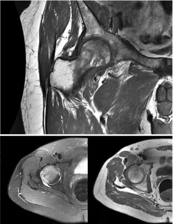
Figure 3. Core decompression and autologous mononuclear bone marrow cell implantation performed. MRI of after 24 months. He was evaluated as stage 0.
Figure 2. Preoperative MRI, stage IC with respect to Steinberg classification.
agents such as electric stimulation. [1-7,10] Lieberman et al. applied allograft and human derived BMP toget-her to the 17 hips of the patients with stage II-A and II-B osteonecrosis. Authors reported that total hip replacement required in three patients and there was no clinical and radiological deterioration 86 % of the patients at the end of 53 months follow-up. The oste-onecrotic area is healed after the core decompression. However, the healing process is usually not be comp-leted due to the decreasing of the amount and activity of the mesenchymal cells located in the proximal
fe-mur.[11] Biological healing is occurred with the
colo-nization of bone cells which differentiated from the hematopoietic stem cells based in necrotic area. Sin-ce 1990’s, Core decompression has been applied with autologous bone marrow since failure of the results
with single use. [7,12]
Bone marrow transplantation initially was per-formed in the treatment of nonunion. After that, it has been utilized for the treatment of osteonecrosis.
[7] There are mesenchymal stem cells in the capacity
of differentiate into the hematologic cells and osteob-lasts in the bone marrow. Stem cells are
undifferenti-ated cells that have regeneration potential. [13,14]
Diffe-rent stem cells have been isolated from embryo, fetal
tissues and cord blood. [14] Fetal cells differentiate to
the adult type stem cells with the maturation. Adult stem cells produce the cells of tissue where they lo-cated. Stem cells can differentiate to the specific cell where he derived. On the other hand it can also diffe-rentiate to another cell in case of a suitable biological stimulus.[14]
We used allograft bone plug for closing the access hall of the tunnel and preventing the leaking out of the concentrated CD34 cells. The reason for the using of allograft were to avoid the donor site complicati-ons and the possibility of the affecting the results by the autologous bone graft. There is a debate on inc-reasing of the intraosseos pressure due to the closure with the bone plug of the tunnel after injection of the
concentrated CD34 in it. [7,10] Some of the CD34 leak
out from the tunnel just after the injection. Therefore the pressure does not increase after the plugging of
the tunnel.[7,10,12] The leaking out of the whole
con-centrated CD34 was also advocated. But the radi-onuclide studies revealed that, the most part of the
injected cell solution stay in the bone.[7,10,12] In current
study we could not measure the intraosseos
pressu-re. Nevertheless, there was no decreasing of oxygen saturation, abnormal changes of heart rate and blood pressure per operatively. It was observed that oste-onecrosis signs were improved during the follow-up examinations both clinically and radiologically.
Camp and Colwell[15] reported 10% fracture
comp-lication within the patients performed only core de-compression. Most of the studies reported between 8-12 mm tunnel diameters for the core
decompres-sion. [1,3,5] Israelite et al. [5] advocated two tunnels in
6 mm diameter stopped 5 mm to the joint cartilage. Full weight bearing was allowed between six weeks and 3 months according to these studies. We used 4.5 mm diameter drill bit in our series of the patient. We allowed full weight bearing in postoperative third week because of the low risk of fracture. We did not encounter fracture complication in these cases.
Her-nigou and Beaujean [10] applied core decompression
with bone marrow injection to the 189 hips. In this study, of the patients 145 were stage I and II and total hip replacement was required for only nine of them at the end of seven years of follow-up. Gangji et al.
[16] performed core decompression and bone marrow
injection to the 13 patients with grade I and II oste-onecrosis of the FH. Pain and WOMAC osteoarthri-tis index of the patients improved within two years follow-up and they did not need to do total hip repla-cement for any of the patients.
Our results are promising for the concentrated bone marrow transplantation with core decompressi-on in the cases without collapse of the femoral head although the number of the cases and follow-up dura-tion are relatively low. VAS score and WOMAC os-teoarthritis index of the patients improved and Harris Hip score increased postoperatively. There were no osteonecrosis in the control MRI of the patients at the 24. month follow-up. Core decompression with the autologous bone marrow injection is the treatment of choice for the early stage of the osteonecrosis of the femoral head because of its lower cost and mor-bidity.
In conclusion, bone marrow transplantation pre-vents the subchondral fracture due to the inhibition of progression of disease and hip pain related to os-teonecrosis. Concentrated mononuclear bone marrow injection is an encouraging treatment method for the non traumatic osteonecrosis of FH because of its suc-cess rate.
References
1. Keizer SB, Kock NB, Dijkstra PD, Taminiau AH, Nelissen RG. Treatment of avascular necrosis of the hip by a non-vascularised cortical graft. J Bone Joint Surg [Br] 2006; 88:460-6.
2. Petrigliano FA, Lieberman JR. Osteonecrosis of the hip: novel approaches to evaluation and treatment. Clin Orthop Relat Res 2007;465:53-62.
3. Lieberman JR, Conduah A, Urist MR. Treatment of os-teonecrosis of the femoral head with core decompression and human bone morphogenetic protein. Clin Orthop Relat Res 2004;(429):139-45.
4. Dailiana ZH, Toth AP, Gunneson E, Berend KR, Urbaniak JR. Free vascularized fibular grafting following failed core decompression for femoral head osteonecrosis. J Arthro-plasty 2007;22:679-88.
5. Israelite C, Nelson CL, Ziarani CF, Abboud JA, Landa J, Steinberg ME. Bilateral core decompression for osteone-crosis of the femoral head. Clin Orthop Relat Res 2005; 441:285-90.
6. Etienne G, Mont MA, Ragland PS. The diagnosis and treat-ment of nontraumatic osteonecrosis of the femoral head. Instr Course Lect 2004;53:67-85.
7. Hernigou P, Poignard A, Manicom O, Mathieu G, Rouard H. The use of percutaneous autologous bone marrow trans-plantation in nonunion and avascular necrosis of bone. J Bone Joint Surg [Br] 2005;87:896-902.
8. Mont MA, Marulanda GA, Jones LC, Saleh KJ, Gordon N, Hungerford DS, et al. Systematic analysis of classification systems for osteonecrosis of the femoral head. J Bone Joint Surg [Am] 2006;88 Suppl 3:16-26.
9. Anderson JG, Wixson RL, Tsai D, Stulberg SD, Chang RW. Functional outcome and patient satisfaction in total knee patients over the age of 75. J Arthroplasty 1996;11:831-40. 10. Hernigou P, Beaujean F. Treatment of osteonecrosis with
autologous bone marrow grafting. Clin Orthop Relat Res 2002;(405):14-23.
11. Hernigou P, Beaujean F, Lambotte JC. Decrease in the mesenchymal stem-cell pool in the proximal femur in cor-ticosteroid-induced osteonecrosis. J Bone Joint Surg [Br] 1999;81:349-55.
12. Hernigou P, Manicom O, Poignard A, Nogier A, Filippini P, De Abreu L. Core decompression with marrow stem cells. Operative Techniques in Orthopaedics 2004;14:68-74. 13. Ural AU. Kök hücreler. TOTBİD Dergisi 2006;5:140-5. 14. Kömürcü M, Özkan H. Mezenkimal kök hücre ve
ortope-dide kullanımı. TOTBİD Dergisi 2006;5:130-9.
15. Camp JF, Colwell CW Jr. Core decompression of the femo-ral head for osteonecrosis. J Bone Joint Surg [Am] 1986; 68:1313-9.
16. Gangji V, Hauzeur JP, Matos C, De Maertelaer V, Toungouz M, Lambermont M. Treatment of osteonecrosis of the femo-ral head with implantation of autologous bone-marrow cells. A pilot study. J Bone Joint Surg [Am] 2004; 86:1153-60.
![Table 1. Steinberg Classification for the osteonecrosis of femoral head [8]](https://thumb-eu.123doks.com/thumbv2/9libnet/4304966.69890/3.854.449.800.811.1039/table-steinberg-classification-osteonecrosis-femoral-head.webp)