ABSTRACT
In this study we describe a case with interface fluid syndro-me developing after laser in situ keratomileusis (LASIK) result from steroid-induced intraocular pressure (IOP) elevation.The aim of this study was to emphasize the im-portance of ocular manifestations and clinical course of interface fluid syndrome which leads to inaccurately low central applanation tonometry measurements that obscure the diagnosis of steroid-induced glaucoma. Early recogni-tion and treatment of this syndrome are essential to avoid complications associated with prolonged elevated IOP. Key Words: Flep interface fluid syndrome, Lasik.
ÖZ Bu çalışmada laser in situ keratomileusis (LASIK) cerrahisi sonrası gelişen, steroidle indüklenmiş göz içi basınç (GİB) artışının neden olduğu flep ara yüzey sıvı birikimi sendrom-lu bir hasta tariflenmiştir. Çalışmamızın amacı aplanasyon tonometresi ile yalancı olarak düşük GİB ölçümüne neden olan ve bu yüzden steroidle uyarılan glokomu maskeleyen flep ara yüzey sıvı birikimi sendromunun oküler bulguları- nın ve klinik seyrinin önemine vurgu yapmaktır. Bu sendro- mun erken tanı ve tedavisi artmış GİB’na bağlı komplikas-yonlardan kaçınmak için esastır.
Anahtar Kelimeler: Flep ara yüzey sıvı birikimi sendromu, Lasik.
Glo-Kat 2010;5:114-116
INTRODUCTION
Interface fluid syndrome may occur secondary to steroid-induced elevation of intraocular pressure (IOP) after laser in situ keratomileusis (LASIK).1-5 Interface fluid
syndrome after LASIK is sometimes misdiagnosed as dif-fuse lamellar keratitis (DLK) and treated improperly with topical steriods which may lead to worsening of the con-dition. High IOP may cause damage to the optic nerve, leading to vision loss.2,4
Received : March 04, 2010 Accepted : April 16, 2010 1- M.D. Assistant Professor, İstanbul Bilim University, Avrupa Florance Nighthingale Hospital Eye Clinic İstanbul/TURKEY ARTUNAY Ö., [email protected] YUZBASIOGLU E., [email protected]
2- M.D., İstanbul Bilim University, Avrupa Florance Nighthingale Hospital Eye Clinic İstanbul/TURKEY
RASIER R., [email protected]
3- M.D. Assistant, İstanbul Bilim University, Avrupa Florance Nighthingale Hospital Eye Clinic İstanbul/TURKEY
SENGUL A., [email protected]
4- M.D. Professor, İstanbul Bilim University, Avrupa Florance Nighthingale Hospital Eye Clinic İstanbul/TURKEY
BAHÇECİOĞLU H., [email protected] Correspondence: M.D.Assistant Professor, Özgür ARTUNAY
İstanbul Bilim University, Avrupa Florance Nighthingale Hospital Eye Clinic İstanbul/ TURKEY Geliþ Tarihi : 04/03/2010 Kabul Tarihi : 16/04/2010 1- İstanbul Bilim Üniversitesi, Göz Hastalıkları A.D., İstanbul, Yrd. Doç. Dr. 2- İstanbul Bilim Üniversitesi, Göz Hastalıkları A.D., İstanbul, Uz. Dr. 3- İstanbul Bilim Üniversitesi, Göz Hastalıkları A.D., İstanbul, Asist. Dr. 4- İstanbul Bilim Üniversitesi, Göz Hastalıkları A.D., İstanbul, Prof. Dr.
A Glaucoma Case with Masked Increasing Intraocular
Pressure due to Flep Interface Fluid Accumulation
After Refractive Surgery
Refraktif Cerrahi Sonrası Gelişen Flep Ara Yüzey Sıvı Birikimi Nedeniyle
Göz İçi Basınç Artışı Maskelenen Glokom Olgusu
Özgür ARTUNAY1, Rifat RASİER2, Alper ŞENGÜL3, Erdal YÜZBAŞIOĞLU1, Halil BAHÇECİOĞLU4
Glo-Kat 2010;5:114-116 Artunay et al. 115
CASE PRESENTATION
A 24-year-old man was referred to us underwent bi-lateral LASIK surgery for correction of mild myopic astig-matism using 160 µm flaps: -3.00 (175º-0.75) in the right eye and -3.50 (175º-1.50) in the left eye with best corrected visual acuity of 20/100 in each eye. Preopera-tive pachymetry revealed a 550 µm thickness in the right eye and a 540 µm thickness in the left eye. IOP measured by aplanation tonometry was 18 mmHg in the right eye and 17 mmHg in the left eye preoperatively. After un-eventful LASIK prosedure, prednisolone acetate, 1%, and ciprofloxacin hydrochloride, 0.3% (Ciloxan, Alcon Labo-ratories, Ft. Worth, TX), eye drops were started four times daily as part of the normal postoperative regimen. By 12 days postoperatively, trace interface debris inferotempo-rally and stage 1 DLK-like appearance was present in the right eye. Left eye was normal. Uncorrected visual acuity was 20/20, and prednisolone has been continued. IOP measured by aplanation tonometry was 19 mmHg in the right eye and 14 mmHg in the left eye postoperatively. At the postoperative second week visit, uncorrected visual acuity had decreased to 20/30 in right eye, and the DLK-like appearance had worsened. Prednisolone drops were increased to every 2 hours around the clock. There was no improvement on the higher steroid regimen, so the flap was lifted and irrigated at postoperative third week. Steroid drops were continued to every 2 hours. Four days later neither the clinical examination nor the symptoms had improved. IOP measured by aplanation tonometry was 20 mmHg in the right eye and 15 mmHg in the left eye at this moment. Slit-lamp biomicroscopy revealed an optically clear fluid-filled interface between the flap and stromal bed with peripheric microcystic corneal edema
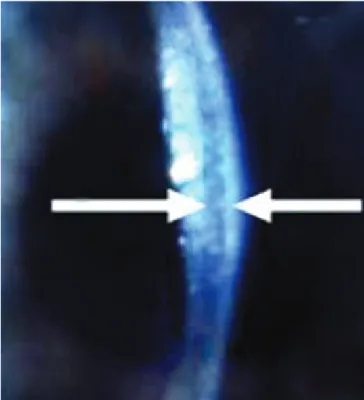
(Figure 1). Anterior segment OCT showed that the fluid accumulation was exist under the LASIK flap (Figure 2). After this observation we decided to measured IOP by schiotz tonometry. It was 40 mmHg. We measured IOP again by aplanation tonometry at corneal periphery to minimize altering effects of interface fluid on intraocular pressure. It was 44 mmHg at this time. This observation had led us to believe that steroid-induced elevation of IOP has occurred with masking interface fluid. Oral ac-etazolamide, 250mg three times daily, on dorzolamide hydrochloride 2%/timolol maleate 0.5% (Cosopt,Merck Laboratories, West Port, PA) twice daily, were started and prednisolone drops were tapered to three times daily. Best-corrected visual acuity had improved to 20/20 by the third day and the interface fluid had nearly resolved (Figure 3). Resolution of the interface fluid was seen on repeated OCT. (Figure 4). IOP using aplanation tonom-etry was 13 mmHg centrally and 16 mmHg peripherally. Topical steroids and antiglaucomatous therapy were gradually discontinued after three weeks. Visual field testing 6 months postoperatively revealed mild superior nasal field defects in the right eye.
DISCUSSION
The first report of interface fluid developing after LASIK was reported by Lyle and Jin.1 This case featured
steroid-induced IOP elevation and falsely low IOP read- ing from applanation tonometry. Subsequently, Najman-Vainer et al. reported a case of interface fluid after LASIK surgery in a patient with DLK without epithelial ingrowth who went on to have severe optic neuropathy develop from steroid-induced elevated glaucoma.2 McLeod
et al however, reported two cases of corneal interface flap edema associated with anterior uveitis after LASIK.3
These cases were documented as interface fluid collec-tion and flap edema represent result from complicacollec-tions attributable to uveitis after LASIK.
Normally, highly elevated IOP causes transudation of fluid across the endothelium. This fluid typically col-lects in the basal epithelium, presenting clinically as mi-crocystic edema.6 In the post-LASIK cornea, however, the
central stroma has a potential space created by the mi-crokeratome incision. In contrast to the epithelium, which is tightly adherent, this potential space requires little force to open and is commonly opened iatrogenically to per-form LASIK enhancement surgery. It has been suggested that the fluid migrates forward through the stoma, it
col-Figure 1: Diffuse lamellar keratitis DLK- like appearance. Figure 2: Slit-lamp biomicroscopy revealed an optically clear fluid-fil-led interface between the flap and stromal bed (white arrows).
116 A Glaucoma Case with Masked Increasing Intraocular Pressure due to Flep Interface Fluid Accumulation...
lects in this potential space, because it is easier to open than are the tight junctions between corneal epithelial cells.3-5 This fluid collects in the interface and expands it,
causing the fluid that we observed clinically.
DLK occasionally follows LASIK and is seen with dif-fuse mild inflammation confined to the lamellar inter-face. The treatment of choice for DLK remains topical corticosteroids.7,8 Çagıl et al investigated the frequency
of steroid-induced IOP elevation after laser-assisted sub-epithelial keratectomy for myopia and they sugessted that due to the high frequency of steroid-induced IOP elevation, corrected IOP levels must be closely monitored after LASEK procedures.9 As regard as our case,
cortico-steroids were continued to treat spuriously inflammation. We believe that the apparent long-term inflammation was actually corneal edema related to the interface fluid rather than DLK. We hypothesized that the fluid resulted from transudation of aqueous across the stromal bed, resulting from steroid-induced elevated intraocular pres-sure. The role of endothelial function in developing inter-face fluid associated with elevated IOP is uncertain. How-ever, It has been postulated that post-LASIK eyes with en-dothelial dysfunction may have interface fluid develop at a lower IOP than normal eyes.4 It has been also
hypoth-esized that the uveitis related to LASIK may also have led to elevated IOP thus increasing demand on endothelial pump function.3 As a result of all these reasons, fluid
col-lected in the potential space between the flap and bed, causing erroneous applanation tonometry readings. The interface fluid caused the falsely low central Goldmann applanation tonometry measurement. In our case, el-evated IOP was misdiagnosed, because the standard
central measurement of IOP by Goldmann tonometry was erroneously very low than the actual..If elevated IOP was misdiagnosed, high IOP may cause damage to the optic nerve, leading to vision loss. Beacuse of this reason refractive surgeon must be aware of this clinical entity. A patient who has been on topical steroids for treatment of DLK and has not had resolution in 10 days should be carefully evaluated for interface fluid. In addition, a diagnosisof DLK made for the first time more than 10 days after the surgery should prompt an examination for interface fluid. Because of the Goldmann applanation tonometer in theory will give a falsely low reading of the pressure of the fluid in the interface. On the other hand one should carefully examine the peripheral cornea for the presence of microcystic edema. In any suspicious case, IOP should be measured by different types of to-nometers and/or the the aplanation tonometry is should be applied at the corneal periphery to minimize altering effects of the interface fluid on intraocular pressure. We believe this complication warrants early recognition and special attention to IOP measurement to avoid potentially devastating vision loss.
REFERENCES/KAYNAKLAR
1. Lyle WA, Jin GJC.: Interface fluid associated with diffuse lamellar keratitis and epithelial ingrowth after laser in situ keratomileusis. J Cataract Refract Surg. 1999;25:1009-1012.
2. Najman-Vainer J, Smith RJ, Maloney RK.: Interface fluid after LASIK: misleading tonometry can lead to end-stage glaucoma [letter]. J Cataract Refract Surg. 2000;26:471-472.
3. McLeod SD, Mather R, Hwang DG, et al.: Uveitis-associated flap edema and lamellar interface fluid collection after LASIK. Am J Ophthalmol. 2005;139:1137-1139.
4. Hamilton DR, Manche EE, Rich LF, et al.: Steroid-induced glau-coma after laser in situ keratomileusis associated with interface fluid. Ophthalmology. 2002;109:659-665.
5. Protellinha W, Kuchenbuk M, Nakano K, et al.: Interface fluid and diffuse corneal edema after laser in situ keratomileusis. J Refract Surg. 2001;17:192-195.
6. Klyce SD, Beuerman RW.: Structure and function of the cornea. In: Kaufman HE, Barron BA, McDonald MB, Waltman SR, eds. The Cornea, New York: Livingstone. 1988;38-47.
7. Clark AF.: Steroids, ocular hypertension, and glaucoma. J Glau-coma. 1995;4:354-369.
8. Glaucoma. Basic and Clinical Science Course 2000-2001. Sec-tion 10. San Francisco: American Academy of Ophthalmology. 2000;20.
9. Çagil N, Çakmak HB, Özalp PK, et al.: Steroid-Induced Intraocu-lar Pressure Elevation After LASEK. J Glaucoma-cataract. 2008;3: 117-123.
Figure 3: Anterior segment OCT showing the fluid accumulation under the LASIK flap (white arrow).
Figure 4: The interface fluid had nearly resolved and recovery of cor-neal transparency is seen obviously after treatment.