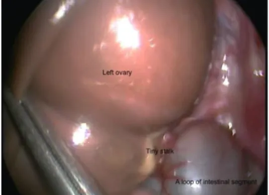
Laparoscopic Management of Synchronous Bilateral Ovarian Torsion in a Neonate
Tam metin
Şekil
Benzer Belgeler
UĞUZ A., GÜRCÜ B., KAZIMİ M., NART D., ÖZKÖK S., GÖKER E., TEKEŞİN O., YILMAZ BARBET F., ZEYTUNLU M., ÇOKER A. Ulusal Cerrahi Kongresi 2008, Türkiye, 28 - 31 Mayıs
The presence of hyponatremia has significant association with fever, increased acute- phase reactants, need for hospitalization, mortality, length of hospital stay (LOS), need
1 İstanbul University Faculty of Medicine, Department of Pediatric Intensive Care, İstanbul, Turkey 2 İstanbul University Faculty of Medicine, Department of Pediatric
Based on these studies, we aimed to describe demograph- ics, clinical characteristics, severity of the disease, and out- comes of confirmed and probable COVID-19 patients ad- mitted
The data gathered from the patients admitted with complaints of substance abuse and patients who had substances detected in their urine using screening tests despite having
Pediatric Bilateral Pheochromocytoma and Experience of Laparoscopic Cortical Sparing Adrenalectomy.. DO I:
Aim: The aim of this study was to evaluate the proportion of pediatric surgeons who committed medical malpractice (MM) while on duty in hospitals, whether this proportion
2 Department of Pediatric Surgery, Faculty of Medicine, Dokuz Eylül University İzmir, Turkey,.. 3 Department of Pediatric Oncology, Faculty of Medicine, Dokuz Eylül University
