Orthopaedic manifestations of glutaric acidemia
Type 1
Ahmet Imerci
1Kevin A. Strauss
2Geovanny F. Oleas-Santillan
3Freeman Miller
3 AbstractPurpose: Glutaric acidemia type 1 (GA1), a rare hereditary metabolic disease caused by biallelic mutations of GCDH, can result in acute or insidious striatal degeneration within the first few years of life. We reviewed the orthopaedic sequelae and management of 114 neurologically injured patients with a confirmed molecular diagnosis of GA1.
Methods: We performed a retrospective chart review spanning 28 years identifying 114 GA1 patients, most from the Old Or-der Amish population of Lancaster County, Pennsylvania, who were homozygous for a pathogenic founder variant of GCDH (c.1262C>T). We collected demographics, medical comorbidi-ties, muscle tone patterns, Gross Motor Function Classification System level, gastrostomy tube status, seizure history, inpatient events, orthopaedic diagnoses and operative characteristics. Results: Over an average follow-up of 4.7 ± 3.4 years, 24 (21%) of 114 patients had musculoskeletal problems requir-ing orthopaedic consultation. Scoliosis (n = 14), hip dislo-cation (n = 8/15 hips), hip subluxation (n = 2/three hips), and windswept hip deformity (n = 2) in the spine and hip joint were most common. In total, 35 orthopaedic surgeries were performed in 17 (71%) patients. The most common pri-mary operations were one-stage procedures with proximal femoral varus derotation osteotomy and/or pelvic osteotomy (n = 8/14 hips) for subluxation or dislocation. In all, 11 pa-tients had posterior spinal fusion for severe scoliosis. With the recommended metabolic management, there were no dis-ease-specific complications in this cohort.
Conclusions: Children with GA1 who have static striatal le-sions are at risk for musculoskeletal complications, especially
1 Department of Orthopaedics and Traumatology, Faculty of
Medicine, Mugla Sitki Kocman University, Mugla, Turkey
2 Clinic for Special Children, Strasburg, Pennsylvania, USA 3 Nemours/Alfred I. duPont Hospital for Children, Wilmington,
Delaware, USA
Correspondence should be sent to Freeman Miller, Department of Orthopaedics, Nemours/Alfred I. duPont Hospital for Children, 1600 Rockland Road, Wilmington, DE 19803, USA.
E-mail: [email protected]
scoliosis and hip dislocation, and appropriate operative management requires consultation with a metabolic spe-cialist with specific considerations for fluid management and nutrition.
Level of Evidence: IV
Cite this article: Imerci A, Strauss KA, Oleas-Santillan GF, Miller F. Orthopaedic manifestations of glutaric acidemia Type 1. J Child Orthop 2020;14:473-479. DOI: 10.1302/1863-2548.14.200059
Keywords: glutaric acidemia; scoliosis; hip surgery;
operative management
Introduction
Glutaric acidemia type 1 (GA1; OMIM #231670) is a dis-order of systemic and cerebral organic acid metabolism caused by biallelic variants of glutaryl-CoA dehydrogenase (GCDH), which encode the mitochondrial flavin-depen-dent GCDH that mediates degradation of lysine, hydrox-ylysine and tryptophan.1 The birth incidence of GA1 is approximately one per 90 000 worldwide,2 but is much higher among certain endogamous groups such as the Old Order Amish of North America,3 a modern religious sect descended from a few hundred Swiss Anabaptists who immigrated to Pennsylvania during the eighteenth century.4 Within certain Amish demes, a pathogenic
GCDH c.1262C>T founder mutation has reached carrier
frequencies of approximately 10%,5 resulting in disease incidence rates as high as one per 400 births. High-risk
GCDH founder alleles are also found in other endogamous
populations such as the Oji-Cree natives of Ontario6 and ‘Travelers’ of Ireland.7
Neuronal GCDH deficiency results in proximal accu-mulation of glutaryl-CoA and its neurotoxic derivatives glutaric (GA) and 3-hydroxyglutaric (3HGA) acids,8 which become concentrated in brain tissue.9-13 Without presymp-tomatic detection and appropriate treatment, cerebral
GCDH deficiency predisposes to sudden, histologically
selective, and developmentally restricted degeneration of medium spiny neurons within the lentiform nuclei.14 More than 80% of untreated children develop striatal lesions,15 which typically strike within the first two years of life.15-17 These encephalopathic crises most often manifest as sud-den motor regression during an acute infectious illness but can occur in the absence of an apparent trigger and may even happen in utero.16,18
Outcomes for GA1 have improved considerably over the last two decades; with the combination of newborn screening for glutarylcarnitine (C5DC), adherence to a lysine-restricted/arginine-enriched prescription diet,19,20 and inpatient therapy during intercurrent illnesses,18,21 fewer than 10% of GA1 patients develop brain injury.19 Those who remain neurologically healthy until their sec-ond birthday face an excellent long-term prognosis.22,23 Nevertheless, the risk for striatal degeneration remains high for patients born in resource-limited settings who do not have access to tandem mass spectrometry-based new-born screening or prescription medical foods.8,24
Regardless of their timing or mechanism, static stria-tal lesions result in a complex extrapyramidal movement disorder that is the principal determinant of clinical out-come.8,16-19,22 Severe, generalized dystonia is the most common motor pattern observed among neurologically injured GA1 patients, and entrains serious gastrointesti-nal, pulmonary and musculoskeletal complications that exact a heavy disease burden.22,25 The orthopaedic sur-geon can play a critical role in alleviating this burden. Here, we review orthopaedic complications and their surgical management in a large cohort of GA1 patients treated at a single tertiary care centre.
Methods
Following institutional review board approval, we retro-spectively collected data on 114 patients found within our institutional database who had a confirmed diag-nosed of GA1. For subjects born between 1988 and 1994, the diagnosis was based on a characteristic clinical phe-notype paired with detection of GA and 3HGA in urine by gas chromatography-mass spectroscopy. Detection of a pathognomonic metabolite (C5DC) using tandem mass spectrometry was incorporated into Pennsylvania newborn screening in 1994, and detection of the GCDH c.1262C>T founder allele was introduced as a reflex sec-ond-tier screening test in 1999. Sanger sequencing of
GCDH was performed to confirm neonatal screening
results or corroborate a clinical diagnosis of GA1 in older symptomatic patients.
Using our institution’s inpatient and outpatient elec-tronic medical records system, we extracted data about the method and age of diagnosis, current age, Gross Motor Function Classification System (GMFCS) score, medical comorbidities, gastrostomy tube status, seizure history, orthopaedic diagnoses, surgical interventions and postoperative follow-up. The large majority of physical examinations and operative decisions were conducted by a single senior pediatric orthopaedic surgeon (FM).
Indications for reconstructive surgery of a subluxed or dislocated hip included severe movement restriction,
difficulty in perineal care, or pain with ambulation, transfers or sitting. The primary surgical technique was proximal femoral varus derotation osteotomy (VDRO) performed in a single stage. The reconstructive proce-dure combined routine varus shortening osteotomy with soft-tissue lengthening26 and acetabular reconstruction with peri-ilial pelvic osteotomy27 commonly used in chil-dren with cerebral palsy. No hip spicas were used. Phys-ical therapy commenced on the first postoperative day and hip movement was allowed as much as the patient could tolerate. A smooth perioperative transition typically required aggressive management of both pain and the movement disorder using epidural blocks, oral and intra-venous analgesics, and high doses of diazepam.
When structural scoliosis was evident on physical examination, patients were evaluated with sitting whole spine radiographs; spinal curvature ≥ 60 degrees was typically considered an indication for fusion. The strategy for posterior spinal fusion (PSF) followed rules of scolio-sis secondary to cerebral palsy, meaning all curves were fused from T1 or T2 to the pelvis. These patients similarly needed careful postoperative monitoring to control pain and exacerbation of extrapyramidal movements while also mitigating any risk for metabolic instability.
Statistical analysis
Parametric and nonparametric analyses were performed. Descriptive and frequencies statistics were used to describe the population by mean and standard deviation. Statistical analysis was performed using SPSS v25 (IBM, Armonk, New York).
Results
Our retrospective review included a total of 114 children (50% female) diagnosed with GA1 during a 28-year period from 1988 to 2018 (Table 1). Mean age at follow-up was 11.9 ± 9.0 years (range six months to 40 years). In all, 24 (21%) GA1 patients had significant orthopaedic pathol-ogy on physical examination. The most common prob-lems were severe scoliosis (n = 14) and abnormalities of the hip joint, including dislocation (n = eight patients/15 hips), subluxation (n = two patients/three hips), wind-swept deformity (n = 2), and dysplasia (n = 1) (Table 2). Although 48% of the patients in this cohort were GMFCS I (normal motor function) and 22% had milder motor problems (GMFCS II or III), primarily patients with severe impairments (GMFCS IV-V) developed significant ortho-paedic deformities requiring surgical treatment (Table 1).
A total of 35 surgeries were performed in 17 (71%) of 24 patients with musculoskeletal pathology. The mean age at first operation was 13.8 ± 4.8 (range six months to 25 years) and mean postoperative follow-up was 4.7
± 3.4 years (range six months to 10 years). Nine (38%) individuals required multiple procedures (two surgeries (n = 3) three surgeries (n = 4), five surgeries (n = 2)) and the mean interval between the first and second operation was 3.4 ± 1.4 years (range nine months to five years). The most common primary surgical procedures were PSF for severe scoliosis (n = 11) and one-stage VDRO and/or
pelvic osteotomy for subluxation or dislocation of the hip (n = eight patients/14 hips) (Table 3). Three patients expe-rienced significant postoperative complications: blade plate prominence caused skin irritation requiring removal in three hips of two patients and one individual (patient 5, Table 3) underwent revision VDRO due to recurrent hip dislocation.
Table 1 Demographic characteristics of overall cohort of patients with glutaric aciduria type 1 (GA1) Medical comorbidities Total number of patients with orthopaedic aspects
(n = 24) Total number of patients with GA1(n = 114)
Normal muscle tone, n (%) 0 (0) 51 (45)
Hypotonic type, n (%) 1 (4) 15 (13) Dystonic type, n (%) 15 (63) 27 (21) Mixed type, n (%) 8 (33) 31 (27) Pattern type (%) Diplegic, n 0 (0) 6 (5) Hemiplegic, n 2 (8) 3 (3) Quadriplegic, n 22 (92) 37 (32) Type of GMFCS, n (%) I 0 (0) 55 (48) II 1 (4) 19 (17) III 5 (21) 6 (5) IV 3 (13) 11 (10) V 15 (63) 23 (20) Seizure history, n (%) 9 (38) 19 (17) Feeding tube, n (%) 16 (67) 27 (24)
GMFCS, Gross Motor Function Classification System
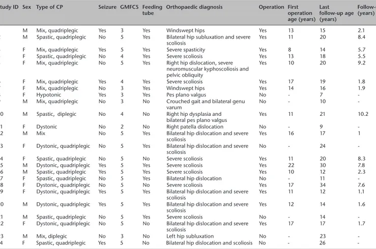
Table 2 Case list of patients with significant musculoskeletal pathology Study ID Sex Type of CP Seizure GMFCS Feeding
tube Orthopaedic diagnosis Operation First operation age (years) Last follow-up age (years) Follow-up (years)
1 M Mix, quadriplegic Yes 3 Yes Windswept hips Yes 13 15 2.1
2 M Spastic, quadriplegic No 5 Yes Bilateral hip subluxation and severe
scoliosis Yes 11 20 8.4
3 F Mix, quadriplegic Yes 5 Yes Severe spasticity Yes 8 14 5.7
4 F Spastic, quadriplegic No 4 Yes Severe scoliosis Yes 13 18 5.5
5 F Mix, quadriplegic No 5 Yes Right hip dislocation, severe neuromuscular kyphoscoliosis and pelvic obliquity
Yes 10 20 9.2
6 F Mix, quadriplegic Yes 4 Yes Severe scoliosis Yes 17 19 1.8
7 F Mix, quadriplegic No 3 Yes Windswept hips Yes 14 16 1.9
8 F Hypotonic Yes 3 Yes Pes plano valgus No - 7
-9 M Mix, quadriplegic No 3 No Crouched gait and bilateral genu
varum No - 10
-10 M Spastic, diplegic No 4 No Right hip dysplasia and
bilateral pes plano valgus Yes 11 21 10.2
11 F Dystonic No 2 No Right patella dislocation No - 9
-12 M Mix No 5 Yes Bilateral hip dislocation and severe
scoliosis Yes 16 17 1
13 F Dystonic, quadriplegic No 5 Yes Bilateral hip dislocation and severe
scoliosis No - 24
-14 F Spastic, quadriplegic No 5 No Severe scoliosis Yes 11 20 8.3
15 M Dystonic, quadriplegic Yes 5 Yes Severe scoliosis Yes 22 30 7.8
16 M Spastic, quadriplegic Yes 5 Yes Severe scoliosis Yes 10 12 2.3
17 F Spastic, quadriplegic No 5 Yes Bilateral hip dislocation No - 11
-18 F Dystonic, quadriplegic No 5 No Severe scoliosis Yes 17 34 7.6
19 F Dystonic, quadriplegic Yes 5 Yes Bilateral hip dislocation and severe
scoliosis Yes 11 12 1.1
20 M Dystonic, quadriplegic Yes 5 Yes Bilateral hip dislocation and severe
scoliosis Yes 12 14 1.6
21 M Spastic, quadriplegic No 5 Yes Severe scoliosis No - 14
-22 F Dystonic, quadriplegic No 5 No Bilateral hip dislocation and severe
scoliosis Yes 17 17 1.7
23 M Mix, diplegic No 3 No Left hip subluxation No - 23
-24 F Spastic, quadriplegic Yes 5 No Bilateral hip dislocation and scoliosis No - 26
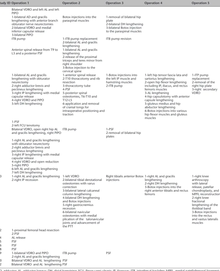
Table 3 List of orthopaedic procedures performed
Study ID Operation 1 Operation 2 Operation 3 Operation 4 Operation 5
1 Bilateral VDRO and left AL and left PIPO
2 1-bilateral AD and gracilis lengthening with anterior branch obturator nerve neurectomies 2-bilateral VDRO and medial inferior capsular release 3-bilateral PIPO
Botox injections into the
paraspinal muscles 1-removal of bilateral hip plate 2-bilateral DH lengthening 3-bilateral Botox injection to the paraspinal muscles
3 ITB pump 1-ITB pump replacement
2-bilateral AL and gracilis lengthening
ITB pump revision 4 Anterior spinal release from T9 to
L3 and a posterior PSF 1-bilateral AL and gracilis lengthening 2-release of the proximal triceps and teres minor from right shoulder
3-Botox injection to the cervical spine 5 1-bilateral AL and gracilis
lengthening with obturator neurectomy
2-right adductor brevis and pectineus lengthening
3-right IP lengthening with medial capsular release
4-right VDRO and PIPO 5-left DH lengthening
1-anterior spinal release 2-T10 thoracotomy and rib resection 3-thoracotomy tube 4-PSF 5-posterior spinal osteotomies, T6-T10 and T12-L5
6-application and removal of cranial tongs for
intraoperative positioning and traction
1-Botox injections into the left IP muscle and hamstring muscles 2-ITB pump
1-left hip tensor fascia lata and sartorius lengthening 2-open hip flexor lengthening including IP, iliacus, and rectus femoris muscles
3-AL lengthening
4-hip capsulotomy with anterior capsule lengthening
5-gluteus medius and hip abductor lengthening 6-Botox injections into various hip flexor muscles and gluteus muscles
1-ITP pump replacement 2-removal of the right hip plate 3-right secondary VDRO
6 1-PSF
2-left FCU tenotomy
7 Bilateral VDRO, open right hip AL
and gracilis lengthening, right PIPOITB pump 1-PSF2-removal of bilateral hip plates
9 1-right AL and gracilis lengthening with obturator neurectomy 2-right adductor brevis and pectineus lengthening
3-right IP lengthening with medial capsular release
4-right VDRO and open reduction 5-right PIPO
6-left AL and gracilis lengthening 7-left DH lengthening
10 1-right AL and gracilis lengthening
2-right IP recession 1-left VDRO 2-bilateral tibial derotational osteotomies with varus correction
3-bilateral lateral calcaneal column lengthening 4-bilateral DH lengthening and Botox injections 5-right gastrocnemius recession
6-bilateral navicular osteotomies with medial plication of the talonavicular joints and advancement of the PTT
Right tibialis anterior Botox
injections 1-right AL and gracilis lengthening 2-right DH lengthening 3-Botox injections into the right anterior tibialis and rectus femoris 1-right knee arthroscopy with lateral release, patellar chondroplasty, and MPFL reconstruction 2-right knee fractional lengthening of the iliotibial band 3-Botox injections into the rectus and vastus lateralis muscles
12 1-proximal femoral head resection 2-PSF
14 AL release
15 PSF
16 PSF
18 PSF
19 1-bilateral VDRO and PIPO
2-right AL and gracilis lengthening ITB pump PSF 20 Bilateral VDRO and AL lengthening PSF
22 Bilateral VDRO and AL lengthening PSF
AD, adductor; AL, adductor longus; DH, distal hamstring; FCU, flexor carpi ulnaris; IP, iliopsoas; ITB, intrathecal baclofen; MPFL, medial patellofemoral ligament; PIPO, peri-iliac pelvic osteotomy; PSF, posterior spinal fusion; PTT, posterior tibial tendon; VDRO, varus derotation osteotomy
An intrathecal baclofen (ITB) pump was implanted in four patients to palliate severe, medically intractable dys-tonia. The catheter tip was positioned at the low cervi-cal-high thoracic spinal cord level and average ITB usage time was 4.1 years (range seven months to ten years). The ITB pump was replaced three times in two patients, twice due to expired battery life and once due to dysfunction. (Table 3). Four patients had one or more botulinum toxin injections in paraspinal (n = 2) or lower extremity (n = 2) muscles for transient relief of focal dystonia.
Discussion
When the diagnosis of GA1 is made after an acute enceph-alopathic crisis, irreversible degeneration of striatal neu-rons leaves patients with a dystonic movement disorder irrespective of GCDH genotype.9,22,28-32 This pattern of motor disability is consistent across GA1 cohorts, as reported in two large natural history studies from 2003 (n = 77)22 and 2006 (n = 279),33 which document incident brain injury rates of 77% and 66%, respectively, among a genetically diverse group of GA1 patients. In the modern era, fewer than 10% of GA1 patients suffer neurological lesions,8,16,33,34 attributable to the combined benefits of newborn screening,16,18 timely inpatient neuroprotective therapies14,35,36 and more widespread use of lysine-free, arginine-enriched medical formulas.19,20 However, disabil-ity rates remain high among GA1 patients born in nations that do not screen newborns for elevated C5DC.9,24,37
We found a relatively high incidence of neurological injury among individuals in our cohort (Table 1), most of whom were GCDH c.1262C>T homozygotes born prior to the advent of statewide newborn screening (ca.1994).38,39 Only 36% of our patients had normal motor function (GMFCS I) whereas 40% had severe functional motor disability (GMFCS III–V). Among this latter group, 62% with GMFCS V developed major musculoskeletal compli-cations40,41 (Table 1). The outcome of the scoliosis man-agement with spinal fusion allowed patients to improve seating alignment with no recorded reoperations or post-operative infections. This is consistent with our outcomes of a much larger cohort of children with cerebral palsy. 42,43
The outcomes of treating dysplastic and dislocated hips in which one revision for recurrent dislocation and three hardware removals were required are also similar to our outcomes in children with cerebral palsy. 44,27 All treated hips were located and pain free at last follow-up. Based on this experience, the outcome of hip and spine treatment in children with GA1 should produce similar results to the treatment methods used for children with cerebral palsy. However, since the predominant motor pattern is dysto-nia, there were fewer significant contractures or spastic deformities requiring surgical management typically seen in patients with spastic cerebral palsy.
Because GA1 manifests clinically as a static rather than progressive encephalopathy, the orthopaedic approach is similar to that for cerebral palsy, but with a critical dis-tinction: surgical planning in patients with GA1 should include a detailed anticipatory strategy to support inter-mediary metabolism during fasting and surgical stress.45 We recommend that elective procedures be planned in consultation with a metabolic specialist, who can coop-erate with an anesthesiologist to develop a perioperative treatment protocol that safeguards against metabolic complications45-48 (Table 4).
For any patient with severe dystonia, medical provid-ers should also recognize risks for pulmonary aspiration, post-extubation laryngeal dystonia and adverse reactions to paralytic agents.48 During the postoperative period, effective analgesia is especially important to prevent a self-reinforcing cycle of pain, anxiety and worsening dystonia that can escalate to life-threatening status dys-tonicus.25,39 High intravenous doses of analgesic and anx-iolytic medications are typically required to control such ‘dystonic storms’. Recognizing the risk for this and other serious complications, we prefer to correct all musculo-skeletal deformities in a single surgical session.
The literature includes reports of botulinum toxin injec-tion and ITB for treatment of the dystonia associated with GA1.31,49 In four patients with focal or generalized dysto-nia, Burlina and colleagues found that botulinum toxin was particularly beneficial for the upper extremities but had minimal impact on craniocervical dystonia.49 Kyller-man et al31 used ITB to successfully treat two patients with
Table 4 Management guidelines for elective surgery with glutaric acidemia type 1
Preoperative precautions 2 to 3 hours intravenous infusion of dextrose 10% normal saline (D10/NS) prior to general anesthesia at a rate 1 to 1.5 times maintenance fluid requirement
During surgery Continue to hydrate with D10/NS – DO NOT USE RINGER’S LACTATE
Postoperative management 1. Maintain D10/NS infusion until enteral or gastrostomy tube feeding is well established. Once feeding is well established, decrease D10/NS rate to half rate for several hours before discontinuing. Expect that the patient will be in the hospital longer than a normal individual with the same procedure.
2. Administer intravenous L-carnitine starting with the first dose prior to anesthesia and continue every eight hours until hospital discharge. Dosage: children < 20 kg – 100 mg/kg/dose, children over 20 kg – 2000 mg/dose.
3. If total parenteral nutrition is necessary, total daily ‘intact’ protein intake should be 0.5 to 1.0 g/kg/day. (Intralipids and lipid-based general anesthetics can be used safely in children with glutaric acidemia type 1.)
4. We do NOT recommend the use of benzodiazepine reversal agents for patients who are chronically exposed to high doses of benzodiazepines. Rather, if patients require postoperative doses of anxiolytics and analgesics that suppress respiratory drive, we recommend assisted forms of ventilation and oxygenation until recovery of spontaneous respiration to avoid dystonic crisis.
severe dystonia, and found that botulinum toxin injection of the cervical paraspinal and lower extremity muscles controlled focal dystonia following PSF. In four patients from our cohort, ITB provided relatively effective palliation for intractable dystonia but required close follow-up for pump refills and management of mechanical problems.
In conclusion, severe dystonia and its attendant mus-culoskeletal complications are common among GA1 patients who develop static stiatal lesions during the first few years of life. Encephalopathic crisis strikes fewer than 10% of affected children in the modern era of newborn screening and appropriate prospective care,8,9 but this outcome still remains tragically high in resource-limited settings.24,37 Among neurologically injured patients with GA1, scoliosis and hip dislocation are the predominant indications for orthopaedic intervention, and all elective surgeries should be executed with a perioperative strategy to minimize metabolic stress (Table 4) and a postoperative plan to control the cycle of pain and anxiety that can cul-minate in life-threatening status dystonicus.25,39
Received 3 April 2020; accepted after revision 14 July 2020
COMPLIANCE WITH ETHICAL STANDARDS FUNDING STATEMENT
No benefits in any form have been received or will be received from a commercial party related directly or indirectly to the subject of this article.
OA LICENCE TEXT
This article is distributed under the terms of the Creative Commons Attribution-Non Commercial 4.0 International (CC BY-NC 4.0) licence (https://creativecommons.org/ licenses/by-nc/4.0/) which permits non-commercial use, reproduction and distribu-tion of the work without further permission provided the original work is attributed.
ETHICAL STATEMENT
Ethical approval: The data in this article was obtained after getting review approv-al from our institutionapprov-al review board for retrospective charts.
Informed consent: The institutional review board stated that consent was not re-quired for this work.
ICMJE CONFLICT OF INTEREST STATEMENT
None declared.
AUTHOR CONTRIBUTIONS
AI: Substantial contributions to the conception or design of the work; Acquisition, analysis or interpretation of data for the work; Drafting of the work; Final approval of the version to be published; Agreement to be accountable for all aspects of the work in ensuring that questions related to the accuracy or integrity of any part of the work are appropriately investigated and resolved
KAS: Acquisition, analysis or interpretation of data for the work; Revising it critical-ly for important intellectual content; Final approval of the version to be published; Agreement to be accountable for all aspects of the work in ensuring that questions related to the accuracy or integrity of any part of the work are appropriately investi-gated and resolved
GFO: Substantial contributions to the conception or design of the work; Drafting of the work; Final approval of the version to be published; Agreement to be accountable for all aspects of the work in ensuring that questions related to the accuracy or integ-rity of any part of the work are appropriately investigated and resolved
FM: Substantial contributions to the conception or design of the work; Acquisition, analysis or interpretation of data for the work; Drafting of the work; Revising it criti-cally for important intellectual content; Final approval of the version to be published; Agreement to be accountable for all aspects of the work in ensuring that questions related to the accuracy or integrity of any part of the work are appropriately investi-gated and resolved
REFERENCES
1. Saudubray J, Baurmgartner M, Walter J, eds. Inborn metabolic
diseases: diagnosis and treatment. Berlin, Heidelberg: Springer, 2016.
2. Therrell BL Jr, Lloyd-Puryear MA, Camp KM, Mann MY. Inborn errors of metabolism identified via newborn screening: ten-year incidence data and costs of nutritional interventions for research agenda planning. Mol Genet Metab 2014;113:14-26. 3. Strauss KA, Puffenberger EG, Morton DH. One community’s effort to control genetic disease. Am J Public Health 2012;102:1300-1306.
4. Kraybill DB, Johnson-Weiner K, Nolt SM. The Amish. Baltimore: Johns Hopkins University Press, 2013.
5. Strauss KA, Puffenberger EG. Genetics, medicine, and the Plain people. Annu Rev Genomics Hum Genet 2009;10:513-536.
6. Haworth JC, Booth FA, Chudley AE, et al. Phenotypic variability in glutaric aciduria type I: report of fourteen cases in five Canadian Indian kindreds. J Pediatr 1991;118:52-58.
7. Naughten ER, Mayne PD, Monavari AA, Goodman SI, Sulaiman G, Croke DT. Glutaric aciduria type I: outcome in the Republic of Ireland. J Inherit Metab Dis 2004;27:917-920.
8. Boy N, Mengler K, Thimm E, et al. Newborn screening: a disease-changing intervention for glutaric aciduria type 1. Ann Neurol 2018;83:970-979.
9. Funk CB, Prasad AN, Frosk P, et al. Neuropathological, biochemical and molecular findings in a glutaric acidemia type 1 cohort. Brain 2005;128:711-722.
10. McMillan TA, Gibson KM, Sweetman L, Meyers GS, Green R. Conservation of central nervous system glutaryl-coenzyme A dehydrogenase in fruit-eating bats with glutaric aciduria and deficient hepatic glutaryl-coenzyme A dehydrogenase. J Biol Chem 1988;263:17258-17261.
11. Sauer SW, Okun JG, Fricker G, et al. Intracerebral accumulation of glutaric and 3-hydroxyglutaric acids secondary to limited flux across the blood-brain barrier constitute a biochemical risk factor for neurodegeneration in glutaryl-CoA dehydrogenase deficiency. J Neurochem 2006;97:899-910.
12. Kölker S, Sauer SW, Surtees RA, Leonard JV. The aetiology of neurological complications of organic acidaemias—a role for the blood-brain barrier. J
Inherit Metab Dis 2006;29:701-704.
13. Ramsay RR, Zammit VA. Carnitine acyltransferases and their influence on CoA pools in health and disease. Mol Aspects Med 2004;25:475-493.
14. Strauss KA, Morton DH. Type I glutaric aciduria, part 2: a model of acute striatal necrosis. Am J Med Genet C Semin Med Genet 2003;121C(1):53-70.
15. Larson A, Goodman S. Glutaric acidemia type 1. GeneReviews. Seattle: University of Washington, 2019.
16. Boy N, Garbade SF, Heringer J, Seitz A, Kölker S, Harting I. Patterns, evolution, and severity of striatal injury in insidious- versus acute-onset glutaric aciduria type 1. J Inherit Metab Dis. 2018 May 2. (Epub ahead of print).
17. Kölker S, Garbade SF, Boy N, et al. Decline of acute encephalopathic crises in children with glutaryl-CoA dehydrogenase deficiency identified by newborn screening in Germany. Pediatr Res 2007;62:357-363.
18. Strauss KA, Lazovic J, Wintermark M, Morton DH. Multimodal imaging of striatal degeneration in Amish patients with glutaryl-CoA dehydrogenase deficiency. Brain 2007;130:1905-1920.
19. Strauss KA, Brumbaugh J, Duffy A, et al. Safety, efficacy and physiological actions of a lysine-free, arginine-rich formula to treat glutaryl-CoA dehydrogenase deficiency: focus on cerebral amino acid influx. Mol Genet Metab 2011;104: 93-106.
20. Kölker S, Boy SP, Heringer J, et al. Complementary dietary treatment using lysine-free, arginine-fortified amino acid supplements in glutaric aciduria type I - A decade of experience. Mol Genet Metab 2012;107:72-80.
21. Strauss KA, Donnelly P, Wintermark M. Cerebral haemodynamics in patients with glutaryl-coenzyme A dehydrogenase deficiency. Brain 2010;133:76-92. 22. Strauss KA, Puffenberger EG, Robinson DL, Morton DH. Type I glutaric aciduria, part 1: natural history of 77 patients. Am J Med Genet C Semin Med
Genet 2003;121C:38-52.
23. Heringer J, Boy SP, Ensenauer R, et al. Use of guidelines improves the neurological outcome in glutaric aciduria type I. Ann Neurol 2010;68:743-752. 24. Wajner M, Coelho DdeM, Ingrassia R, et al. Selective screening for organic acidemias by urine organic acid GC-MS analysis in Brazil: fifteen-year experience. Clin
Chim Acta 2009;400:77-81.
25. Lumsden DE, King MD, Allen NM. Status dystonicus in childhood. Curr
Opin Pediatr 2017;29:674-682.
26. Beauchesne R, Miller F, Moseley C. Proximal femoral osteotomy using the AO fixed-angle blade plate. J Pediatr Orthop 1992;12:735-740.
27. Miller F, Girardi H, Lipton G, Ponzio R, Klaumann M, Dabney KW. Reconstruction of the dysplastic spastic hip with peri-ilial pelvic and femoral osteotomy followed by immediate mobilization. J Pediatr Orthop 1997;17:592-602. 28. Boy N, Mühlhausen C, Maier EM, et al; Additional individual contributors. Proposed recommendations for diagnosing and managing individuals with glutaric aciduria type I: second revision. J Inherit Metab Dis 2017;40:75-101.
29. Morton DH, Bennett MJ, Seargeant LE, Nichter CA, Kelley RI. Glutaric aciduria type I: a common cause of episodic encephalopathy and spastic paralysis in the Amish of Lancaster County, Pennsylvania. Am J Med Genet 1991;41: 89-95.
30. Gitiaux C, Roze E, Kinugawa K, et al. Spectrum of movement disorders associated with glutaric aciduria type 1: a study of 16 patients. Mov Disord 2008;23:2392-2397. 31. Kyllerman M, Skjeldal O, Christensen E, et al. Long-term follow-up, neurological outcome and survival rate in 28 Nordic patients with glutaric aciduria type 1. Eur J Paediatr Neurol 2004;8:121-129.
32. Cerisola A, Campistol J, Pérez-Dueñas B, et al. Seizures versus dystonia in encephalopathic crisis of glutaric aciduria type I. Pediatr Neurol 2009;40:426-431.
33. Kölker S, Garbade SF, Greenberg CR, et al. Natural history, outcome, and treatment efficacy in children and adults with glutaryl-CoA dehydrogenase deficiency. Pediatr Res 2006;59:840-847.
34. Heringer J, Valayannopoulos V, Lund AM, et al; additional individual contributors of the E-IMD consortium. Impact of age at onset and newborn screening on outcome in organic acidurias. J Inherit Metab Dis 2016;39:341-353. 35. Zinnanti WJ, Lazovic J, Housman C, et al. Mechanism of age-dependent susceptibility and novel treatment strategy in glutaric acidemia type I. J Clin Invest 2007;117:3258-3270.
36. Kölker S, Strauss KA, Goodman SI, Hoffmann GF, Okun JG, Koeller DM. Challenges for basic research in glutaryl-CoA dehydrogenase deficiency. J Inherit Metab Dis 2004;27:843-849.
37. Karam PE, Habbal MZ, Mikati MA, Zaatari GE, Cortas NK, Daher RT. Diagnostic challenges of aminoacidopathies and organic acidemias in a developing country: a twelve-year experience. Clin Biochem 2013;46:1787-1792.
38. Morton DH. Through my window—remarks at the 125th year celebration of Children’s Hospital of Boston. Pediatrics 1994;94:785-791.
39. Allen NM, Lin JP, Lynch T, King MD. Status dystonicus: a practice guide. Dev Med Child Neurol 2014;56:105-112.
40. Hoffmann GF, Athanassopoulos S, Burlina AB, et al. Clinical course, early diagnosis, treatment, and prevention of disease in glutaryl-CoA dehydrogenase deficiency. Neuropediatrics 1996;27:115-123.
41. Bjugstad KB, Goodman SI, Freed CR. Age at symptom onset predicts severity of motor impairment and clinical outcome of glutaric acidemia type 1. J Pediatr 2000;137:681-686.
42. Nishnianidze T, Bayhan IA, Abousamra O, et al. Factors predicting postoperative complications following spinal fusions in children with cerebral palsy scoliosis. Eur Spine J 2016;25:627-634.
43. Tsirikos AI, Lipton G, Chang WN, Dabney KW, Miller F. Surgical correction of scoliosis in pediatric patients with cerebral palsy using the unit rod instrumentation. Spine (Phila Pa 1976) 2008;33:1133-1140.
44. Inan M, Gabos PG, Domzalski M, Miller F, Dabney KW. Incomplete transiliac osteotomy in skeletally mature adolescents with cerebral palsy. Clin
Orthop Relat Res 2007;462:169-174.
45. Tsiotou AG, Malisiova A, Bouzelos N, Velegrakis D. The child with glutaric aciduria type I: anesthetic and perioperative management. J Anesth 2011;25:301-304. 46. Kyllerman M, Skjeldal OH, Lundberg M, et al. Dystonia and dyskinesia in glutaric aciduria type I: clinical heterogeneity and therapeutic considerations. Mov Disord 1994;9:22-30.
47. Kamboj M. Clinical approach to the diagnoses of inborn errors of metabolism. Pediatr
Clin North Am 2008;55:1113-1127, viii.
48. Hernández-Palazón J, Sánchez-Ródenas L, Martínez-Lage JF, Collado IC. Anesthetic management in two siblings with glutaric aciduria type 1. Paediatr Anaesth 2006;16:188-191.
49. Burlina AP, Zara G, Hoffmann GF, Zschocke J, Burlina AB. Management of movement disorders in glutaryl-CoA dehydrogenase deficiency: anticholinergic drugs and botulinum toxin as additional therapeutic options. J Inherit Metab