THE GRADUATE SCHOOL OF NATURAL AND APPLIED SCIENCE OF SELÇUK UNIVERSITY
PREPARATION OF CHITOSAN
MICROCAPSULES AND INVESTIGATION OF ITS METAL ADSORPTION PROPERTIES
İdris SARGIN
THESIS OF DEGREE OF DOCTOR OF PHILOSOPHY
Chemistry Department
December-2015 KONYA All Rights Reserved
TEZ BİLDİRİMİ
Bu tezdeki bütün bilgilerin etik davranıĢ ve akademik kurallar çerçevesinde elde edildiğini ve tez yazım kurallarına uygun olarak hazırlanan bu çalıĢmada bana ait olmayan her türlü ifade ve bilginin kaynağına eksiksiz atıf yapıldığını bildiririm.
DECLARATION PAGE
I hereby declare that all information in this document has been obtained and presented in accordance with academic rules and ethical conduct. I also declare that, as required by these rules and conduct, I have fully cited and referenced all material and results that are not original to this work.
Ġdris SARGIN December-2015
i
ÖZET DOKTORA TEZİ
KİTOSAN MİKROKAPSÜLLERİN HAZIRLANMASI VE METAL ADSORPSİYON ÖZELLİKLERİNİN İNCELENMESİ
İdris SARGIN
Selçuk Üniversitesi Fen Bilimleri Enstitüsü Kimya Anabilim Dalı
Danışman: Doç.Dr. Gülşin ARSLAN 2015, 99 Sayfa
Jüri
Danışman: Doç.Dr. Gülşin ARSLAN Prof.Dr. Emine ÖZCAN Prof.Dr. Mustafa ERSÖZ
Prof.Dr. Ali TOR Doç.Dr. Murat KAYA
Biyolojik temelli adsorbanlar ağır metal uzaklaĢtırılmasında yaygın olarak kullanılmaktadır. Doğal polimerleri biyo-adsorban olarak kullanmak geleneksel sentetik adsorbanlara göre daha avantajlıdır. Daha önce yaralanılmamıĢ biyolojik kütlelerden ağır metal uzaklaĢtırılması için kitosan tabanlı biyo-adsorbanların geliĢtirilmesi son on yıl içerisinde çok ilgi çekmiĢtir. Bu çalıĢmada dört kitosan ve iki kitin temelli adsorbanın hazırlanmasını ve ağır metallerin uzaklaĢtırılmasında kullanılması araĢtırılmıĢtır. Kitosan kompozit mikrokapsüller, ticari kitosan (orta molekül ağırlığında) ile dört farklı biyolojik materyal ile hazırlanmıĢtır, bunlar; sporopollenin, mikromantar sporları (Ustilago digitariae ve
Ustilago Maydis) ve ipliksi makroalg Cladophora’dır. Mekanik kararlılığı arttırmak için kompozit
mikrokapsüller gluteraldehit ile çapraz bağlanmıĢtır. Kitin mikro kafesler su piresinin (Daphnia) kıĢ yumurtalarını koruyan ephippium yapılarından elde edilmiĢ ve akabinde manyetit yüklemesi yapılmıĢtır. Mikrokapsüllerin ve mikrokafeslerin karakterizasyonu FT-IR, SEM ve TGA ile yapılmıĢtır. Absorbanlar sulu çözeltilerden Cd(II), Cr(III), Cu(II), Ni(II) ve Zn(II) iyonlarının uzaklaĢtırılmasında test edilmiĢtir. Adsorbanların performansını etkileyen adsorban miktarı, temas süresi, pH, metal iyon konsantrasyonu ve sıcaklık gibi parametreler araĢtırılmıĢ ve adsorbanların adsorpsiyon kapasiteleri karĢılaĢtırılmıĢtır. Metal iyonlarının uzaklaĢtırılmasında belirtilen absorbanlar kullanılabilir: kitosan/sporopollenin mikrokapsüller; Cd(II) ve Cu(II), manyetit yüklü kitin mikrokafesler; Cr(III), kitosan/U. maydis mikrokapsüller; Ni(II) and kitosan/U. digitariae mikrokapsüller; Zn(II).
ii
ABSTRACT
THESIS OF DEGREE OF DOCTOR OF PHILOSOPHY
PREPARATION OF CHITOSAN MICROCAPSULES AND INVESTIGATION OF ITS METAL ADSORPTION PROPERTIES
İdris SARGIN
THE GRADUATE SCHOOL OF NATURAL AND APPLIED SCIENCE OF SELÇUK UNIVERSITY
THE DEGREE OF DOCTOR OF PHILOSOPHY IN CHEMISTRY
Advisor: Assoc.Prof.Dr. Gülşin ARSLAN
2015, 99 Pages
Jury
Advisor: Assoc.Prof.Dr. Gülşin ARSLAN Prof.Dr. Emine ÖZCAN
Prof.Dr. Mustafa ERSÖZ Prof.Dr. Ali TOR Assoc.Prof.Dr. Murat KAYA
Bio-based adsorbents have been widely used in heavy metal removal. Use of natural polymers as biosorbents for heavy metal removal is more advantageous over the conventional synthetic adsorbents. Designing chitosan-based biosorbents from unexploited biomass for heavy metal removal has received much attention over the past decade. This study investigated the preparation of four chitosan-based and two chitin-based adsorbents. Chitosan composite microcapsules were prepared with commercial chitosan (medium molecular weight) and four biological material i.e., sporopollenin, microfungal spores (Ustilago
digitariae and Ustilago Maydis) and filamentous macroalgae Cladophora. The composite microcapsules
were cross-linked with glutaraldehyde to enhance the mechanical strength. Chitin microcages were obtained from the ephippial resting eggs of a microcrustacean (Daphnia; water flea) and subsequently were loaded with magnetite. Characterisation of the microcapsules and magnetite-loaded chitin microcages were performed with FT-IR, SEM and TGA. The adsorbents were tested in removal of Cd(II), Cr(III), Cu(II), Ni(II) and Zn(II) ions from aqueous solutions. The parameters affecting the performance of the adsorbents i.e., adsorbent dosage, contact time, pH, metal ion concentration and temperature were investigated. Comparison of the adsorption capacity of the adsorbents was made. The adsorbents can be used at removal of specified heavy metal contaminants: chitosan/sporopollenin microcapsules; Cd(II) and Cu(II), magnetite loaded chitinous microcages; Cr(III), chitosan/U. maydis microcapsules; Ni(II) and chitosan/U. digitariae microcapsules; Zn(II).
iii
ACKNOWLEDGMENTS
I am very grateful to my supervisor Assoc.Prof.Dr. GülĢin ARSLAN for her guidance, advice and support throughout my studies, and doing the research would be impossible without her valuable suggestions. I must thank Assoc.Prof.Dr. Murat KAYA for his support and encouragement at every step along the way.
I am especially grateful to Lütfiye TAYLAK, BüĢra Ebru ATAOĞLU, AyĢegül YAMAN, Ġlker AKIN and Talat BARAN for their valuable contribution to the analyses.
I am also deeply indebted to Selçuk University Research Foundation for their support to the project BAP-14201082.
I must acknowledge the aid, patience and encouragement of my family.
Ġdris SARGIN KONYA-2015
iv CONTENTS ÖZET ... i ABSTRACT ... ii ACKNOWLEDGMENTS ... iii CONTENTS ... iv 1. INTRODUCTION ... 1 1.1. Literature review ... 2
1.1.1. Adsorption as a versatile tool in water treatment ... 2
1.1.2. A functional biopolymer: Chitosan ... 2
1.1.3. Glutaraldehyde as an agent for cross-linking of chitosan ... 4
1.1.4. Sporopollenin one of the most inert biomaterial in the biosphere ... 4
1.1.5. Microfungal spores from plant pathogens ... 5
1.1.6. Macroalgal bloom biomass: Filamentous green algae Cladophora ... 6
1.1.7. Chitin cages from water flea (Daphnia longispina) ephippia ... 7
1.2. Aim of the study ... 8
2. EXPERIMENTAL ... 9
2.1. Materials ... 9
2.1.1. Smut spore collection and alkaline treatment ... 9
2.1.2. Algal biomass sample collection and bleaching treatment ... 10
2.1.3. Collection of ephippial resting eggs of Daphnia longispina ... 10
2.2. Preparation of the adsorbents ... 10
2.2.1. Determination of optimum amount of cross-linking agent ... 10
2.2.2. Preparation of chitosan microcapsules and plain chitosan microbeads ... 11
2.2.3. Preparation of magnetic microcages from ephippia of D. longispina ... 12
2.3. Characterisation of the adsorbents and the instrumentation ... 13
2.4. Metal sorption experiments ... 13
2.5. Adsorption isotherms ... 14
2.6. Thermodynamic analysis ... 15
3. RESULTS AND DISCUSSION ... 17
3.1. Optimum amount of glutaraldehyde for microcapsules ... 17
3.1.1. Characterisation of CSMCs ... 17
3.1.2. Cu(II) removal studies ... 21
3.1.3. Thermodynamic analysis ... 25
3.1.4. Sorption from binary mixtures ... 26
3.1.5. Desorption studies and the re-usability of the adsorbents ... 27
3.1.6. Isotherm analysis ... 27
3.2. Chitosan/sporopollenin microcapsules and plain chitosan microbeads ... 30
3.2.1. Characterisation of chitosan/sporopollenin microcapsules and plain chitosan microbeads ... 30
v
3.2.3. Thermodynamic analysis ... 38
3.2.4. Isotherms analysis ... 39
3.3. Chitosan/microfungal spore microcapsules ... 42
3.3.1. Characterisation of chitosan/microfungal spore microcapsules ... 42
3.3.2. Metal ion removal studies ... 47
3.3.3. Thermodynamic analysis ... 50
3.3.4. Isotherm analysis ... 51
3.4. Chitosan/Cladophora microcapsules ... 53
3.4.1. Characterisation of chitosan/macroalgal biomass microcapsules ... 53
3.4.2. Metal ion removal studies ... 55
3.4.3. Thermodynamic analysis ... 58
3.4.4. Isotherm analysis ... 59
3.5. Magnetite loaded chitinous microcages ... 61
3.5.1. Characterisation of magnetite loaded chitinous microcages ... 61
3.5.2. Metal ion removal studies ... 67
3.5.4. Isotherm analysis ... 71
3.6. Comparison of the metal ion sorption capacities of the adsorbents ... 74
4. CONCLUSIONS AND SUGGESTIONS ... 75
4.1. Conclusions ... 75
4.2. Suggestions ... 77
REFERENCES ... 78
1. INTRODUCTION
Heavy metal contamination in water bodies is a critical issue. A great deal of water resources have been contaminated with heavy metal ions through effluents of many industries such as metal plating, mining, textile and electric/electronic devices manufacturing (Bilal et al., 2013; Hasmath Farzana and Meenakshi, 2015). The waste effluents from those industrial operations, untreated or even treated, can have significant amounts of heavy metal ions. Especially, disposal of untreated effluents of the plating, mining and textile manufacturing industries into the environment have caused accumulation of heavy metal ions in the ecosystem (Bilal et al., 2013). Many heavy metal ions have been reported to have detrimental impacts on living organisms (Abou El-Reash et al., 2011; Abdolali et al., 2014; Ghasemi et al., 2014). These ions have a recalcitrant nature; that is, they are non-biodegradable and are capable of accumulating in the food chain (Kilic et al., 2013). Unfortunately, it requires much effort to remove them once they have been introduced into the environment. When their detrimental effects and toxicity are considered, removal of heavy metal ions from water bodies is a critical issue to save diminishing water resources.
Many techniques have been employed for heavy metal removal in wastewaters: membrane filtration, precipitation, chelation/complexation, ion-exchange, oxidation/reduction and adsorption. Each method has some advantages over the rest. However, adsorption is still the best option because it makes possible to use low cost, sustainable, high efficient and eco-friendly bioadsorbents with easy of recovery and re-usability (Kobya, 2004; Mellah et al., 2006; Bilal et al., 2013). Materials with biological origin can make better adsorbents due to their non-toxicity, abundance and efficiency. Additionally, metal ions also have a tendency to interact with the surfaces providing coordination or chelating sites to bind; thus, making it possible to use various materials with functionalised surfaces in the removal of heavy metal contaminants (Yavuz et al., 2003). In this sense, adsorption is considered a simple and versatile tool (Ali and Gupta, 2007), but its effectiveness is largely determined by the selection of the adsorbent (Fomina and Gadd, 2014).
1.1. Literature review
1.1.1. Adsorption as a versatile tool in water treatment
There have been increasing interest and efforts to improve conventional techniques to treat the metal contaminated effluents efficiently. Among the conventional physicochemical methods, adsorption has been extensively employed because of its ease of use, effectiveness and feasibility (Yavuz et al., 2003; Bilal et al., 2013).
Selection of sorbent is a key parameter when designing new sorbents for heavy metal removal. In addition to physicochemical characteristics of a sorbent such as its selectivity towards certain species and sorption capacity, its cost and production procedures should be assessed. Many materials with natural or artificial origin such as activated carbon, resins, fly ash, oxides, silicates, clays, zeolites, pine bark and cotton waste have been used as adsorbents and reported in the literature (Babel and Kurniawan, 2003; Ahmaruzzaman, 2008). Those studies have demonstrated the need for more efficient, inexpensive and renewable adsorbents. Bio-based sorbents can fulfil these needs; biomaterials are abundant in nature, and also many functional groups for metal interaction are present on them or they can be easily functionalized.
Also, adsorbents with biological origins are nontoxic and biodegradable (Wang and Chen, 2009). However; biosorbents are not always available in desired form, requiring much chemical modification for fictionalisation or physical treatment to ensure appropriate size and monodispersity (Ngah et al., 2011). In recent years, due to the functional moieties on their cell walls and their uniformity of size, many bacteria, fungi and algae species, living and dead, have been exploited in studies aimed at developing biosorbents for heavy metal removal (Cataldo et al., 2013; Rouhollahi et al., 2014; Won et al., 2014).
1.1.2. A functional biopolymer: Chitosan
Chitosan, the deacetylated derivative of chitin, is a functional biopolymer with biodegradability, biocompatibility and high sorption capacity (Muzzarelli et al., 2012). It can be physically and chemically modified in different forms: membranes (Bayramoglu et al., 2007), fibres (Pillai et al., 2009), hydrogels (Pal et al., 2013), nanoparticles (Wei et al., 2012), beads (Chen et al., 2008), films (Sharmin et al., 2012)
and capsules (Kumar et al., 2004). It has many applications in pharmaceutical (Aranaz et al., 2009), cosmetics (Ong et al., 2008), biotechnology (Cota-Arriola et al., 2013), food industry (Shahidi and Abuzaytoun, 2005) and water treatment (Chen et al., 2008). In metal ion removal, it can be used in raw (Sankararamakrishnan et al., 2007) or modified forms (Vijaya et al., 2008). Also, in recent years, various chitosan composite sorbents have been synthesized with clay minerals (Peng et al., 2013), natural/biomaterials (Liu et al., 2011; Yu et al., 2013), magnetite (Tran et al., 2010), sand (Wan et al., 2010) and synthetic polymers (Janaki et al., 2012).
Chitosan is produced via deacetylation of chitin in high alkaline conditions, (Muzzarelli, 2011; Kyzas et al., 2013) and it has a high affinity for metal ions (Crini and Badot, 2008; Kyzas et al., 2013). The alkaline hydrolysis of chitin exposes free amino groups (–NH2) and gives the polymer unique cationic nature (Ali, 2010). The pendant
amino groups of chitosan are primary cause of its higher metal ion sorption capacity and its solubility in aqueous solutions when compared to chitin. Amino and hydroxyl groups (especially at the C-3 position) on chitosan can serve as electrostatic interaction and complexation sites for metal cations (Guibal, 2004; Yu et al., 2013). This makes chitosan an appropriate sorbent in heavy metal uptake (Mi et al., 2015). Many workers have opted for chitosan composites as sorbent and prepared chitosan composites from natural products (Miretzky and Cirelli, 2009; Wu et al., 2010). There has been extensive research regarding its metal binding nature (Muzzarelli, 2011; Lang et al., 2013; Shukla et al., 2013; An et al., 2014).
In designing composite biosorbents, chitosan carriers is preferred for the immobilisation of fine particles due to the ease of cross-linking (Abdel-Mohsen et al., 2012). Cross-linked chitosan composites have received much attention in recent years due to their efficiency in heavy metal removal (Ngah et al., 2011; Kadouche et al., 2012). Composite material production from renewable sources is a promising area for heavy metal removal thanks to eco-friendly nature of biological waste materials (Wang and Chen, 2009). Chitosan, the by-product of food processing industry, is a functional biopolymer which is desired in heavy metal removal due to its metal ion interacting groups; –OH and –NH2. Also through these functional groups, chitosan polymer can be
easily cross-linked, which makes it an effective matrix for composite material production (Guibal, 2004; Wang et al., 2013; Salaberria et al., 2015).
1.1.3. Glutaraldehyde as an agent for cross-linking of chitosan
Once dissolved in acidic solutions, chitosan can be transformed into insoluble gel form via cross-linking; giving the polymer structural stability in acidic media. Cross-linking also enables incorporation of fine particles into the polymeric matrice. Cross-linking forms three dimensional sites within the networks of chitosan and enhances its metal uptake capacity (Wang et al., 2004). Preparation of chitosan composite sorbents from different sources has been reported earlier. Glutaraldehyde, which forms Schiff bases with amines, is one of the cross-linking agents used in synthesis and modification of these chitosan-based adsorbents (Webster et al., 2007).
Cross-linking makes chitosan not only mechanically stronger but more selective to some metal ions. Various cross-linking agents are used to cross-link the chitosan polymer; epichlorohydrin, ethyleneglycol diglycidyl ether, 1,1,3,3-tetramethoxypropane, β-cyclodextrin polyaldehyde or glutaraldehyde. Free –NH2 and –
OH groups of chitosan polymer play a role in forming coordination complexes with metal ions. But, in case of cross-linking with glutaraldehyde (GA), pendant –NH2
groups on the chitosan polymer are involved in Schiff base formation, which in turn affects its interaction with metal ions by lowering the sorption sites on the polymer (Guibal, 2004). Wang et al. (2013) recently reported that the amount of GA strongly affected Cu(II) adsorption onto chitosan/polyethylene glycol semi-interpenetrating polymer blend. They commented that higher GA dosages led to lower Cu(II) sorption capacity by decreasing number of pendant amino groups on chitosan polymer.
1.1.4. Sporopollenin one of the most inert biomaterial in the biosphere
Sporopollenin is a natural biomacromolecule present in the outer wall (also called exine) of spores and pollens. This polymeric material is highly resistant to chemical and biological attack and can retain its morphology in geological strata over millions of years (Brooks, 1978; Yule et al., 2000). Many analytical techniques have been performed to reveal its chemical nature; however, information available on its chemical structure is still limited and needs clarifying. Nevertheless, some studies indicated that sporopollenin is mainly an aliphatic polymer with phenolic and aromatic groups or conjugated side chains (Ahlers et al., 1999); it is considered as a
macromolecule composed mainly of carotenoid and carotenoid esters (Dominguez et al., 1999).
Sporopollenin particles extracted from Lycopodium clavatum (common club moss) has excellent mechanical strength and are reasonably monodisperse (Binks et al., 2005). Recently, Fraser et al. (2014) reported that sporopollenin from L. clavatum is also resistant to heat and its chemistry does not alter until a threshold of 250–300°C (Fraser et al., 2014). Many researchers have appreciated the unique nature of the sporopollenin and have conducted works with raw or functionalised form of it including metal removal studies (Atkin et al., 2011; Braun and Cardoso, 2012; Gubbuk et al., 2012; Watson et al., 2012).
1.1.5. Microfungal spores from plant pathogens
Fungi are ubiquitous and abundant in the biosphere. Many species have been widely used in heavy metal uptake due to their affinity and selectivity for a wide range of metal ions. However, in literature no attention has been paid to the use of smut fungi genus Ustilago in biosorption studies (Wang and Chen, 2009). These organisms are fungal plant pathogens distributed worldwide. They cause substantial losses in crop yield and biomass (Gallart et al., 2009) by infecting over 4000 species (Ruiz-Herrera et al., 2008). Ustilago maydis and Ustilago digitariae are two common Ustilago species. These biotrophic fungal pathogens cause smut disease in maize (Mueller et al., 2008) and weed, Digitaria, (Gallart et al., 2009).
Designing biosorbent with microfungal spores and chitosan can be beneficial in some ways: (1) These fungal pathogens are abundant and widely distributed in many areas; (2) collection of these smut spores, particularly swollen host tissue of smut on the ears of maize, can help to reduce the area of the infected fields, preventing the spread of the disease and thereby lowering the use of chemical fungicides (Johnson and Baudoin, 1997; Gallart et al., 2009); (3) size (10µ) (Hu et al., 2003) and (4) surface characteristics of the spores (Won et al., 2014) facilitates preparation of chitosan microcapsules without need of any pre-treatments; and, (5) the cell walls of these spores have functional groups which are capable of interacting with metal cations (Won et al., 2014).
1.1.6. Macroalgal bloom biomass: Filamentous green algae Cladophora
Cladophora is genus of filamentous green algae usually found in shallow fresh
and saline waters around the world (Gubelit and Berezina, 2010). In case of excessive input of nutrients into an aquatic system, which is called eutrophication, these organisms thrive in large numbers. In a eutrophic aquatic ecosystem, excessive growth and reproduction of algae (also called as macroalgal bloom) is usually observed. When left in water, the algal biomass is consumed by the bacteria. This bacterial decomposition cause further eutrophication in the system by disrupting the mainly dissolved oxygen level. Accumulation of floating algal mats on the surface prevents sun rays from reaching the deeper, which further deteriorates the conditions for photosynthesis organisms (Guidone and Thornber, 2013; Kim et al., 2014). All these concerns stipulate that macroalgal biomass should be removed from the medium following a bloom of macroalgae, and the utility of algal biomass should be addressed for wide range of applications. Cladophora sp. biomass can be preferred in heavy metal removal studies for a couple of reasons: (1) It is available in huge amounts worldwide, (2) it is low-cost, (3) algal cell walls have functional moieties for interaction of metal ions, and (4) its collection from coastal regions can solve possible eutrophication problem.
Many studies have been conducted for heavy metal removal by using chitosan as a main composite material. In some studies, chitosan composites were produced by blending chitosan solution with inorganic materials such as clay (Bhattacharyya and Gupta, 2008), sand (Wan et al., 2010) and magnetite (Liu et al., 2009) etc. Chitosan composite materials with biological origin such as alginate (Ngah and Fatinathan, 2008), cellulose (Zhao and Mitomo, 2008) and cotton (Zhang et al., 2008) have also been synthesized and tested for heavy metal removal. On the other hand, algal based biosorbents are known to be effective at treatment of wastewater with heavy metal contaminants (Wang and Chen, 2009). However, few studies have been conducted on heavy metal removal by chitosan-algal biomass (Sargassum sp.) composite (Liu et al., 2011; Yang et al., 2011).
1.1.7. Chitin cages from water flea (Daphnia longispina) ephippia
Polymeric matrixes of biological origins have attracted much attention over the past decade due to their unique nature; biocompatibility, biodegradability, abundance and high affinity for charged species (Sankar et al., 2012). Among the biopolymers, chitin, a water-insoluble polysaccharide, and particularly its deacetylated water-soluble form, chitosan, have been extensively exploited to design carriers or matrixes for biomolecules and particles.
Thanks to pendant amino groups in its structure, chitosan dissolves in acidic aqueous solutions, and therefore can be cross-linked easily, making chitosan much more advantageous in polymeric matrix production over chitin. In the literature related to chitin, there have already been numerous studies on chitosan matrixes (Guo et al., 2005; Chen et al., 2010). However, there is relatively less literature available on the use of chitin as matrix for composite production, apparently stemming from the insolubility of chitin in many solvents because of its rigid crystalline structure resulting from the hydrogen bonds (Tamura et al., 2011). Additionally, obtaining chitin with intact three dimensional shape from exoskeleton of organisms is another problem. Therefore, many researchers have had to use chitin powder for production of chitin composites. In those studies, chitin powder or flakes were used after dissolution in dimethylacetamide-lithium chloride solution (Mi et al., 2002), 1,1,1,3,3,3-hexafluoro-2-propanol (Min et al., 2004a), phosphoric acid (Min et al., 2004b) or calcium chloride saturated methanol (Sankar et al., 2012). Even in more recent studies, chitin slurry was prepared in water from chitin powder (Deng et al., 2014).
However, there have been attempts to extract chitin scaffolds from large organisms such as pens of the common squid (Loligo sp.) (Falini et al., 2002),
Verongida sponges (Ehrlich et al., 2010), freshwater sponge Spongilla lacustris (Ehrlich
et al., 2013), marine sponge Aplysina fistularis (Wysokowski et al., 2013) and crab shell (Chen et al., 2014). On the other hand, although it has been long known that chitin occurs in the exoskeleton of freshwater microcrustaceans Daphnia (water flea) (Seidman and Larsen Jr, 1979) and chitin is the main structural part of the robust shell of their resting eggs, called ephippia (Kawasaki et al., 2004), there are few studies on chitin isolation from Daphnia (Cauchie et al., 2002) itself or from its ephippial eggs (Kaya et al., 2013; Kaya et al., 2014b).
In the lights of current attempts in production of chitin matrixes, we hypothesize that chitinous scaffold of a microcladoceran ephippia can be obtained intact, and subsequently the chitin microcages can be used as a carrier for fine particulates. Using the ephippia of Daphnia longispina as a model, it was demonstrated here for the first time that ephippial eggs of a microcladoceran can be used as a source for production micro sized chitin scaffolds, and fine particles of magnetite (Fe3O4) can be incorporated
into the polymeric matrix (vide infra).
1.2. Aim of the study
The present study aimed to provide insights into the production of four chitosan-based adsorbents from sporopollenin, microfungal spores (smut spores; Ustilago maydis and Ustilago digitariae) and filamentous macroalgae (Cladophora sp.), and a novel magnetic adsorbent from ephippial eggs of a microcrustacean (water flea; Daphnia
longispina). Characterisation studies were carried out by Fourier Transform Infrared
spectroscopy (FTIR), thermogravimetric analysis (TGA), scanning electron microscope (SEM), SEM-EDX and light microscopy. Cu(II), Cd(II), Cr(III), Ni(II) and Zn(II) sorption performance of the microcapsules were evaluated and compared to that of plain cross-linked chitosan microbeads without any additional material.
2. EXPERIMENTAL
2.1. Materials
Chitosan from BioLog (Biotechnology and Logistic GmbH) (in flake form with deacetylation degree of 85%, viscosity 2500 mPa´s, ash content of <1%) was used in the preliminary experiments. Medium molecular weight chitosan (Sigma–Aldrich) were used in the all the metal ion sorption experiments. Metal salts Cu(NO3)2.3H2O,
Cr(NO3)3.9H2O, Ni(NO3)2.6H2O, Zn(NO3)2.4H2O were purchased from Merck,
Cd(NO3)2.4H2O was obtained from Sigma–Aldrich. Glutaraldehyde (25% in water, v:v)
and acetic acid were obtained from Merck. Ethanol and NaOH were obtained from Sigma–Aldrich. Methanol was from obtained Analar Normapur. NH3, HCl, FeCl3.6H2O
and FeSO4.7H2O were purchased from Merck. NaClO solution was obtained from a
local supplier and was diluted to 3% prior to its use. Sporopollenin from L. clavatum with 20 µm particle size was purchased from Fluka Chemicals. Double-distilled water purified with Barnstead (Dubuque, IA) ROpure LP® reverse osmosis system or with ELGA DV 25 water purification system was used throughout the study. The metal solutions were prepared by diluting of the stock solution of 1.0 M. Metal solution pH was adjusted with dilute solutions of HCl or NaOH.
2.1.1. Smut spore collection and alkaline treatment
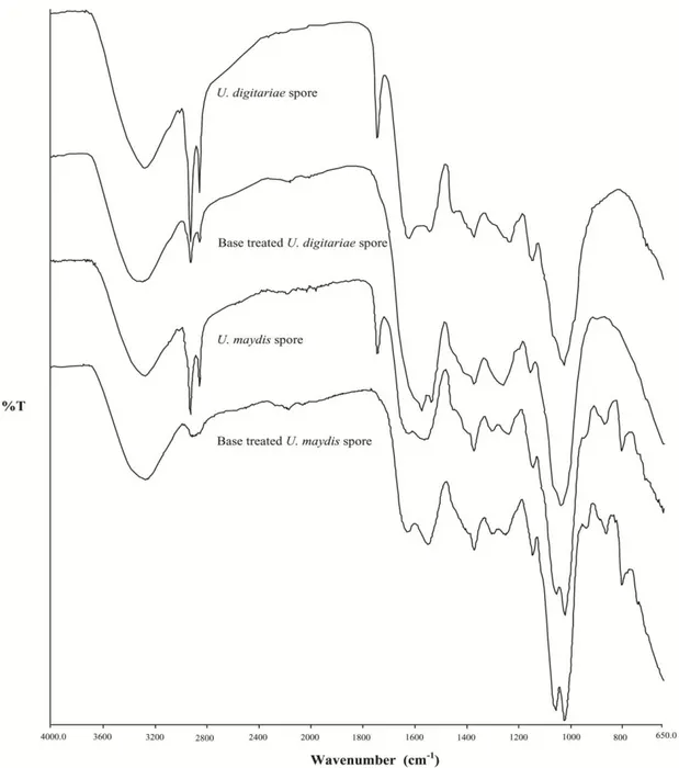
The plants (Digitaria) infected by loose smut (U. digitariae (Kunze) Rabenh) were collected (May 2013; Aksaray, Turkey) and the smutted (U. maydis) maize ears (September 2013; Kayseri, Turkey). The smutted panicles of the weed and ears of the maize were removed and allowed to air-dry. The dried samples were crushed and sieved through a mesh to remove any plant debris. Following the light microscope examination, the isolated spores were rinsed with water and then treated with 0.5 M NaOH solution for 12 h to remove any organic or inorganic residues in the hyphae and mycelial components of the spores. Subsequently, treated spores were rinsed with distilled water to neutrality and were allowed to air-dry. On the other hand, when preparing microcapsules with untreated spores, chitosan solution lost its viscosity and got thinner; this made the formation of spherical microcapsules in coagulation solution
(mixture of water, methanol and NaOH) (infra vide) impossible. Additionally, untreated spores in coagulation solution gave decomposition products by giving the solution dark brown colour. To prevent the chitosan solution from getting thinner and the leakage of decomposition products in the coagulation solution, spores were first subject to the alkaline treatment in NaOH solution and then they were used.
2.1.2. Algal biomass sample collection and bleaching treatment
Cladophora sp. samples were collected from Uluırmak stream (Central
Anatolia) on 17 July 2014 following a macroalgal bloom. The samples were washed and left to dry at room temperature for two weeks. Prior to bleaching treatment, dry algal biomass was ground by using a commercial blender and sieved (with a 100 µm sieve). The powdered algal biomass was treated with the bleaching agent (sodium hypochlorite solution, 4% v:v) at 50°C for 10 minutes. The bleached samples were filtered and rinsed with distilled water until reaching the neutral pH.
2.1.3. Collection of ephippial resting eggs of Daphnia longispina
The floating cladoceran ephippia were collected from the shores of a Mamasın Dam reservoir in Central Anatolia (Aksaray, Turkey) following a peak observed in cladoceran (Daphnia sp.) population (November, 2012). Collection of cladoceran ephippia was accomplished with the use of plankton net. The samples were subsequently shipped to the laboratory for identification by a light microscopy. The samples were washed with water and then ephippial eggs belonging to Daphnia species were identified. Ephippial eggs of D. longispina were picked and used in the experiments.
2.2. Preparation of the adsorbents
2.2.1. Determination of optimum amount of cross-linking agent
Chitosan (1.500 g) was dissolved in 150 mL of acetic acid solution (2% v/v) and stirred for 24 h. Then, sporopollenin grains (1.140 g) were mixed with the chitosan solution. After blending for 2 h, the mixture was transferred into a burette. The mix was
dropped into coagulation solution (methanol-water-NaOH; 300 mL:200 mL:60 g) (Pal et al., 2013). The microcapsules were incubated in the coagulation solution for overnight to ensure the complete gelation. Then, the microcapsules were recovered and rinsed with water to neutral pH. Wet capsules were transferred into cross-linking reaction solution (GA solution in 60 mL of methanol) and refluxed at 70°C for 6 h. In this step, three different amounts of GA solutions were used; 0.3, 0.9 and 1.5 mL. The microcapsules obtained in each cross-linking procedure were named CSMCs1 (chitosan/sporopollenin microcapsules), CSMCs2 and CSMCs3, respectively. The final product was rinsed with ethanol and then with water. Wet cross-linked microcapsules were left to dry at room temperature and kept in sealed containers.
2.2.2. Preparation of chitosan microcapsules and plain chitosan microbeads
Preparation of chitosan microcapsules was carried out as follows: Chitosan solution was prepared by dissolving 3.00 g chitosan in 150 mL of acetic acid solution (2% v/v). The mixture was continuously stirred for 24 h. Subsequently, 1.500 g of particulate matter (sporopollenin, alkali treated U. maydis spores, alkali treated U.
digitariae spores or bleached macroalgal biomass) was added into the chitosan solution
and then the mixture was stirred for 3 h until homogeneity. The mixture was transferred into a burette and dropped into the coagulation solution (a mixture of 200 mL of water, 300 mL of methanol and 60.0 g NaOH). Resulting microcapsules were incubated in the coagulation solution for 24 h to achieve a complete gelation, giving a yellow-brownish colour to the medium. Then, the microcapsules were recovered and rinsed thoroughly with distilled water until the filtrate was of neutral pH and free of coloured decomposition products of the sporopollenin grains. Wet microcapsules (otherwise they could not retain their spherical shape when dried) were recovered by filtration and transferred into cross-linking reaction solution (0.9 mL of glutaraldehyde solution in 90 mL of methanol).The linking solution was refluxed at 70°C for 6 h. Finally, cross-linked microcapsules were recovered and washed thoroughly first with ethanol and then with water to remove any unreacted glutaraldehyde molecules. The cross-linking treatment and the following washing steps were performed in a fume hood to arrest any glutaraldehyde vapour. The microcapsules were allowed drying at room temperature. Plain cross-linked chitosan microbeads were prepared by observing the same method without adding any particulate matter.
2.2.3. Preparation of magnetic microcages from ephippia of D. longispina
Resting eggs of cladoceran D. longispina are encased in chitin shell layers with randomly distributed pits filled with minerals such as calcium phosphate or sulphate (Kawasaki et al., 2004). Under the exterior layer occurs a layer of protein-chitin fibres (Seidman and Larsen Jr, 1979; Cáceres, 1998). A treatment should be done to remove the chitinous structure from the matrix. In this study, chitinous microcages were isolated by removing the content of the eggs through a line of simple treatments at ambient conditions; demineralization with dilute HCl acid solution, deproteinization with NaOH solution and bleaching with NaClO solution. It was previously reported that sodium hypochlorite solution had an effect on the ephippial eggs of Daphnia sp. (Pancella and Stross, 1963; Gray et al., 2006); therefore, sodium hypochlorite solution was preferred as a bleaching agent.
Briefly, the ephippia (10 g) were treated with 500 mL of 0.5 M HCl solution for 1h at 50°C. Then, after rinsing with water until neutrality, the samples were stirred in 500 mL of 0.5 M NaOH solution for 1h at 50°C. The samples were separated from the alkali medium and washed with water. In the final step, the samples were subject to the bleaching treatment in 400 mL of NaClO solution (3%) at 50°C for 6 min. The bleached samples were removed by filtration and washed with 8 L of water. Then, half of the wet microcages were left to dry at room temperature for 48 h. The other half in wet form was allotted for magnetite particle loading.
Synthesis of magnetite particles was conducted following the method reported elsewhere with some modifications (Ozmen et al., 2010). The incorporation of magnetite particles into the microcages was achieved as summarised below. The microcages were transferred into 200 mL of water, and subsequently 0.445 g of FeSO4.7H2O was added; the resulting mixture was stirred at 70°C for 1 h. Then, 0.756 g
of FeCl3.6H2O was added and the mixture was stirred for 1h at the same temperature.
This was followed by addition of 4.0 mL of NH3 solution. The hot reaction medium was
stirred for 1 h. Finally, the microcages with Fe3O4 particles were filtrated out and
washed with water. The magnetite particles loaded microcages were dried at room temperature for 48 h.
2.3. Characterisation of the adsorbents and the instrumentation
Chemical structure of the adsorbents was examined by Fourier transform infrared spectroscopy (Perkin-Elmer 100 FT-IR Spectrometer 2.5 in range of 4000 and 625 cm−1). Thermogravimetric analyses (TGA) of the adsorbents were performed by an EXSTAR S11 7300 (under nitrogen atmosphere; gas flow rate: 20 mL/min and heating rate: 10°C min−1). Surface examination of the adsorbents was carried out with a scanning electron microscope (SEM) (EVO LS 10 ZEISS). Prior to SEM analyses, the samples were coated with gold using Sputter Coater (Cressingto Auto 108). SEM/Energy Dispersive X-Ray Analysis (EDX) analysis was performed to detect the presence of iron atoms in the microcages upon magnetite loading. A light microscopy was used for examination the surface characteristics of the samples. Metal ion sorption experiments were conducted on a shaker (Heidolph Promax 2020) at 200 rpm. A flame atomic absorption spectrometer (ContrAA 300, Analytikjena) was used to determine the metal ion concentration of the solutions from heavy metal ion sorption experiments.
2.4. Metal sorption experiments
Metal sorption experiments were done in a batch system at room temperature on a shaker (Heidolph Promax 2020) at 200 rpm. The adsorbent (0.1500 g) was added into 25 mL of metal solution (10 mg L−1) and then agitated for 4 h at 200 rpm. The sorbent was removed the medium with a filter paper (Whatman, No: 42). The metal ion concentration in the solutions was determined by using a flame atomic absorption spectrometer (ContrAA 300, Analytikjena). To determine the optimum conditions, different experimental parameters were tested; initial metal ion concentration: 2–12 mg L−1; contact time: 60– 480 min; temperature: 25, 35 and 45°C and amount of the sorbent: 0.0500–0.2500 g. pH of the metal solutions varied in a range of 3.0–5.80. The following equation gave the sorption capacity of the adsorbent (qe) in mg g−1.
qe = (Ci–Ce)V/W (1)
where Ci and Ce are the initial and equilibrium liquid-phase concentrations of metal ions
adsorbent (in g). Sorption yield for each of the metal ions was calculated from following equation;
Sorption (%) = [(Ci – Ce)/ Ci] x 100 (2)
where Ci and Ce are the initial and equilibrium concentration of metal ion (mg L−1). The
conditions and the parameters for each of the experiment sets with different adsorbent were detailed in subsequent sections.
2.5. Adsorption isotherms
To make quantitative evaluation of the sorption behaviour of the metal ions, adsorption isotherm model analysis was done. Although binding of metal ions to biosorbents often fits the Langmuir (Langmuir, 1918) and Freundlich models (Freundlich, 1907), two other models, Dubinin–Radushkevich (D–R) (Dubinin and Radushkevich, 1947) and Scatchard plot analysis (Crist et al., 1994), are used to describe the sorption equilibrium as well.
The Freundlich model: log qe = log KF + (1/n) log Ce (3)
with qe,, amount of solute adsorbed in mmol g−1, Ce, the equilibrium concentration of the
adsorbate in mmol L−1 and KF (mmol g-1) and n Freundlich constants denoting
adsorption capacity and intensity of adsorption, respectively.
The Langmuir model: Ce/qe = Ce/Q0 + 1/Q0b (4)
with qe, amount of solute adsorbed in mmol g−1, Ce, the equilibrium concentration of the
adsorbate in mmol L−1, Q0 (in mmol g-1) and b (in L mmol-1) Langmuir constants related to adsorption capacity and energy of adsorption.
The D–R model: ln qe = ln Xm− Kε2 (5)
where ε is Polanyi Potential [RT ln(1 + (1/Ce))], qe is the amount of solute adsorbed per
unit weight of adsorbent (mmol g-1) and Xm is the adsorption capacity (mmol g −1
constant related to the adsorption energy in mol2kJ−2. The values of Xm and K were
calculated from the intercept and slope of the lnqe versus ε2 plots. By using the K values
in the equation below, the mean free energy of adsorption (E) was obtained:
E = (–2K)−0.5 (6)
The Scatchard plot analysis: qe/Ce = Ks (Qs−qe) (7)
where Ce, the equilibrium concentration of the adsorbate in mmol L−1, qe, equilibrium
adsorbate capacity in mmol L−1,Ks (in L mmol-1) and Qs (in mmol g-1) are the Scatchard
isotherm constants.
The parameters and correlation coefficients obtained from the plots of Freundlich (log qe vs. log Ce), Langmuir (Ce/qe vs. Ce) and D–R (ln qe vs. ε2) and the
Scatchard plot analysis (qe/Ce vs. qe) are listed in each section devoted to the sorption
systems.
2.6. Thermodynamic analysis
The linear form of the van’t Hoff equation was used to derive thermodynamics parameters governing sorption behaviour of the metal ions studied onto the microcapsules and the chitosan microbeads. Changes in standard free energy (ΔG°), enthalpy (ΔH°), and entropy (ΔS°), obtained from the linear van’t Hoff plot of log KC versus 1/T, were analysed and discussed.
∆G° RT ln KC (8)
∆G° ∆H°T∆S° (9)
log KC = (∆S°/2.303R) (∆H°/2.303RT) (10)
where KC is the thermodynamic equilibrium constant i.e., the ratio of the equilibrium
concentration of metal ions on the adsorbent to that in the solution and R is universal gas constant, 8.314 J mol−1 K−1 and T is the temperature (K). The value of standard
entropy change, ∆S° and the standard enthalpy, ∆H° were calculated from the slope (∆H°/2.303R) and intercept (S°/2.303R) of van’t Hoff plot, log KC versus 1/T. Gibbs free energy values were obtained for each temperature by operating the equations (Wu and Yu, 2006). Thermodynamic parameters for the adsorption of Cu(II), Cd(II), Cr(III), Ni(II) and Zn(II) on the adsorbents are presented in each section.
3. RESULTS AND DISCUSSION
3.1. Optimum amount of glutaraldehyde for microcapsules
3.1.1. Characterisation of CSMCs
SEM images
Surface characteristics of CSMCs1, CSMCs2 and CSMCs3 microcapsules are depicted in the SEM images taken in different magnifications (Figure 3.1). The figures confirm that entrapment of the sporopollenin grains was carried out successfully and they were embedded randomly in the polymer matrice. The images exhibit porous and rough surface morphology of the microcapsules. CSMCs3 microcapsule, which had the highest cross-linker/chitosan ratio, was more spherical in shape and its surface was smoother and less sporopollenin grains exposed to the surface.
During the synthesis, the microcapsules were subjected to chemical treatments; gelation in methanol-water-NaOH mixture and cross-linking in methanol-GA mixture. The effects of these treatments on sporopollenin grains can be seen in the SEM images. As presented in Figure 3.1, although their surface morphologies were partially disrupted and the walls of the cavities were folded or destroyed, the grains could retain their structural integrity to some extent.
Figure 3.1. SEM images of chitosan/sporopollenin microcapsules; CSMCs1 (a), CSMCs2 (b) and CSMCs3 (c).
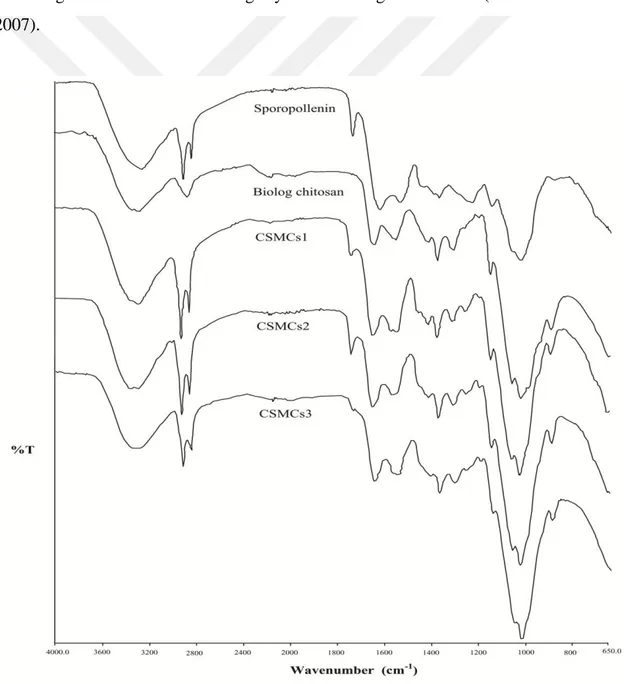
FT-IR analysis
The infrared spectra of the sporopollenin, chitosan and CSMCs are given in Figure 3.2. Chitosan has a typical broad band (O–H and N–H stretching) at 3293 cm−1. In the spectrum of chitosan, the bands appearing at about 1556 and 1375 cm−1 could be assigned to the bending vibrations of N–H and C–H. The strong peak at 1151 cm−1 can be corresponded to the C–O stretching. In the spectrum of raw sporopollenin, the band at 3279 cm−1 is assigned to the stretching of –OH, the bands at 2923 and 2853 cm−1 to
C–H, the band at 1744 cm−1 to C=O, the band at 1141 cm−1 to C–N stretching vibrations and the one at 1417 cm−1 to CH2 and CH3 groups (Gubbuk, 2011). The bands at around
1655 and 1417 cm−1 for –CO and –NH– groups were also observed. These observations in FT-IR spectra indicate condensation of GA onto chitosan polymer. In the spectra of CSMCs, the band observed at 1741 (CSMCs1), 1743 (CSMCs2) and 1743 cm−1 (CSMCs3) can be assigned to the stretching of carbonyl group of sporopollenin. The bands the bands appearing at 1648 (CSMCs1), 1650 (CSMCs2) and 1649 cm−1 (CSMCs3) can be attributed to imine (C=N) groups formed in via condensation of aldehydes of GA with –NH2 groups of chitosan. Additionally, the bands at 1579
(CSMCs1), 1573 (CSMCs2) and 1558 cm−1 (CSMCs3) can be corresponded to C–N stretching and these bands can signify cross-linking of chitosan (Altun and Cetinus, 2007).
Thermogravimetric analysis
Chitosan is a highly crystalline polymer due to the strong intermolecular hydrogen bonds between its strands. Chemical modifications (i.e., imine formation via Schiff bases reaction) that disrupt these hydrogen bonds act on its crystallinity and also reduce its thermal stability (Tirkistani, 1998; Guibal, 2004). Maximum decomposition temperature of chitosan has been reported to be 302°C (Kaya et al., 2014a). A gradually lower decomposition temperature for GA cross-linked chitosan is predicted as the GA/chitosan ratio increases. As for sporopollenin, a recent study by Fraser et al. (2014) revealed that this macromolecule was heat resistant and its chemical structure did not alter until a threshold of 300°C.
Three decomposition steps were observed in the thermograms of the CSMCs (Figure 3.3). The first one (at around 110°C) could be resulted from the evaporation of water molecules present within the polymeric network rather than degradation or decomposition of chitosan and sporopollenin. It seems that decomposition of cross-linked chitosan occurred in the second step at lower temperatures when compared to pristine chitosan. Mass losses in these steps were observed to be in the order of CSMCs3>CSMCs2>CSMCs1, representing the same trend as in GA/chitosan ratio of the CSMCs. On the other hand, the decomposition temperatures in the first and the second steps decreased as the GA/chitosan ratio increased. The last decomposition step could be attributed mainly to the degradation of heat resistant sporopollenin grains. It appears that sporopollenin grains incorporated in the polymeric network did not contribute to the thermal stability of cross-linked chitosan. However, sporopollenin itself mainly decomposed at around 440°C.
Figure 3.3. TG-DTG curves of chitosan/sporopollenin microcapsules; CSMCs1 (a), CSMCs2 (b) and CSMCs3 (c).
3.1.2. Cu(II) removal studies
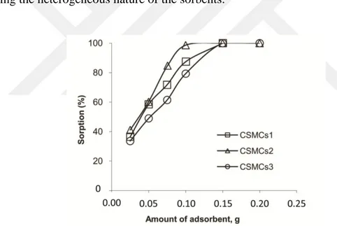
Effect of adsorbent dosage
The amount of adsorbent was studied in varying amounts of CSMCs1, CSMCs2 and CSMCs3 microcapsules (i.e. 0.0250–0.1500 g) (Figure 3.4). Increase in the amount of the microcapsules led to higher percentage of Cu(II) ion removal at a certain point (~0.1000 g). After this point, increasing amount of the adsorbent did not significantly contribute to the percentage of the metal removal once sorption sites on the microcapsules were saturated (Ngah and Fatinathan, 2008). It appeared that 0.1 g of microcapsules was sufficient for 25 mL of metal solution (dosage 4 g L−1). This amount was not high when compared to the adsorbent dosages reported in the earlier literature. In a review paper on copper ion removal from industrial wastewaters, Bilal et al. (2013) compared the efficiency of biosorbents from various sources and the authors listed adsorbent doses (in g L−1) in wider ranges: activated carbon with different origins; 0.1–
25, fungal biomass; 0.2–15, algal biomass; 0.1–8, bacterial biomass; 0.2–32, yeasts biomass; 1–15 and agricultural/forest wastes adsorbents; 0.1–50 g L−1.
It was observed that sorption equilibrium was achieved in lower dosages for CSMCs2 than CSMCs1 and CSMCs3, indicating its higher sorption efficacy. As reported in earlier studies, cross-linking of chitosan with GA reduced metal sorption capacity of chitosan sorbents (Guibal, 2004; Wang et al., 2013). Ngah and Fatinathan (2008) synthesized chitosan-alginate composites sorbents for Cu(II) removal and they used higher adsorbent dosage for chitosan–alginate beads cross-linked with GA (chitosan beads; 0.125, chitosan–GA 1:1 ratio beads; 0.200 and chitosan–GA 2:1 ratio beads; 0.500 g). However, here, the sorbent synthesized with the lowest GA amount did not have the highest sorption capacity. This can be attributed to the sporopollenin contents of the sorbents rather than the cross-linking ratio. As discussed in SEM analysis, CSMCs sorbents were composite materials with different size and shapes, showing the heterogeneous nature of the sorbents.
Figure 3.4. Effect of adsorbent dosage on the sorption of Cu(II) by chitosan/sporopollenin microcapsules (initial concentration of Cu(II): 10 mg L−1; pH of the solution: 5.6; temperature: 25±1°C; shaking speed:
200 rpm).
Effect of contact time
The effect of contact time was studied to determine the time required to reach the adsorption equilibrium (Figure 3.5). In first 60 minutes, there was relatively rapid Cu(II) ion uptake for all the adsorbents. Equilibrium was attained nearly in 240 min for
CSMCs1 and CSMCs2, but it took CSMCs3 about 240 min more time to reach the plateau value. This may be attributed to the physical features of the microcapsules (surface morphology, size and porosity). As discussed in the section 3.1.1, CSMCs3 microcapsules had less sporopollenin grains exposed to the surface and therefore less pores and less binding sites on them. As a result, Cu (II) ions needed more time to diffuse into CSMCs3 capsules. In an earlier study on copper ion sorption by sporopollenin grains (Unlu and Ersoz, 2006), the sorption kinetics was fast and the equilibrium was reached within in the first 30 min. In another study (Gubbuk, 2011), functionalized sporopollenin (first with 3-aminopropyltrimethoxysilane then with glutaraldehyde) exhibited slower adsorption kinetics for copper ions and the equilibrium was attained in 90 min. It seems that sporopollenin grains entrapped in polymeric matrix show slower adsorption kinetics for copper ions. This can explain the longer contact time observed in this study.
Figure 3.5. Effect of contact time on the sorption of Cu(II) by chitosan/sporopollenin microcapsules (initial concentration of Cu(II): 10 mg L−1; pH of the solution: 5.6; amount of CSMCs: 0.10 g;
temperature: 25±1°C; shaking speed: 200 rpm).
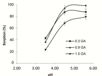
Effect of pH
The effect of solution pH on Cu (II) ion removal was investigated at pH; 3.50, 4.50 and 5.60 (Figure 3.6). Maximum Cu(II) sorption was achieved at pH:5.60. Sorption studies could not be carried out at pH higher than 6.0 to avoid possible precipitation of copper hydroxide (Wang et al., 2013). The amount of Cu (II) ion sorbed
by the microcapsules was highly pH-dependent. Both metal ions and the corresponding adsorbent were greatly affected by alterations in the solution pH. Hydroxyl and amine functional groups on chitosan backbone were protonated at low pH, increasing positively charged adsorption sites on the adsorbent. This could lead to lower metal removal percentage because of the enhanced repulsion between the metal cations and the binding sites (Wan et al., 2010). The CSMCs3 sorbent, the one with highest GA ratio, was less affected by the changes in pH when compared to the others. The cross-linking agent, GA, reacted with amino groups on chitosan via Schiff’s base formation and lowered the number of free amino groups. Therefore, CSMCs3 microcapsules had less free amino groups and exhibited a lower performance in Cu(II) removal. Other than amino groups, hydroxyl groups at the C-3 position on chitosan can interact with metal ions through electrostatic interaction and complexation (Guibal, 2004). On the other hand, the sporopollenin itself has polymeric nature and the metal ion chelating moieties (aromatic, hydroxyl, carbonyl/carboxyl and ether functional groups) on it are also subject to protonation acidic medium. In an earlier study, the highest Cu(II) sorption on the sporopollenin grains occurred at pH 5.0 (Unlu and Ersoz, 2006). Another study reported Cu(II) ion adsorption on modified sporopollenin and the author commented that higher the maximum adsorption was at pH 5.5 (Gubbuk, 2011). In a recent study by Wang et al. (2013), the authors synthesized a semi-penetrating polymeric hydrogels with chitosan and they observed that Cu(II) sorption capacity decreased at higher pHs as the GA/chitosan ratio was increased. The authors concluded that the swelling ratio of the hydrogels decreased at pH higher than 5, which in turn led to the reduction of internal volumes and Cu(II) sorption capacity. It seems that Cu(II) sorption on CSMCs has pH depending nature stemming from the cross-linking ratio of the microcapsules and its content; chitosan and sporopollenin.
Figure 3.6. Effect of pH on the sorption of Cu(II) by chitosan/sporopollenin microcapsules (initial concentration of Cu(II): 10 mg L−1; contact time: 240 min; amount of CSMCs: 0.10 g; temperature:
25±1°C; shaking speed: 200 rpm).
3.1.3. Thermodynamic analysis
As it is presented in Figure 3.7, Cu (II) uptake was improved with the increase in the temperature for all three types of adsorbents. Cu(II) ion sorption percentage of CSMCs2 microcapsules increased by about 2% (i.e., from 98.5% to 99.9%), but rising temperature enhanced the sorption capacity of CSMCs1 and CSMCs3 by around 13 and 11%. This observation may be explained by the expanding of channels/pores on the capsules with temperature and thus easier diffusion of Cu (II) ions through the pores. This could indicate more flexible and swelling nature of the least cross-linked one, CSMCs1 microcapsules.
Figure 3.7. Effect of temperature on the sorption of Cu(II) by chitosan/sporopollenin microcapsules (initial concentration of Cu(II): 10 mg L−1; contact time: 240 min; pH of the solution 5.6; amount of
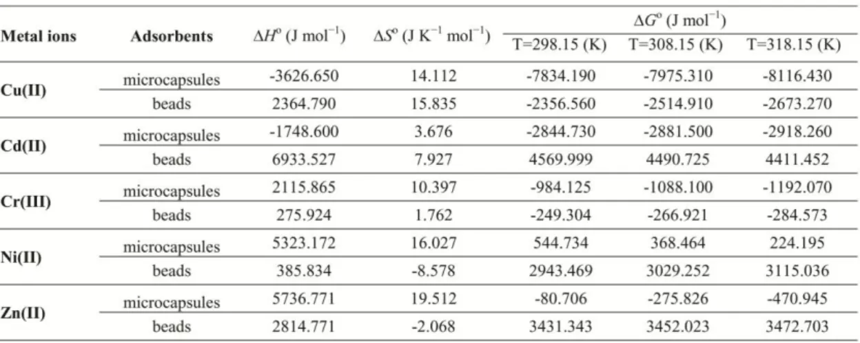
Thermodynamic parameters for the adsorption of Cu(II) on CSMCs1, CSMCs2 and CSMCs3 microcapsules were calculated (Table 3.1). The positive values of ΔHo indicated that the sorption of Cu(II) on the three microcapsule types was of endothermic nature. The negative values of change in ΔGo
revealed that the sorption of Cu(II) onto the microcapsules was spontaneous and thermodynamically feasible for each temperature in the studied conditions (Unlu and Ersoz, 2006; Gubbuk, 2011). Further, as seen from Table 3.1, there was an increase in negative values of Gibbs free energy for each type of sorbents at higher temperatures. The positive value of ΔSo indicated the increased randomness at the system during the sorption, and this can be attributed to the replacement of adsorbed species by copper ions as suggested by some workers or release of water molecules coordinated to the metal cation (Gubbuk, 2011). However, in recent paper, Liu and Lee (2014) reviewed copper ion adsorption studies with thermodynamic analysis and argued that further experimental studies on the thermodynamic parameters and surface chemistry should be done to reveal exact nature of the sorption.
Table 3.1. Thermodynamic parameters for the adsorption of Cu(II) on chitosan/sporopollenin microcapsules.
3.1.4. Sorption from binary mixtures
Cu(II) sorption capacity of CSMCs1, CSMCs2 and CSMCs3 microcapsules in presence of competitive ions (Zn(II), Cd(II), Ni(II) and Cr(III)) were determined and are listed in Table 3.2. The copper ion adsorption capacity of the sorbents decreased in the binary mixtures when compared to that of in copper solutions; 1.40, 1.58 and 1.27 mmol g−1. In single component Cu(II) solutions, CSMCs2 had the highest sorption capacity. However, in the presence of competitive ions, CSMCs1 exhibited better performance in Cu(II) sorption than CSMCs2 and CSMCs3. Cu(II) ion sorption capacities of the microcapsules were found to be in order of
CSMCs1>CSMCs2>CSMCs3 in all the binary metal solutions. All the microcapsules showed higher affinity for Cu(II) ions over Cr(III), Zn(II), Cd(II) and Ni(II) ions in binary solutions. Ionic radius order of these ions is Cr(III)>Cd(II)>Zn(II)>Cu(II)>Ni(II). The number of accessible coordination sites formed within the polymeric matrix, ionic radius and the charge density of the ions could have a role in this behaviour.
Table 3.2. Cu(II) sorption capacities of chitosan/sporopollenin microcapsules in the presence of competitive ions (adsorbent dose: 0.1 g; temperature: 25°C; pH:5.6; volume of metal solution: 25mL).
3.1.5. Desorption studies and the re-usability of the adsorbents
Desorption percentage of the Cu(II) ions in dilute HCl solution was observed to be significantly lower than in EDTA solution. Desorption in HCl solutions were found to be 3.88, 0.52 and 6.21 % for the CSMCs1, CSMCs2 and CSMCs3, respectively. On the other, desorption of Cu(II) ions by the chelating agent EDTA was more effective; 84.27, 10.12 and 94.65 %. The findings indicated that CSMCs1 and CSMCs3 can be regenerated in dilute EDTA solution.
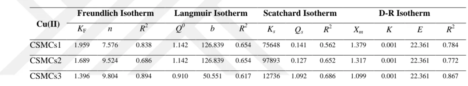
3.1.6. Isotherm analysis
When the regression coefficients are considered, it seems that sorption equilibrium of Cu(II) ions onto all the sorbents can be better explained by Freundlich isotherm model (Table 3.3). The better fitting to Freundlich isotherm model indicated the heterogeneous nature of the microcapsules and the presence of the adsorption sites with differing energy. The values of n > 1 indicated the favourability of adsorption conditions (Wan et al., 2010). Additionally, the deviation from linearity observed in the Scatchard plot analysis showed the presence of different types of the sorption sites on the microcapsules (Kucukosmanoglu et al., 2006). The mean free energy of adsorption,
E values, obtained in D–R isotherm model analysis can provide information on the
nature of the copper ion sorption; chemical, ion-exchange or physical adsorption. When
E is higher than 16 kJ mol−1, the nature of sorption can be considered to be chemisorption, whereas E values in a range of 8–16 kJ mol−1 show an ion-exchange mechanism, and if E< 8 kJ mol−1, the sorption process is supposed to be of physical nature (Tuzen and Sarı, 2010). The higher E values (22.361 kJ mol−1) for copper sorption by the microcapsules showed that the sorption proceeded most likely through chemisorption rather than ion-exchange mechanism or physisorption.
Table 3.3. Parameters of Freundlich, Langmuir, Scatchard and D–R isotherms for sorption of Cu(II) ion on chitosan/sporopollenin microcapsules (adsorbent dosage: 0.1 g; temperature: 25°C; pH:5.6; volume of Cu(II) solution: 25mL).
Cu(II)
Freundlich Isotherm Langmuir Isotherm Scatchard Isotherm D-R Isotherm
KF n R 2 Q0 b R2 Ks Qs R2 Xm K E R2 CSMCs1 1.959 7.576 0.838 1.142 126.839 0.654 75648 0.141 0.562 1.379 0.001 22.361 0.784 CSMCs2 1.689 9.524 0.686 1.142 126.839 0.654 97893 0.127 0.652 1.317 0.001 22.361 0.772 CSMCs3 1.396 9.804 0.894 0.910 50.551 0.617 12736 1.092 0.686 1.099 0.001 22.361 0.867
3.2. Chitosan/sporopollenin microcapsules and plain chitosan microbeads
3.2.1. Characterisation of chitosan/sporopollenin microcapsules and plain chitosan microbeads
SEM images
SEM images clearly demonstrated the almost spherical shape of the microcapsules and the entrapment of the sporopollenin particles in chitosan polymeric matrice (Figure 3.8). The images also showed the effects chemical treatment on the surface morphology of the sporopollenin grains. As depicted in Figure 3.8, prior to introduction into the chitosan network, the sporopollenin grains had high monodispersity and consistency of size. After coagulation and cross-linking treatments, the grains could retain their structural integrity to some extent; but their surface characteristics were partially disrupted; and as a result, the cavities were compressed or disappeared.
Partial decomposition of sporopollenin grains in the gelation solution, which was high alkaline methanol solution, took place in the production process. Bubert et al. (2002) conducted studies on acidic methanolysis of sporopollenin of Typhaangustifolia. They exposed the sporopollenin grains to HCl in methanol in a stepwise manner; and they analysed the decomposition products using spectroscopic techniques. They observed substantial weight losses of the sporopollenin grains at the end of each step. Similarly, we observed 16.8 % mass loss on average upon drying the cross-linked microcapsules with comparison to the chitosan microbeads without sporopollenin. We can conclude that the higher mass losses observed in preparation of the chitosan/sporopollenin microcapsules can be attributable to the decomposition of the sporopollenin grains; indicating the act of methanolic alkali solution on sporopollenin grains incorporated in the chitosan microcapsules during the gelation treatment. However, this phenomenon needs clarifying; a detailed analysis of the decomposition products released from the sporopollenin grains during the incubation in the basic media should be done.
Figure 3.8. SEM images of the sporopollenin grains (Fig.1a-d) (magnification: a: 1000x, b: 3000x, c: 5000x and d: 10000x) and chitosan/sporopollenin microcapsules (Fig.1e-h): (magnification: e: 80x, f:
250x, g: 1000x and h: 5000x).
FT-IR spectra analysis
The analysis of the FT-IR spectra of sporopollenin (Figure 3.9a), methanol-NaOH treated sporopollenin (Figure 3.9b), chitosan (Figure 3.9c), chitosan/sporopollenin microcapsules (Figure 3.9d) and metal ion loaded microcapsules (Figure 3.10) could provide insights into the nature of the metal ion-microcapsule interactions. As seen from Figure 3.9, after the basic methanolysis of the sporopollenin grains, the OH– stretching vibrations at 3378 cm−1 did not shift, but the 1709 cm−1 band that can be assigned to carboxylic/ketone stretching shifted to 1706 cm−1 with lesser intensity, and the sharp peak at 1561 cm−1 attributable to the presence of aromatic C=C group appeared. The C–N stretching vibration band (1187 cm−1) was observed at 1194 cm−1. The band at 1606 cm−1, assigned to aromatic C=C ring stretching showed weak absorbance at 1603 cm−1 as a shoulder. The band at 1260 cm−1 representing aromatic ether groups appeared at 1258 cm−1 with strong absorbance (Steemans et al., 2010). In the FT-IR spectrum of chitosan, the absorption band at 1589 cm−1, which is corresponded to the NH2 groups stretching, disappeared after the cross-linking with
glutaraldehyde, and the band appearing at 1640 cm−1 can be corresponded to the imine stretching (Kandile et al., 2015). The absorption bands, observed at 1656, 1640, 1575 and 1549 cm−1 in the spectrum of chitosan/sporopollenin microcapsules were shifted after forming complexes with metal ions. Additionally, the alterations in the intensity of
CH2 scissoring bands at 1418 cm−1 could be attributed to the contribution of CH2OH
side chains in metal interaction (Figure 3.10) (Hebeish et al., 2013). Here, the characteristic broader band at 3293 cm−1 (assigned to stretching vibration of N–H and axial vibration of OH groups present in chitosan) did not shift after cross-linking. But, the band at 2873 cm−1 (assigned to intermolecular hydrogen bonds of chitosan and the axial C–H stretching (Antony et al., 2013) was shifted and two peaks, at 2919 and 2849 cm−1 (symmetric and asymmetric modes of CH2 group vibration) appeared in spectrum
of cross-linked chitosan/sporopollenin microcapsules. These two peaks were also observed in the presence of metal ions but with some alterations. These observations demonstrated that the interaction of metal ions with the microcapsules occurred mainly via amino, hydroxyl and phenolic groups. Earlier workers studied interactions of metal ions with raw and modified sporopollenin. These workers reported that sporopollenin itself had an affinity for the metal ions due to the functional hydroxyl, carbonyl/carboxyl and ether moieties on it. Based on findings reported (Unlu and Ersoz, 2006; Erzengin et al., 2011) and the analysis of FT-IR spectra of pristine and treated sporopollenin (Figure 3.9) in this study, it can be commented that functional groups on sporopollenin particles could also contribute to the sorption of the metal ions.
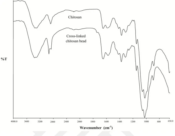
Figure 3.9. FT-IR spectra of sporopollenin (a), methanol-NaOH treated sporopollenin (b), chitosan (c) and chitosan/sporopollenin microcapsules (c).
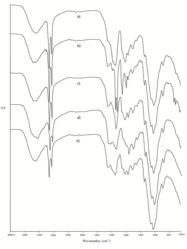
Figure 3.10. FT-IR spectra of chitosan/sporopollenin microcapsules loaded with copper(II) (a), zinc(II) (b), cadmium(II) (c), chromium(III) (d), nickel(II) (e).
Thermogravimetric analysis
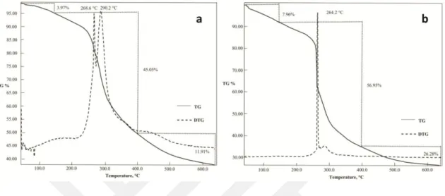
The thermal stability of the microcapsules and the microbeads was evaluated by TGA (Figure 3.11). In the thermogram of the microcapsules, three decomposition steps
were observed. The first decomposition step could be resulted from the evaporation of water molecules (Baran et al., 2015); the others can be ascribed to the degradation of the chitosan polymer and the sporopollenin grains within the matrice. The microcapsules exhibited two maximum decomposition temperatures (DTGmax) (272.4 and 278.8°C);
whereas the chitosan microbeads one at lower temperature 269.3°C. In a recent paper by Fraser et al. (2014), it has been reported that the sporopollenin grains are heat-resistant and the grains start to decompose at around 300°C. Therefore, the higher DTGmax,
(278.8°C) could be corresponded to the degradation of sporopollenin grains. It appears that sporopollenin grains contributed to the thermal stability of the microcapsules.
Figure 3.11. TG-DTG curves of chitosan/sporopollenin microcapsules (a) and chitosan microbeads (b).
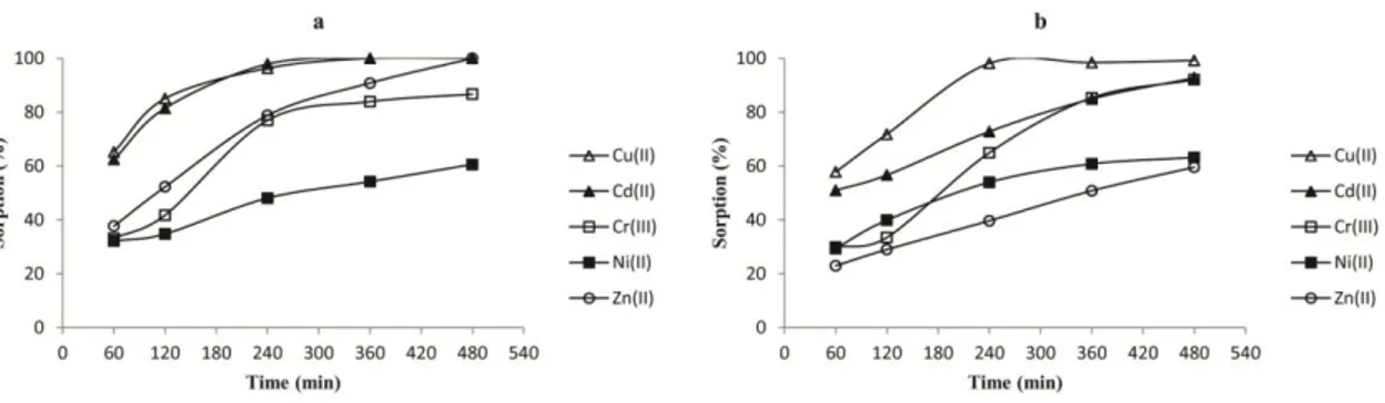
3.2.2. Metal ion removal studies
Effect of the adsorbent dosage
The effect of the amount of chitosan/sporopollenin microcapsules and the chitosan microbeads on metal sorption was investigated for each metal ion (Figure 3.12).As the sorbent dose was increased, so did the amount of metal ions sorbed by the microcapsules to a certain extent. Sorption saturation point, where increase in sorbent dosage did not contribute much to the metal sorption, was different for all the metal ions studied. As seen, such saturation point was reached at about 0.1500 g of microcapsules or the microbeads.