·Original article·
Efficacy of radial keratotomy in the optical
rehabilitation of mild to moderate keratoconus cases
Ibrahim Kocak
1,2, Ali Aydin
1,2, Faruk Kaya
1, Hakan Baybora
1, Salih Bozkurt
31Department of Ophthalmology, Nisa Hospital, Istanbul
34196, Turkey
2Ophthalmology Clinic, School of Medicine, Medipol
University, Istanbul 34214, Turkey
3Inci Eye Hospital, Sakarya 54100, Turkey
Correspondence to: Ibrahim Kocak. 魻zel Nisa Hastanesi, G觟z Hastaliklari Servisi, Fatih Caddesi, Yenibosna, Bahcelievler, Istanbul 34196, Turkey. ibrahimkocak@ msn. com
Received:2012-09-11摇 摇 Accepted:2015-01-16
放射状角膜切开术在轻中度圆锥角膜病例中视
觉康复的疗效
Ibrahim Kocak1,2, Ali Aydin1,2, Faruk Kaya1, Hakan
Baybora1, Salih Bozkurt3
(作者单位:134196 土耳其,伊斯坦布尔,Nisa 医院眼科;234214
土耳其,伊斯坦布尔,Medipol 大学医学院临床眼科;354100 土耳
其,萨卡里亚,Inci 眼科医院)
通讯作者:Ibrahim Kocak. ibrahimkocak@ msn. com
摘要 目的:探讨放射状角膜切开术在轻中度圆锥角膜治疗中光 学和视力康复的效果。 方法:回顾性分析应用放射状角膜切开术治疗圆锥角膜的 病例22 例 31 眼并进行了至少 12mo 的随访。 测量并分析 术前术后裸眼视力,最佳矫正视力,自动屈光计值,角膜曲 率,角膜不规则指数以及并发症。 结果:在最后一次随访中,平均裸眼视力(logMAR)由 0. 86依 0. 34 显著提升至 0. 30依0. 29 (P<0. 0001),平均最佳矫正 视力由0. 47依0. 21 提升至 0. 17依0. 23(P<0. 0001)。 平均 角膜曲 率 由 48. 69 依 3. 68D 降 低 至 44. 33 依 3. 09D ( P < 0郾 0001)。 自动屈光计测得平均等效球镜值由-5. 61 依2. 85D 显著提升至-2. 29依1. 95D(P<0郾 0001)。 在整个随访 过程中,中央角膜厚度和 3mm,5mm 区域的角膜不规则指 数均无变化。 术中和术后没有观察到严重并发症。 结论:在本组病例中,放射状角膜切开术是轻中度圆锥角 膜视觉康复的有效治疗方法。 关键词:放射状角膜切开术;圆锥角膜;视觉重建
引用:Kocak I, Aydin A, Kaya F, Baybora H, Bozkurt S. 放射状
角膜切开术在轻中度圆锥角膜病例中视觉康复的疗效. 国际眼
科杂志2015;15(4):572-576
Abstract
誗AIM: To investigate the efficacy of radial keratotomy
(RK) for the optical and visual rehabilitation of mild to moderate keratoconus.
誗METHODS: Thirty- one eyes of 22 keratoconus patients
with at least 12mo follow-up were recruited and reviewed retrospectively. Uncorrected visual acuity ( UCVA), best spectacle corrected visual acuity (BSCVA), autorefractometer values, keratometric values, corneal irregularity indexes, and complications were analyzed pre- and post-operatively.
誗RESULTS: At the last follow - up visit, mean UVCA
(logMAR) improved significantly from 0. 86依0. 34 to 0. 30依 0. 29 (P<0. 0001), and mean BSCVA (logMAR) improved from 0. 47 依 0. 21 to 0. 17 依 0. 23, ( P < 0. 0001 ). Mean keratometric values decreased from 48. 69依 3. 68 to 44. 33依 3. 09 diopters ( P < 0. 0001 ). Mean spherical equivalent measured by autorefractometer was also improved significantly from - 5. 61 依 2. 85 to - 2. 29 依 1. 95 at the last follow-up visit (P<0. 0001). Central corneal thickness and corneal irregularity index of 3mm and 5mm zones did not change during the overall follow - up time. No serious
intraoperative or postoperative complication was
observed.
誗CONCLUSION: RK is assessed as an effective treatment
method for the optical and visual rehabilitation of mild to moderate keratoconus cases, according to our results in a selected group of keratoconus patients.
誗KEYWORDS: radial keratotomy; keratoconus; optical
rehabilitation
DOI:10. 3980 / j. issn. 1672-5123. 2015. 4. 02
Citation:Kocak I, Aydin A, Kaya F, Baybora H, Bozkurt S. Efficacy of radial keratotomy in the optical rehabilitation of mild to moderate keratoconus cases. Guoji Yanke Zazhi(Int Eye Sci) 2015; 15(4):572-576
INTRODUCTION
K
eratoconus is a progressive disorder in which the cornea assumes a conical shape secondary to paraxial stromal thinning and protrusion[1]. This ectatic corneal disorder hasan incidence of approximately 1/ 2000[2]. In advanced keratoconus
with severe consequences such as irregular astigmatism, corneal scarring, and excessive thinning, patients ultimately require corneal transplantation surgery[3]. In early cases,
spectacles and soft contact lenses may provide adequate vision correction. However, rigid contact lenses are required for higher degrees of irregular astigmatism to render a regular refracting surface[4,5]. When these patients show intolerance
to contact lens, they are faced with the need of various surgical treatment modalities including incisional keratotomy, photorefractive keratectomy (PRK), epikeratoplasty, intrastromal ring implantation, and Athens protocol in which collagen
cross-linking is combined with topography guided PRK to maintain functional vision[6-12].
In the past, radial keratotomy ( RK ) had been used for correction of myopic refractive errors until excimer laser became mainstay of treatment[13]. In addition to treatment of
myopia, it has been also used for optical rehabilitation of mild to moderate keratoconus[14-17]. In our clinic, we have been
treating keratoconus in selected cases using RK, since 2007, based on the technique reported by Utine et al[14]. In this
study, we retrospectively assessed our results to analyze the efficacy and safety of RK in keratoconus treatment.
SUBJECTS ANDMETHODS
Subjects摇 Subjects enrolled in the study were consecutive keratoconus patients who underwent RK at the Nisa Hospital, Istanbul, Turkey, from January 2007 to November 2010. Informed consent approved by the local ethics committee was signed by all patients and the study was conducted in accordance with the Declaration of Helsinki. Patients who had met the following criteria were scheduled for surgery: keratoconus with a central corneal thickness ( CCT ) over 400滋m, poor corrected visual acuity (臆0. 4 at Snellen visual acuity charts), contact lens intolerance, no apical scarring, no ocular or systemic problem other than keratoconus. Although mild to moderate keratoconus was defined as keratoconus with an average keratometry of 臆 54 diopters (D), patients with an average keratometry of >54D were also included if they did not have corneal apical scarring, prominent Descemet蒺s striae, or a CCT of <400滋m.
Contact lens wear was discontinued 1mo before the initial examination in all patients. All patients had a full ophthalmologic examination, including the objective refraction, uncorrected and best spectacle corrected Snellen visual acuity ( UCVA and BSCVA), slit lamp examination, posterior segment examination, intraocular pressure measurement, corneal thickness measurement by using an ultrasonic pachymeter ( DGH 4000B, DGH Technology, Inc. , Frazer, PA, USA), and corneal topography (Orbscan II, Bausch&Lomb, Rochester, NY, USA ), at the preoperative and postoperative visits. Postoperative follow up visits were scheduled at the 1st, 7th, 30th day and the 6th
month after surgery, and then yearly afterward. Patients with at least 1y follow-up were included in the study.
Methods
Surgical procedure 摇 All the surgical treatments were performed under topical anesthesia. Radial incisions were made from limbus to center with a Russian type 30毅 RK diamond blade. Central optical zone marking was made with Thornton optical zone markers ranging from 3mm to 5mm. Profile markers with 4-10 lines were used for corneal marking before radial incisions. Depth of radial incisions was adjusted to 95% of CCT. The target correction was planned according to the preoperative objective cycloplegic refraction measurement. Ten to 12 radial incisions were made for correction of 5 or more diopters of myopia, and 8 incisions for 3 - 5D, 6 incisions for 1 -3D, 4 incisions for one or less diopter were
made. Fine adjustment was achieved by optical zone assessment. Astigmatic corrections were made by oblique optical zone markers which provide longer and more central incisions at the steep axis. No arcuate incision was used for astigmatic correction. Astigmatic correction was not intended if the myopia was more than 8D. If the myopia was more than 10D, peripheral re-deepening was performed 7mm from the center of the cornea with 600滋m incisions. Radial incisions were irrigated with a 27 gauge blunt tip cannula at the end of surgery.
Follow- up 摇 During the first postoperative week, patients were treated with topical antibiotic, artificial tear, and corticosteroid drops. At the end of the first week, topical antibiotic drops were discontinued. During the first postoperative week, patients were treated with topical antibiotic, artificial tear, and corticosteroid drops. At the end of the first week, topical antibiotic drops were stopped. Topical corticosteroids were stopped at the end of the first month with gradual tapering (4伊1 in first two weeks, 2伊1 in third week, and 1伊1 in the last week). In the case of emmetropia (spherical equivalent: 0. 00依 1. 00D ) or overcorrection by the end of the 1st month,
corticosteroid was stopped. If there was undercorrection corticosteroid was continued 1 伊 1 for 3mo. Artificial tear substitutes were also used during the first month after surgery. Statistical Analysis摇 Paired two sample t-test and Wilcoxon matched pairs test were used to compare parameters before and after surgery. Two tailed distribution outcomes were accepted for P values. All analyses were performed with Statplus software ( Analysoft, USA). P values less than 0. 05 were considered to be statistically significant.
RESULTS
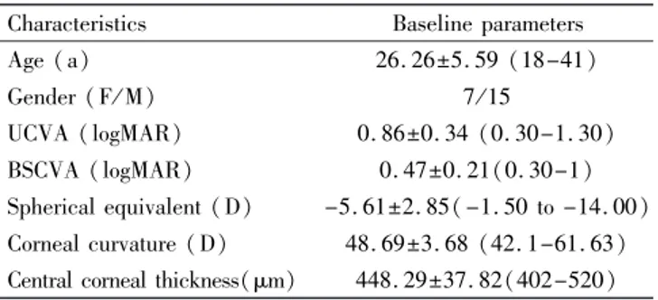
Fifty - five eyes of 36 patients with keratoconus had RK procedure at the Nisa Hospital between 2007 and 2010. Thirty-one eyes of 22 patients with at least Thirty-one year follow-up were qualified for this study. The mean age 依 standard deviation ( SD) of patients was 26. 26依5. 59y (range 18-41y). Fifteen of 22 patients were male and seven were female. Mean follow-up time was 18. 16 依 10. 83mo ( range 12 - 52mo ). Patient characteristics and baseline parameters are presented in Table 1. All of the patients were operated under topical anesthesia. Mean number of incisions was 9. 35 依2. 03 ( range 5 -12). Seventeen ( 55% ) of eyes were treated with 10 or more incisions, 13 (42% ) with six or more, and one (3% ) with less than six incisions. Oblique zone markers were used in 24 (77% ) of eyes. In the remaining seven eyes circular optical zone markers were used. Mean optical zone diameter of these seven eyes was 3. 29 依0. 39mm. No serious intraoperative or postoperative complication was observed. Microperforation occurred in two eyes and treated with therapeutic contact lens application.
At the last follow - up visit, mean UCVA and BSCVA were both increased compared to values of preoperative examination (P < 0. 0001 for both ) ( Table 2 ). Mean BSCVA was improved by the first day of RK procedure and remained stable
3 7 5
Table 1 摇 Patient characteristics and baseline refractive
parameters (
x依s
軃
,range)Characteristics Baseline parameters
Age (a) 26. 26依5. 59 (18-41) Gender (F/ M) 7 / 15 UCVA (logMAR) 0. 86依0. 34 (0. 30-1. 30) BSCVA (logMAR) 0. 47依0. 21(0. 30-1) Spherical equivalent (D) -5. 61依2. 85(-1. 50 to -14. 00) Corneal curvature (D) 48. 69依3. 68 (42. 1-61. 63)
Central corneal thickness(滋m) 448. 29依37. 82(402-520)
till the last follow-up visit. Box-plot graphical presentation of the group蒺s BSCVA changes was shown in Figure 1. Mean Snellen lines gained at the last follow - up visit was 3. 71 依 1郾 89 (range 0-6) for BSCVA, and 4. 08依2. 37 (range 1-9) for UCVA. BSCVA improved in 29 of 31 eyes. In the remaining two eyes BSCVA did not change. None of the eyes showed worsening of BSCVA or UVCA. Mean spherical equivalent measured by autorefractometer was also significantly decreased at the last follow-up visit compared to preoperative values (P<0. 0001). There was a slight decrease in mean cylindrical refractive error, but it was not statistically significant. Objective refraction changes are presented in Table 2.
Mean keratometric values decreased significantly from 48. 69依 3. 68D to 44. 33 依 3. 09D ( P < 0. 0001 ), and corneal astigmatism from 4. 00依1. 61D to 2. 75依1. 96D (P = 0. 0002). CCT, and corneal irregularity index of 3mm and 5mm zones did not change significantly during the overall follow-up time (P= 0. 4189, P = 0. 3975 and P = 0. 6498, respectively). Corneal changes were shown in Table 3. Keratometric values were decreased by the first day of RK procedure, and remained stable during the follow-up. The change of keratometric values is shown by using a box plot presentation in Figure 2. Corneal topography of a demonstrative case treated with RK was shown in Figure 3.
DISCUSSION
In this study, we reviewed and analyzed the outcomes of our RK experience in mild to moderate keratoconus patients with a CCT greater than 400滋m. Patients unwilling or intolerant to contact lens wear were recruited for RK procedure. Mild keratoconus patients that have satisfactory visual acuity with spectacles and advanced cases requiring penetrating keratoplasty were not eligible for the treatment. All the eyes showed improvement in UCVA, and 29 of 31 eyes showed improvement in BSCVA. Differences between last follow -up visit and preoperative state were statistically significant for both UCVA and BSCVA. Myopic refractive error, mean keratometric values, and corneal astigmatism all decreased significantly after RK procedure.
These findings were consistent with other studies in which RK was used for the optical rehabilitation of mild to moderate keratoconus, and keratoconus suspects[14-17]. Grandon and
Weber screened 1368 eyes treated with RK using corneal
Figure 1摇 Box-plot graphic of the BSCVA change from before to after RK.
Figure 2 摇 Box - plot graphic of keratometric change from before to after RK.
Figure 3 摇 Corneal topography of a keratoconus case treated
with RK摇 Preoperative UCVA was 0. 3 and BSCVA was 0. 5 with
-4. 00 (170毅 -3. 25) diopters. Mean corneal power calculated with Orbscan II was 46. 3 依3. 1D ( A). By the end of the first year of RK, UCVA increased to 0. 8 and BSCVA to 1. 0 with (60毅 -0. 75) D. Mean corneal power decreased to 42. 9依3. 5D (B).
topography, and found that 14 eyes of nine patients exhibited topographic patterns of keratoconus suspect status before the surgery[15]. They reported that refraction and UCVA improved
in all 14 of these eyes at 4-19mo postoperatively. In a similar study, Bowman et al[16] reviewed the records of 67
摇 摇 摇 摇 摇 Table 2摇 Visual acuity and objective refraction values before and after radial keratotomy (
x依s
軃
,range)Parameters Preoperative Postoperative P
UCVA 摇 LogMAR 0. 86依0. 34 (0. 30-1. 30) 0. 30依0. 29 (0-1) <0. 0001 Decimal 0. 1 0. 5 BSCVA 摇 LogMAR 0. 47依0. 21 ( 0. 30-1) 0. 17依0. 23 (0-1) <0. 0001 Decimal 0. 3 0. 7 Spherical equivalent (D) 5. 61依2. 85 (1. 50-14. 00) 2. 29依1. 95 (0. 25-8. 00) <0. 0001 Cylinder (D) 3. 15依1. 45 (0. 50-6. 50) 2. 57依1. 64 (0. 00-7. 00) 0. 0737
摇 摇 摇 摇 摇 UCVA:Uncorrected visual acuity; BSCVA: Best spectacle corrected visual acuity.
摇 摇 摇 摇 摇 Table 3摇 Corneal parameters before and after radial keratotomy (
x依s
軃
,range)Parameters Preoperative Postoperative P
Averagekeratometry (D) 48. 69依3. 68 (42. 1-61. 63) 44. 33依3. 09 (37. 75-52. 00) <0. 0001
Corneal astigmatism (D) 4. 00依1. 61 (1. 50-8. 25) 2. 75依1. 96 (0. 50-8. 00) 0. 0002
Central corneal thickness (滋m) 448. 29依37. 82 (402-520) 445. 94依40. 76 (384-518) 0. 4189
Irregularity ( 3mm zone) 5. 36依1. 85 (2. 6依9. 2) 5. 16依1. 73 (1. 5-8. 9) 0. 3975
Irregularity (5mm zone) 5. 54依1. 69 (2. 8-9. 2) 5. 66依1. 98 (1. 7-12. 6) 0. 6498
frequency of keratoconus suspects by using videokeratography, and reported that three patients were identified as keratoconus suspect. All of these eyes achieved postoperative UCVA of 20 / 30 or better. In the large and detailed study of Utine et al[14], authors presented their experience of RK for the
optical rehabilitation of 96 patients with mild to moderate keratoconus. They reported that RK procedure yielded significant increase in UCVA and BSCVA as the result of corneal flattening and reduction of spherical - astigmatic refractive errors in keratoconus patients. This improvement was stable and remained relatively unchanged throughout the follow-up, as we observed in our patients.
In some previous studies, several uncommon late complications of RK are reported in cases with keratoconus. Sharma et al[18] reported an acute hydrops development in a
patient with subclinical keratoconus who underwent RK eight years ago. Panda et al[19] presented three cases that had
incisional dehiscence following blunt trauma 10 - 13y after RK. Durand et al[20] also reported a similar dehiscence two
years after RK. We did not observe such late complications in our series. Utine et al[14] reported that they had observed
microperforation in four eyes (2. 2% ) and perforation in two eyes (1. 2% ), with an incision depth of 100% of CCT. In our study, microperforation occurred in two eyes during the operation. No perforation was observed in our patients. This may be related to our more conservative incision depth that we assigned as 95% of CCT. Besides, we have a limited case number compared to Utine et al蒺s[14]study.
Late hyperopic shift following RK due to progressive corneal flattening is reported in some previous studies[14, 21].
Additionally, Saragoussi et al[22,23] suggested that subclinical
keratoconus might be a possible cause of progressive corneal flattening following RK. In our study, myopic refractive error (spherical equivalent) decreased from -2. 26依2. 16D (at the
6thmonth after RK) to -1. 96 依2. 30D at the last follow-up
visit ( P = 0. 0262). Mean keratometry also decreased from 44. 64依3. 33D (at the 6thmonth after RK) to 44. 33依3. 09D
at the last follow - up visit ( P = 0. 0204 ). These findings reveal the presence of late hyperopic shift due to progressive corneal flattening that shows consistence with previous studies. We believe that hyperopic shift of less than 1D after RK would not create a significant problem in cases with keratoconus who are mostly left with some degree of myopic error following RK operation.
CCT and irregularity index of 3mm, and 5mm zone did not change during the overall follow-up time in our study. These results can be interpreted as RK limits the progression of keratoconus disease at least during follow - up time of our study. We think that fibrotic activation promoted by RK incisions may strengthen the biomechanical structure of cornea and limit progression of disease. It has been shown that perpendicular incisions could trigger a more significant corneal wound healing process compared to lamellar incisions. Additionally, epithelial plugs above the RK incisions cause fibrotic activation even after years[24, 25].
The visual rehabilitation issue of contact lens intolerant cases with mild to moderate keratoconus is still controversial. Authors have suggested different surgical treatment modalities including incisional keratotomy, photorefractive keratectomy, epikeratoplasty, intrastromal ring implantation, and Athens protocol to treat these patients[6-12]. The procedures that
attenuate biomechanical structure of cornea by thinning may increase the progression of keratoconus. However, combining collagen cross linking with surgical procedures such as PRK gives promising outcomes as in the case of Athens protocol. In the study of Kanellopoulos[26], UCVA ( logMAR) improved
from 0. 96依0郾 2 to 0. 3依0. 25, and BSCVA from 0. 39依0. 3 to 0. 11依0. 16 in keratoconus cases treated with Athens protocol,
5 7 5
similar to our results. Intracorneal ring segment implantation is another current option for visual rehabilitation of keratoconus patients. In the study of Kaya et al[27], patients
with keratoconus gained (0. 18 依0. 04) lines of UCVA and (0. 21依0. 05) lines of BSCVA with Intacs rings; and gained (0. 21 依0. 09) lines of UCVA, and (0. 26 依0. 08) lines of BSCVA with Ferrara ring. Visual acuity improvement seems better with RK than ring implantation when these results compared to ours. Besides, some complications related to ring implantation such as severe corneal vascularization, anterior chamber perforation, ring exposure, and dense corneal infiltrates were reported[28,29].
Nowadays, corneal collagen cross - linking has become the standard therapy for stabilizing the progressive keratoconus. It would be interesting to compare the effects of RK and corneal cross-linking on keratoconus in a large progressive study. In conclusion, our study showed that RK is an effective method for the visual and optical rehabilitation of selected cases with mild to moderate keratoconus. However, these results should be interpreted with caution due to the small number of patients and limited follow - up period, and potential biases related to the retrospective and observational nature of our study.
REFERENCES
1 Feder RS, Kshettry P. Noninflammatory ectatic disorders. In: Krachmer JH, Mannis MJ, Holland EJ. eds. Cornea 2nd ed. Elsevier inc; 2005:955-974
2 Rabinowitz YS. Keratoconus. Surv Ophthalmol 1998;42(4):297-319 3 Zadok D, Schwartz S, Marcovich A, Bakana Y, Morad Y, Eting E, Avni I. Penetrating keratoplasty for keratoconus: long - term results. Cornea 2005;24(8):959-961
4 Szczotka LB, Barr JT, Zadnik K. A summary of the findings from the Collaborative Longitudinal Evaluation of Keratoconus ( CLEK) Study. CLEK Study Group. Optometry 2001;72(9):574-584
5 Edrington TB, Szczotka LB, Barr JT, Achtenberg JF, Burger DS, Janoff AM, Olafsson HE, Chun MW, Boyle JW, Gordon MO, Zadnik K, The CLEK Study Group. Rigid contact lens fitting relationships in keratoconus. Collaborative Longitudinal Evaluation of Keratoconus (CLEK) Study Group. Optom Vis Sci 1999;76(10):692-699
6 Kasparova EA, Kasparov AA. Six-year experience with excimer laser surgery for primary keratoconus in Russia. J Refract Surg 2003;19 (2 Suppl):250-254
7 Krumeich JH, Daniel J, Knulle A. Live - epikeratophakia for keratoconus. J Cataract Refract Surg 1998;24(4):456-463
8 Bilgihan K, Ozdek SC, Konuk O, Akata F, Hasanreiso姚glu B. Results of photorefractive keratectomy in keratoconus suspects at 4 years. J Refract Surg 2000;16(4):438-443
9 Siganos D, Ferrara P, Chatzinikolas K, Bessis N, Papastergiou G. Ferrara intrastromal rings for the correction of keratoconus. J Cataract Refract Surg 2002;28(11):1947-1951
10 Siganos SC, Kymnionis GD, Kartakis N, Theodorakis MA, Astyrakakis N, Pallikaris IG. Management of keratoconus with Intacs.
Am J Ophthalmol 2003;135(1):64-70
11 Spitznas M, Eckert J, Frising M, Eter N. Long-term functional and topographic results seven years after epikeratophakia for keratoconus. Graefes Arch Clin Exp Ophthalmol 2002;240(8):639-643
12 Krueger RR, Kanellopoulos AJ. Stability of simultaneous topography-guided photorefractive keratectomy and riboflavin/ UVA cross-linking for progressive keratoconus: case reports. J Refract Surg 2010;26(10):827-832 13 Sanders DR, Marks RG. Prospective clinical study of radial keratotomy. Ophthalmology 1982;89(11):1292-1293
14 Utine CA, Bayraktar S, Kaya V, Kucuksumer Y, Eren H, Perente I, Yilmaz OF. Radial keratotomy for the optical rehabilitation of mild to moderate keratoconus: more than 5 years蒺 experience. Eur J Ophthalmol 2006;16(3):376-384
15 Grandon SC, Weber RA. Radial keratotomy in patients with atypical inferior steepening. J Cataract Refract Surg 1994;20(6):670-671 16 Bowman CB, Thompson KP, Stulting RD. Refractive keratotomy in keratoconus suspects. J Refract Surg 1995;11(3):202-206
17 Lombardi M, Abbondanza M. Asymmetric radial keratotomy for the correction of keratoconus. J Refract Surg 1997;13(3):302-307 18 Sharma N, Sachdev R, Jindal A, Titiyal JS. Acute hydrops in keratectasia after radial keratotomy. Eye Contact Lens 2010;36(3):185-187 19 Panda A, Sharma N, Kumar A. Ruptured globe 10 years after radial keratotomy. J Refract Surg 1999;15(1):64-65
20 Durand L, Monnot JP, Burillon C, Assi A. Complications of radial keratotomy: eyes with keratoconus and late wound dehiscence. Refract Corneal Surg 1992;8(4):311-314
21 Waring GO 3rd, Lynn MJ, McDonnel PJ. Results of the prospective evaluation of radial keratotomy ( PERK) study 10 years after surgery. Arch Ophthalmol 1994;112(10):1298-1308
22 Saragoussi JJ, Pouliquen YJ. Does the progressive increasing effect of radial keratotomy ( hyperopic shift ) correlate with undetected early keratoconus? J Refract Corneal Surg 1994;10(1):45-48
23 Saragoussi JJ. Progressive hyperopia after radial keratotomy. A new etiological hypothesis early keratoconus. J Fr Ophtalmol 1993;16(10): 499-500
24 Cameron JD. Corneal reaction to injury. In: Krachmer JH, Mannis MJ, Holland EJ. eds. Cornea 2nd ed. : 2005 Elsevier Inc. ; 2005: 115-131
25 Tuli S, Goldstein M, Schultz GS. Modulation of corneal wound healing. In: Krachmer JH, Mannis MJ, Holland EJ. eds. Cornea 2nded. Elsevier inc. ; 2005:133-150
26 Kanellopoulos AJ. Comparison of sequential vs same - day simultaneous collagen cross - linking and topography - guided PRK for treatment of keratoconus. J Refract Surg 2009;25(9):812-818 27 Kaya V, Utine CA, Karakus SH, Kavadarli I, Yilmaz OF. Refractive and visual outcomes after Intacs vs ferrara intrastromal corneal ring segment implantation for keratoconus: a comparative study. J Refract Surg 2011;27(12):907-912
28 Kymionis GD, Kontadakis GA. Severe corneal vascularization after intacs implantation and rigid contact lens use for the treatment of keratoconus. Semin Ophthalmol 2012;27(1-2):19-21
29 Kanellopoulos AJ, Pe LH, Perry HD, Donnenfeld ED. Modified intracorneal ring segment implantations (INTACS) for the management of moderate to advanced keratoconus: efficacy and complications. Cornea 2006;25(1):29-33
6 7 5