Expanding the Phenotypic Spectrum of
Lupus Erythematosus in Aicardi-Goutie
`res Syndrome
Georgia Ramantani,
1Ju
¨rgen Kohlhase,
2Christoph Hertzberg,
3A. Micheil Innes,
4Kerstin Engel,
1Susan Hunger,
1Wiktor Borozdin,
2Jean K. Mah,
5Kristina Ungerath,
6Hartmut Walkenhorst,
7Hans-Helmut Richardt,
7Johannes Buckard,
8Andrea Bevot,
9Corinna Siegel,
10Celina von Stu
¨lpnagel,
11Chrysanthy Ikonomidou,
12Kara Thomas,
13Virginia Proud,
13Frank Niemann,
14Dagmar Wieczorek,
15Martin Ha
¨usler,
16Pascal Niggemann,
17Volkan Baltaci,
18Karsten Conrad,
19Pierre Lebon,
20and Min Ae Lee-Kirsch
1 Objective. Aicardi-Goutie`res syndrome (AGS) isan early-onset encephalopathy resembling congenital viral infection that is characterized by basal ganglia calcifications, loss of white matter, cerebrospinal fluid (CSF) lymphocytosis, and elevated interferon-␣ levels in the CSF. Studies have shown that AGS is an autosomal-recessive disease linked to mutations in 5 genes,
encod-ing the 3ⴕ-repair DNA exonuclease 1 (TREX1), the 3 subunits of ribonuclease H2 (RNASEH2A–C), and ster-ile alpha motif domain and HD domain–containing protein 1 (SAMHD1). In this study we further charac-terized the phenotypic spectrum of this disease.
Methods. Clinical and laboratory data were
ob-tained from 26 patients fulfilling the clinical diagnostic criteria for AGS. Genomic DNA was screened for mu-tations in all 5 AGS genes by direct sequencing, and sera were analyzed for autoantibodies.
Results. In 20 patients with AGS, 20 mutations,
12 of which were novel, were identified in all 5 AGS genes. Clinical and laboratory investigations revealed a high prevalence of features (some not previously de-scribed in patients with AGS) that are commonly seen in patients with systemic lupus erythematosus (SLE), such as thrombocytopenia, leukocytopenia, antinuclear anti-bodies, erythematous lesions, oral ulcers, and arthritis, which were observed in 12 (60%) of 20 patients with AGS. Moreover, the coexistence of AGS and SLE, was for the first time, demonstrated in 2 patients with molecularly proven AGS.
Conclusion. These findings expand the
pheno-typic spectrum of lupus erythematosus in AGS and provide further insight into its disease mechanisms by
Supported by the Deutsche Forschungsgemeinschaft (DFG grant LE 1074/3-1).
1Georgia Ramantani, MD (current address: University of
Freiburg, Freiburg, Germany), Kerstin Engel, Susan Hunger, Min Ae Lee-Kirsch, MD: Technische Universita¨t Dresden, Dresden, Germany;
2Ju¨rgen Kohlhase, MD, Wiktor Borozdin, PhD: Center of Human
Genetics Freiburg, Freiburg, Germany; 3Christoph Hertzberg, MD:
Vivantes Klinikum Neuko¨lln, Berlin, Germany;4Micheil Innes, MD,
FRCP, FCCMG: University of Calgary, Calgary, Alberta, Canada;
5Jean K. Mah, MD, MSc, FRCPC: Alberta Children’s Hospital,
Calgary, Alberta, Canada; 6Kristina Ungerath, MD: Altonaer
Kinderkrankenhaus, Hamburg, Germany; 7Hartmut Walkenhorst,
MD, Hans-Helmut Richardt, MD: Evangelisches Krankenhaus Be-thanien, Iserlohn, Germany;8Johannes Buckard, MD: Evangelisches
Krankenhaus Du¨sseldorf, Du¨sseldorf, Germany;9Andrea Bevot, MD:
Universita¨tsklinik fu¨r Kinder- und Jugendmedizin, Tu¨bingen, Ger-many;10Corinna Siegel, MD: Technische Universita¨t Mu¨nchen,
Mu-nich, Germany;11Celina von Stu¨lpnagel, MD: Klinikum Harlaching,
Munich, Germany;12Chrysanthy Ikonomidou, MD, PhD: University
of Wisconsin School of Medicine and Public Health, Madison;13Kara
Thomas, MS, CGC, Virginia Proud, MD: Children’s Hospital of The King’s Daughters and Eastern Virginia Medical School, Norfolk, Virginia;14Frank Niemann, MD: Kinder- und Jugendklinik
Gelsen-kirchen, GelsenGelsen-kirchen, Germany;15Dagmar Wieczorek, MD:
Univer-sita¨tsklinikum Essen, Essen, Germany;16Martin Ha¨usler, MD:
Uni-versita¨tsklinikum Aachen, Aachen, Germany; 17Pascal Niggemann,
MD: Privatpraxis fu¨r Kernspintomographie, Ko¨ln-Rodenkirchen, Ger-many; 18Volkan Baltaci, MD: Ufuk University, Ankara, Turkey; 19Karsten Conrad, MD: Technische Universita¨t Dresden, Dresden,
Germany; 20Pierre Lebon, MD: Hoˆpital St. Vincent de Paul, Paris,
France.
Drs. Ramantani and Kohlhase contributed equally to this work.
Dr. Proud has received a clinical education support grant from Genzyme Therapeutics (less than $10,000).
Address correspondence and reprint requests to Min Ae Lee-Kirsch, MD, Klinik und Poliklinik fu¨r Kinder- und Jugendmedi-zin, Technische Universita¨t Dresden, Fetscherstrasse 74, D-01307 Dresden, Germany. E-mail: minae.lee-kirsch@uniklinikum-dresden. de.
Submitted for publication September 30, 2009; accepted in revised form January 20, 2010.
showing that activation of the innate immune system as a result of inherited defects in nucleic acid metabolism could lead to systemic autoimmunity.
Aicardi-Goutie`res syndrome (AGS) was first de-scribed in 1984 in 8 children who presented with an early-onset progressive encephalopathy, characterized by basal ganglia calcifications, white matter abnormali-ties, and chronic cerebrospinal fluid (CSF) lymphocyto-sis (1). Familial occurrence of the disease suggested that it has an autosomal-recessive inheritance. Furthermore, the finding of raised levels of the antiviral cytokine interferon-␣ (IFN␣) in the CSF of affected children implies that there are similarities between AGS and congenital viral infection, in spite of a lack of evidence to support common prenatal infections (2,3).
Affected children typically present with irritabil-ity, inconsolable crying, dystonia, hypotonia, and sei-zures. Symptoms occur in an episodic manner starting as early as the first day of life, leading to progressive microcephaly and spastic quadriplegia, with severe de-velopmental delay. Extraneurologic manifestations in-clude hepatosplenomegaly, thrombocytopenia, and chil-blain lesions, which are inflammatory cutaneous lesions at acral locations (4). On the basis of symptoms de-scribed in 3 case reports, it was first suggested that there is clinical overlap of AGS with the autoimmune disease lupus erythematosus, as observed in patients who pre-sented with lupus-like rash and antinuclear antibodies, in addition to progressive encephalopathy with intracra-nial calcifications (5–7). However, in a recent large study that included 123 patients with AGS, no further lupus-associated symptoms were observed, and only 6 patients were reported to have an abnormal antibody profile (8). AGS is a genetically heterogeneous disorder, and to date, biallelic mutations have been identified in 5 genes, encoding the 3⬘-repair DNA exonuclease 1 (TREX1; in locus AGS1), the 3 subunits of the ribonu-clease H2 complex (RNASEH2B, RNASEH2C, and RNASEH2A in loci AGS2, AGS3, and AGS4, respec-tively), and sterile alpha motif domain and HD domain– containing protein 1 (SAMHD1; in locus AGS5), which is a putative regulator of the innate immune system (9–11). HomodimericTREX1 functions as an intracell-ular DNA exonuclease with high specificity for single-stranded DNA. The RNASEH2 complex constitutes an endonuclease that cleaves RNA with RNA:DNA hy-brids or single ribonucleotides embedded within DNA duplexes. These findings suggest that an inappropriate activation of the innate immune response that occurs as
a result of defects in nucleic acid metabolism may underlie the pathogenesis of AGS.
Heterozygous mutations in TREX1 have also been described in patients with familial chilblain lupus, an autosomal-dominant form of cutaneous lupus ery-thematosus manifesting in early childhood (12–14), and in patients with autosomal-dominant retinal vasculopa-thy with cerebral leukodystrophy, an adult-onset disor-der characterized by central nervous system degenera-tion, retinal vasculopathy, and nephropathy (15). Furthermore, we identified heterozygous mutations in TREX1 in patients with sporadic systemic lupus ery-thematosus (SLE), further expanding the phenotypic spectrum ofTREX1 mutations (16). This is an intriguing finding, in view of the fact that both diseases, AGS and SLE, are characterized by the activation of interferon pathways (2,3,17,18).
TREX1 deficiency has been shown to impair DNA damage during granzyme A–mediated apoptosis (13). Thus, improper clearance of altered DNA may induce, through as yet unidentified intracellular sensors and signaling pathways, an immune-mediated inflamma-tory response. Moreover, recent evidence suggests that intracellular accumulation of single-stranded DNA in TREX1-deficient cells may be attributable to defects in the degradation of nucleic acids derived from chronic cell-cycle checkpoint activation or from endogenous retroviruses (19,20). Collectively, these findings under-pin the importance of defects in intracellular nucleic acid metabolism as a possible pathogenic mechanism of systemic autoimmunity.
In this report, we present clinical and molecular data from 20 patients with AGS who were found to carry mutations in all 5 of the AGS genes known to date. Our results provide evidence that phenotypic features of lupus erythematosus are more prevalent in AGS than have hitherto been appreciated.
PATIENTS AND METHODS
Patients. Patients with a clinical presentation
sugges-tive of AGS underwent molecular analysis of all 5 AGS genes. Patients were included in this study based on the following diagnostic criteria: 1) neurologic symptoms of an encephalop-athy, 2) intracranial calcification, 3) absence of findings of common prenatal infections, and/or 4) a CSF white cell count ofⱖ5 white cells/mm3or raised levels of IFN␣ (⬎2 IU/liter) in
the CSF. IFN␣ levels were measured using a cytopathic effect inhibition assay with vesicular stomatis virus. All mutation-negative patients and patients in whom only 1 mutation could be identified also fulfilled the diagnostic criteria outlined above.
Portuguese, Sinti, Hindi, or Moroccan origin were screened. All patients from Canada or the US were of European descent. After the patients provided their informed written consent, blood samples were obtained from all patients and, if available, also from their parents and siblings. Patients and their families were seen by the primary investigators or were referred to us by the pediatric neurologist responsible for the care of the patient. Clinical, neuroimaging, and laboratory data were obtained from the medical records, magnetic resonance imag-ing (MRI) scans, and computed tomography scans. All patients for whom serum was available were tested for autoantibodies (antinuclear antibodies [ANAs], anti–extractable nuclear anti-gen, anti–double-stranded DNA, anti–single-stranded DNA, anti-C1q, and anticardiolipin) and for their levels of comple-ment (C3 and C4). Autoantibodies and complecomple-ment were determined at the diagnostic laboratory of the Institute of Immunology, Technical University Dresden, according to stan-dard procedures.
Mutation analysis. Genomic DNA and RNA were
isolated from peripheral blood leukocytes using the QIAamp Blood Mini Kit and the RNeasy mini kit, respectively (Qiagen). Polymerase chain reaction (PCR) amplification of all coding exons and flanking intronic regions of TREX1/AGS1 (314 amino acids), RNASEH2A/AGS4 (299 amino acids), RNASEH2B/AGS2 (308 amino acids), RNASEH2C/AGS3 (164 amino acids), andSAMHD1/AGS5 (626 amino acids) was performed using gene-specific oligonucleotide primers (se-quences available from the corresponding author upon re-quest). Following verification of the PCR product size by agarose gel electrophoresis, amplicons were column purified and sequenced in both directions using fluorescently labeled dideoxy nucleotides on ABI 3100 or 3730 genetic analyzers (Applied Biosystems). Sequencing data were analyzed with Vector NTI software (Invitrogen) or Seqpilot software (JSI
Medical Systems). Peripheral blood leukocytes from at least 100 unrelated healthy individuals were also sequenced, to confirm the absence of newly identified missense changes in healthy individuals.
RESULTS
Molecular findings. Of the 26 patients fulfilling the clinical diagnostic criteria for AGS, 20 patients from 18 families were found to harbor mutations in 1 of the 5 AGS genes known to date. Altogether, we identified 20 mutations in AGS1–5, 12 of which were novel mutations (Table 1). Seven families (39%) carried mutations in TREX1/AGS1, and 3 of these were either homozygous or compound heterozygous for a common substitution (R114H) (9). One patient carried a novel heterozygous missense mutation, D200H, that was confirmed to be de novo based on parental genotypes. In this patient, the presence of a second wild-type allele could be confirmed based on the finding of heterozygosity for a common single-nucleotide polymorphism (SNP) at c.531 in TREX1.
Five families (28%), of various ethnic origins, carried mutations in RNASEH2B/AGS2, including a novel missense mutation, S229P, and a recurrent substi-tution, A177T (9). Two families (11%) harbored muta-tions in RNASEH2C/AGS3. One patient, of Indian origin, was homozygous for an R69W substitution, which was previously reported to occur on a common
haplo-Table 1. Mutations in patients with AGS*
Family/ancestry Consanguinity Locus/gene Nucleotide change Protein change 1/Turkish No AGS1/TREX1 c.150_151delTC⫹ c.907A⬎C Q51fsX100⫹ T303P
2/Turkish Yes AGS1/TREX1 c.341G⬎A, hom R114H
3/European American No AGS1/TREX1 c.341G⬎A ⫹ c.500delG R114H⫹ S166fsX179 4/German No AGS1/TREX1 c.341G⬎A ⫹ c.635delC R114H⫹ P212fsX276
5/Turkish Yes AGS1/TREX1 c.592G>A, hom E198K
6/German No AGS1/TREX1 c.598G>C, het, de novo† D200H
7/Lebanese Yes AGS1/TREX1 c.859_876del18, hom L287_G292del
8/Portuguese Yes AGS2/RNASEH2B c.529G⬎A, hom A177T
9/Sinti Yes AGS2/RNASEH2B c.529G⬎A, hom A177T
10/German No AGS2/RNASEH2B c.529G⬎A, het‡ A177T
11/European American No AGS2/RNASEH2B c.529G⬎A, het‡ A177T 12/European Canadian No AGS2/RNASEH2B c.529G⬎A ⫹ c.685T>C A177T⫹ S229P 13/German No AGS3/RNASEH2C c.115G>T⫹ c.343_344delGA D39Y⫹ D115fsX169
14/Indian Yes AGS3/RNASEH2C c.205C⬎T, hom R69W
15/European Canadian No AGS4/RNASEH2A c.4G>T;8T>C⫹ c.716_717dupGC D2Y;L3P⫹ T240fsX316
16/Moroccan Yes AGS4/RNASEH2A c.556C⬎T, hom R186W
17/Turkish Yes AGS5/SAMHD1 c.499C>T, hom H167Y
18/German No AGS5/SAMHD1 c.869G>A⫹ c.1642C⬎T R290H⫹ Q548X
* Novel mutations in patients with Aicardi-Goutie`res syndrome (AGS) are indicated in boldface. For complementary DNA numbering, ⫹1 corresponds to the A of the ATG initiation codon.TREX1 ⫽ 3⬘-repair DNA exonuclease 1; hom ⫽ homozygous; het ⫽ heterozygous; RNASEH2B ⫽ ribonuclease H2 subunit B;SAMHD1 ⫽ sterile alpha motif domain and HD domain–containing protein 1.
† Mutation was confirmed to be a de novo change according to genetic analyses. ‡ Only a single mutation was identified in AGS1–5.
Table 2. Phenotypic characteristics of the patients with AGS*
Family/age/sex Presentation Laboratory findings Additional features 1/4 years/M Dystonia, hypotonia, MC, SZ, death at 4
years
Thrombocytopenia Septicemia
2/1 year/M Neonatal SZ, HSM, dystonia, MC ALFT, thrombocytopenia None 3/1 year/M Multiple purpuric lesions at birth, HSM,
MC, dystonia, newborn SZ, death at 14 months
Thrombocytopenia, leukocytopenia, normal levels of IFN␣ and PT in CSF
ASD, hearing loss, retinal hemorrhages, corneal clouding
4/3 months/F Dystonia, hypotonia, MC, SZ Elevated levels of IFN␣, protein, and PT in CSF, ALFT,
thrombocytopenia
Nystagmus, hearing loss
5/19 years/M Marked startle reaction, dystonia, MC, refractory SZ, SE
Elevated lactate levels, protein levels, and cell count in CSF, ANA 1:1,280, anti-ENA, decreased C3 levels
Nystagmus, duplicated kidney, contractures, neuropathic scoliosis, chilblain lesions on feet, oral ulcers
6/2 years/F Dystonia, hypotonia, MC ALFT, ANA 1:1,280, anti-dsDNA, anti-C1q, anticardiolipin IgG
Chilblain lesions on fingers/toes/nose, oral ulcers
7/14 years/M Dystonia, severe DD NA None
8/26 years/F† Dystonia, severe DD, MC NA None 8/20 years/F† Dystonia, moderate DD, MC NA None 9/3 years/F Recurrent fever, dystonia, MC, SZ, SE,
moderate DD (evident after vaccination)
CSF lymphocytosis, normal IFN␣ and PT levels in CSF, leukocytopenia, anticardiolipin IgM
None
10/4 years/M Dystonia, severe DD, MC NA None 10/12 years/F Dystonia, severe DD, MC NA None
11/3 years/M Dystonia, severe DD NA None
12/1 year/M SZ, spastic quadriparesis NA None 13/1 year/M MC, dystonia, marked startle reaction
(evident after vaccination)
Elevated IFN␣ levels, lactate levels, cell count, and PT levels in CSF, anti-ssDNA
None
14/6 months/M Newborn SZ, hypotonia, dystonia, MC Thrombocytopenia Blindness, nystagmus, hearing loss, arrhythmia
15/19 years/M† Hypotonia, refractory SZ, spastic quadriparesis, MC
CSF lymphocytosis None
15/20 years/M† Moderate DD, no MC CSF lymphocytosis Chilblain lesion on toe 16/6 months/F HSM, newborn SZ, dystonia, MC Elevated IFN␣ levels and
lymphocytosis in CSF, ALFT
Intestinal atresia, VSD
17/8 months/F Preterm, intrauterine brain calcification, HSM, hypotonia, dystonia, MC
Thrombocytopenia, elevated cell count, elevated IFN␣ and PT levels in CSF
None
18/11 years/M Newborn SZ, refractory SZ, SE, dystonia, MC, marked startle reaction, recurrent fever, spastic quadriparesis
Thrombocytopenia, leukocytopenia, elevated IFN␣ levels and lymphocytosis in CSF, normal PT levels in CSF, ANA 1:160
Cortical blindness, hearing loss, arthritis in knees/finger, chilblain lesions on finger/toes, oral ulcers
19/1 year/F Hypotonia, dystonia, MC Elevated IFN␣ levels and
lymphocytosis in CSF, decreased C4 levels
None
20/8 months/F Hypotonia, dystonia, severe DD, SZ, MC
CSF lymphocytosis,
thrombocytopenia, leukocytopenia
None
21/3 years/F Hypotonia, dystonia, moderate DD, MC Elevated IFN␣ levels in CSF, ALFT, thrombocytopenia, ANA, 1:1,250, anti-ENA, anti-dsDNA
Arthritis
22/2 months/M Hypotonia, dystonia, MC Elevated IFN␣ levels in CSF, thrombocytopenia, leukocytopenia
None
23/3 months/M HSM, hypotonia, dystonia, death at 3 months
CSF lymphocytosis, ALFT,
thrombocytopenia, leukocytopenia
Septicemia
24/1 year/F Hypotonia, dystonia, SZ, MC Unremarkable None
* All patients with Aicardi-Goutie`res syndrome (AGS) presented with basal ganglia calcifications and various degrees of white matter defects and brain atrophy. In patients from families 19–24, no mutations in any of the 5 AGS genes were identified. MC⫽ microcephaly; SZ ⫽ seizures; HSM ⫽ hepatosplenomegaly; ALFT⫽ abnormal liver function test (results); IFN␣ ⫽ interferon-␣; PT ⫽ pterins; CSF ⫽ cerebrospinal fluid; ASD ⫽ atrial septal defect; SE ⫽ status epilepticus; ANA ⫽ antinuclear antibodies; anti-ENA ⫽ autoantibodies against extractable nuclear antigen; anti-dsDNA⫽ autoantibodies against double-stranded DNA; DD ⫽ developmental delay; NA ⫽ not available; anti-ssDNA ⫽ autoantibodies against single-stranded DNA; VSD⫽ ventricular septal defect.
type in Pakistani patients (9). A further patient, of German origin, was compound heterozygous for 2 novel mutations, including the first frameshift change (D39Y ⫹ D115fs) observed in RNASEH2C. Mutations inRNASEH2A/AGS4 were detected in 2 families (11%). In 1 family, 2 novel missense changes on the same allele were found, along with a previously described 2-basepair
duplication on the other allele (D2Y;L3P ⫹
T240fsX316). One patient carried a homozygous mis-sense change, R186W, which has previously been re-ported as a single mutation in AGS4 in a patient with AGS (8). Two patients (11%) harbored mutations in SAMHD1/AGS5, including a Turkish patient with a novel homozygous missense change, H167Y, and 1 German patient who was compound heterozygous for a novel missense mutation, R290H, and a previously re-ported nonsense mutation in SAMHD1 (11). In the 3 families in which only 1 mutation could be detected, mutations in the other known AGS genes were excluded. None of the newly identified missense changes was annotated as an SNP in the public databases, and all of the mutations were absent in at least 200 control alleles from healthy individuals. With the exception of the 1 case in which a heterozygous de novo TREX1 mutation was identified, all parents for whom there were available data were heterozygous for a single mutation. In 3 of the 6 patients in whom no mutations could be identified on a genomic level (families 20, 22, and 23 in Table 2), sequencing of the complementary DNA of all 5 AGS genes did not provide any evidence of aberrant messenger RNA as a result of undetected intronic variants affecting splicing.
Clinical findings. With 1 exception, all patients carrying mutations in AGS1–5 were born at term. One child with intrauterine growth retardation and brain calcification was born at 35 weeks of gestation. In most cases, the birth weight and head circumference were within normal limits. Five children (2 with TREX1 mutations and 1 each with mutations in RNASEH2C, RNASEH2A, or SAMHD1) presented, within the first days of life, with neonatal seizures, hepatosplenomegaly, thrombocytopenia, or feeding problems (Table 2). All other patients experienced, within the first weeks to 6 months of life, a sudden-to-subacute onset of severe encephalopathy, characterized by irritability, inconsol-able crying with insomnia, and a loss of motor skills during the acute episode. These episodes usually lasted several weeks and were followed by a more stable phase, without further deterioration. Prior to the onset of symptoms, all patients thrived and developed normally. In 2 cases, symptoms became evident within 1–2 weeks
after the first vaccination with a combination vaccine against diphtheria, pertussis, Haemophilus influenzae type b, tetanus, polio, and hepatitis B, occurring at the age of 3 months in both patients.
Neurologic signs were characterized by periph-eral spasticity with paroxysmal dystonic movements, truncal hypotonia, and lack of head control. Some patients developed severe contractures secondary to spastic quadriparesis. Seizures were reported in 11 (55%) of 20 patients with molecularly proven AGS, and these comprised either neonatal seizures, febrile sei-zures, refractory epilepsy, or status epilepticus. Two children demonstrated a marked startle reaction to sudden acoustic stimuli, and 2 presented with intermit-tent sterile pyrexias. Severe developmental delay was seen in almost all of the patients and was associated with progressive microcephaly.
Apart from 2 children with biallelicTREX1 mu-tations who died at 14 months and 4 years of age, respectively, all patients with molecularly proven AGS are currently alive, 8 of whom are older than 10 years of age, with the oldest being 26 years. Teenaged or adult patients were relatively more common among those with RNASEH2B/AGS2 or RNASEH2A/AGS4 mutations. These patients also tended to be older at the time of symptom manifestation. Although developmental delay was an invariable finding, it appeared less severe in these patients, and at least 2 children withRNASEH2B muta-tions and 1 with RNASEH2A mutations were able to walk and talk. In addition, a significant phenotypic variability was noted in the 3 families with 2 affected children; one of these families had 1 child with severe impairment and the other child with only moderate developmental delay or no microcephaly.
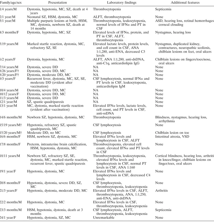
Hearing loss was reported in 4 cases, and oph-thalmologic findings, varying from retinal hemorrhages with corneal clouding to cortical blindness, were noted in 3 patients. Chilblain lesions were observed in 4 patients (20%), of whom 2 carried mutations inTREX1 (Figures 1C–E), 1 carried mutations inRNASEH2A, and 1 carried mutations in SAMHD1. Three patients pre-sented with intermittent oral ulcers. These lesions were mainly located in the buccal mucosa but also extended into the pharyngeal region in 1 patient. A lesional biopsy was performed in 1 patient, with the findings revealing nonspecific signs of inflammation. Additional cutaneous findings included purpuric lesions due to severe throm-bocytopenia, occurring at birth in 1 patient. One patient with SAMHD1 mutations developed, at the age of 6 years, recurrent rheumatoid factor–negative arthritis of the knees and proximal and middle phalangeal joints,
which responded to therapy with prednisone and aza-thioprine. Infrequent features included congenital heart defects, renal malformation, and intestinal atresia. The clinical features of the 6 mutation-negative patients are also presented in Table 2.
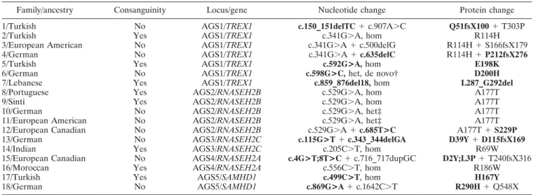
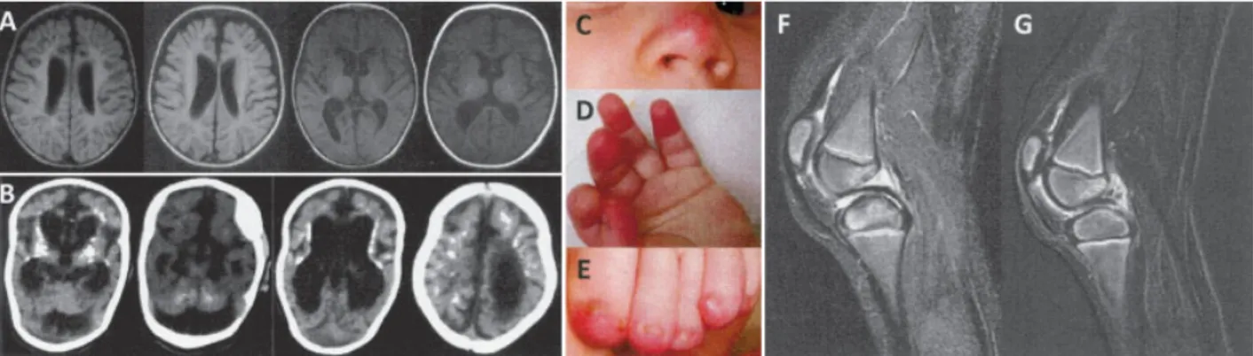
Imaging findings. Intracranial calcifications in-volving the basal ganglia and loss of white matter, as well as atrophic changes, were observed in all patients, albeit with great variability (Figures 1A and B). In some cases, calcifications were evident in the newborn period, pre-senting as a few calcified spots or confluent symmetric lesions extending into the deep white matter. White matter abnormalities ranged from discrete hyperintensi-ties on T2-weighted MRI to severe frontotemporal leukodystrophy. In 1 patient withSAMHD1 mutations, bitemporovacuolar lesions were also observed. While cortical atrophy was a common feature, several children demonstrated marked brain atrophy with brainstem and cerebellar atrophy, dilatation of ventricles, and thinning of the corpus callosum. In 1 patient with SAMHD1 mutations and recurrent arthritis, MRI of the knee demonstrated effusion and thickening of the synovia, consistent with the characteristics of nonerosive arthritis (Figures 1F and G).
Laboratory findings. All patients whose CSF was analyzed underwent lumbar puncture within 3 months of initial presentation of the leukencephalopathy and/or seizures. An elevated white cell count in the CSF was found in 8 (80%) of the 10 patients tested (Table 2), and
when mutation-negative patients were included, 11 (73%) of 15 showed an elevated white cell count in the CSF. Levels of intrathecal IFN␣ were raised in 5 (71%) of the 7 patients tested; with inclusion of mutation-negative patients, 8 (80%) of 10 had elevated IFN␣ levels in the CSF. In 1 severely affected child with biallelic TREX1 mutations, findings in the CSF were unremarkable. Four children were found to have lym-phocytosis in the absence of elevated IFN␣ levels, or vice versa. Elevated pterin levels were recorded in 3 (50%) of 6 patients. Thrombocytopenia (⬍100,000/l) was observed in 7 (35%) of 20 patients, and leukocyto-penia (⬍4,000/l or counts lower than the age-specific third percentile on 2 or more occasions) was observed in 3 (15%) of 20 patients. With mutation-negative patients included in these analyses, 11 (42%) of 26 patients showed thrombocytopenia, and 6 (23%) of 26 presented with leukocytopenia.
Five (71%) of 7 patients who were tested for serologic signs of systemic autoimmunity were found to have ANAs or autoantibodies against extractable nu-clear antigens, double-stranded or single-stranded DNA, C1q, and cardiolipin or to have reduced complement levels (Table 2). When mutation-negative patients were included, 7 (78%) of 9 patients were autoantibody positive or showed reduced complement levels (Table 2). In all patients, titers of autoantibodies were deter-mined beyond the first acute neurologic phase, and the
Figure 1. Imaging findings and cutaneous signs in patients with Aicardi-Goutie`res syndrome (AGS). A, Magnetic resonance imaging of an AGS patient with mutations in ribonuclease H2 subunit C (RNASEH2C)/AGS3 (patient 13), showing extensive white matter abnormalities with an anteroposterior gradient, in addition to cortical atrophy. B, Computed tomography imaging of an AGS patient withRNASEH2A/AGS4 (patient 16), demonstrating marked brain atrophy with prominent cerebellar atrophy, as well as symmetric calcifications of the basal ganglia and periventricular region extending throughout the cerebrum. C–E, Typical chilblain lesions on the nose, fingertips, and toes of an AGS patient with a heterozygous de novo 3⬘-repair DNA exonuclease 1 (TREX1) mutation (patient 6), presenting as painful erythematous swellings. F, Sagittal STIR sequence through the left knee of an AGS patient with mutations in sterile alpha motif domain and HD domain–containing protein 1 (SAMHD1)/AGS5 (patient 18), showing effusion and thickened synovia without surrounding soft tissue edema or bone erosion. G, Subtracted T1-weighted scan, after contrast media application, of the left knee of an AGS patient withSAMHD1/AGS5 (same patient as in F), showing marked contrast enhancement of the synovia, especially adjacent to the posterior cruciform ligament, indicating features compatible with a diagnosis of nonerosive arthritis.
oldest patient was 19 years of age at the time of the examinations.
DISCUSSION
In this report, we present clinical and molecular data from a cohort of patients with AGS that further define the phenotypic and genotypic spectrum of AGS and establish a firm link between AGS and lupus erythematosus at the clinical, molecular, and biochemi-cal levels. The most common AGS subtype in our series wasTREX1/AGS1, followed by RNASEH2B/AGS2. The remaining subtypes, RNASEH2C/AGS3, RNASEH2A/ AGS4, and SAMHD1/AGS5, were observed with equal frequencies. Although a potential role of pathogenic mutations in regulatory regions or larger deletions af-fecting gene expression or splicing could not be fully excluded, the lack of detection of mutations in any of the known AGS genes in the 6 patients with a clinical presentation highly suggestive of AGS may point to the existence of at least 1 additional AGS gene.
Consistent with previous reports, the neurologic phenotype was characterized by the subacute onset of severe encephalopathy with irritability, dystonia, hypo-tonia, and seizures, resulting mostly in severe develop-mental delay and microcephaly (1). All children in our cohort presented with this phenotype within the first year of life. In most cases, symptoms occurred after a period of apparently normal development, without evi-dence of an environmental trigger, while for some, the onset of symptoms was associated with a vaccination. Although no further disease progression beyond the encephalopathic period was observed in most patients, several children were reported to experience continued episodes of irritability, inconsolable crying, and insom-nia during later years. Overall, the clinical features among patients with AGS1–5 were indistinguishable, although neonatal onset or more severe abnormal neu-rologic findings were more frequent among patients with TREX1/AGS1 and SAMHD1/AGS5, whereas patients with RNASEH2B/AGS2 tended to be less severely af-fected.
On the basis of 3 case reports, it was first suggested that there is clinical overlap of AGS with lupus erythematosus, as observed in patients who pre-sented with lupus-like rash and ANAs, in addition to progressive encephalopathy with intracranial calcifica-tions (5–7). However, in the largest case series on AGS to date, which involved 123 patients, thrombocytopenia was observed in only 15 patients (12%), and only 6 patients (⬍5%) were reported to have an abnormal
antibody profile, without further details as to the anti-gens involved or the number of patients examined for the presence of autoantibodies (8). In this study, apart from chilblain lesions, no further symptoms associated with lupus erythematosus were described (8). Likewise, in 3 previous studies of patients with AGS (involving 21 patients, 11 patients, and 10 patients, respectively), no further lupus-associated symptoms were noted, and in 2 of these studies, the results of testing for ANA or complement deficiency were reported to be negative (21–23). This is in contrast to the results reported here, in which 60% of the patients (12 of 20) presented with clinical findings (lupus rash, arthritis, oral ulcers) or laboratory findings (ANAs, autoantibodies to extract-able nuclear antigens, reduced complement levels, thrombocytopenia, leukocytopenia) commonly seen in patients with lupus erythematosus.
If seizures are taken into account as the pheno-typic expression of cerebral lupus, the proportion of patients showing signs of lupus erythematosus in this study would rise to 75% (15 of 20 patients). In fact, the neurologic phenotypes of AGS and SLE show intriguing similarities. Thus, seizures, which are commonly ob-served in patients with AGS, constitute a diagnostic criterion for SLE. Moreover, neuroimaging findings in patients with SLE include calcifications, white matter changes, and atrophy, which are typically observed in patients with AGS (24–26). One may therefore specu-late about common pathogenic mechanisms underlying the neurologic phenotype of AGS and that of cerebral lupus, although this notion requires further investiga-tion.
Novel lupus-associated manifestations that were observed, but previously not described in patients with AGS, included leukocytopenia, oral ulcers, and arthritis, which constitute some of the diagnostic criteria for SLE. In fact, 2 patients, 1 of whom was carrying a heterozy-gous de novo TREX1 mutation and 1 of whom was carrying biallelicSAMHD1 mutations, presented with at least 4 of the 11 diagnostic criteria formally required to establish the diagnosis of SLE, demonstrating, for the first time, the coexistence of AGS and SLE in patients with molecularly proven AGS (27). Of note, nonerosive arthritis was observed in a patient with biallelic SAMHD1 mutations. However, determining whether arthritis represents a manifestation specific for the AGS5 subtype must await further studies.
It is of interest, in this context, that familial chilblain lupus, a monogenic form of cutaneous lupus erythematosus manifesting in early childhood, is also caused by heterozygous mutations in TREX1 (12–14).
Chilblain lupus is characterized by erythematous lesions at acral locations, which are precipitated by cold expo-sure and resemble the chilblain lesions seen in AGS (4,12). However, unlike patients with AGS, patients with familial chilblain lupus do not show any neurologic phenotype, even at old age (12,28). Heterozygous mu-tations inTREX1 have also been described in patients with autosomal-dominant retinal vasculopathy with ce-rebral leukodystrophy, an adult-onset disorder charac-terized by central nervous system degeneration, retinal vasculopathy, and nephropathy (15). Furthermore, we identified rare variants in TREX1 conferring a high relative risk for developing SLE in patients with sporadic SLE, further expanding the phenotypic spectrum of systemic autoimmunity due toTREX1 mutations (16).
In 1 patient, the symptoms of AGS and SLE were attributed to a heterozygous de novoTREX1 mutation (D200H), which affects 1 of 4 magnesium-coordinating residues required for catalytic function (29). Interest-ingly, the only other heterozygous de novo TREX1 mutation reported so far in a patient with AGS affects the same amino acid residue (D200N) (14). These findings may suggest a distinct role for this residue, presumably during recognition, binding, or processing of nucleic acid, and may indicate that these de novo mutations act in a dominant-negative manner.
Our findings suggest that serologic parameters indicative of systemic autoimmunity, such as ANAs, may represent valuable diagnostic markers for AGS. This is of particular relevance during the initial diagnostic eval-uation of an infant who presents with an encephalopathy suggestive of AGS, because early diagnosis of AGS may potentially have a significant impact on clinical manage-ment with immune-modulating agents. With all due caution regarding the neurotoxicity of immunosuppres-sive agents, one may speculate that early immune-modulating intervention during the encephalopathic phase may be helpful in reducing permanent brain damage.
The nucleasesTREX1 and RNASEH2, as well as SAMHD1, constitute putative components of the innate immune system. Although their exact molecular func-tions remain not fully understood, current data suggest that they are involved in the removal of nucleic acid species produced during apoptosis, chronic cell-cycle checkpoint activation, or propagation of endogenous retroviruses, and that a failure of these processes results in an inappropriate activation of the innate immune system (11,13,19,20). Thus, the elucidation of the genetic causes of AGS has revealed a novel functional relation-ship between intracellular nucleic acid metabolism,
nu-cleic acid recognition, and the activation of an innate immune response, underscoring the importance of nu-cleic acid metabolism for mechanisms of antiviral im-mune defense and tolerance. Elevated levels of the antiviral cytokine IFN␣ in the CSF are a hallmark of AGS, and emerging molecular and genetic evidence suggests an important role of interferon pathways in lupus pathogenesis (17,18,30). The coexistence of AGS and SLE in the same patient strongly supports common pathogenic mechanisms for these 2 diseases. Thus, the phenotypic spectrum of lupus erythematosus in AGS defines this monogenic disorder as a novel facet of the immunological disease continuum (31), which paradig-matically highlights the interplay between the innate and the adaptive immune systems in the pathogenesis of systemic autoimmunity.
ACKNOWLEDGMENTS
We thank the patients and their families for their participation in this study.
AUTHOR CONTRIBUTIONS
All authors were involved in drafting the article or revising it critically for important intellectual content, and all authors approved the final version to be published. Dr. Lee-Kirsch had full access to all of the data in the study and takes responsibility for the integrity of the data and the accuracy of the data analysis.
Study conception and design. Ramantani, Lee-Kirsch.
Acquisition of data. Ramantani, Kohlhase, Hertzberg, Innes, Engel,
Hunger, Borozdin, Mah, Ungerath, Walkenhorst, Richardt, Buckard, Bevot, Siegel, von Stu¨lpnagel, Ikonomidou, Thomas, Proud, Niemann, Wieczorek, Ha¨usler, Niggemann, Baltaci, Conrad, Lebon, Lee-Kirsch.
Analysis and interpretation of data. Ramantani, Kohlhase, Borozdin,
Niggemann, Conrad, Lee-Kirsch.
REFERENCES
1. Aicardi J, Goutieres F. A progressive familial encephalopathy in infancy with calcifications of the basal ganglia and chronic cere-brospinal fluid lymphocytosis. Ann Neurol 1984;15:49–54. 2. Lebon P, Badoual J, Ponsot G, Goutieres F, Hemeury-Cukier F,
Aicardi J. Intrathecal synthesis of interferon-␣ in infants with progressive familial encephalopathy. J Neurol Sci 1988;84:201–8. 3. Goutieres F, Aicardi J, Barth PG, Lebon P. Aicardi-Goutieres syndrome: an update and results of interferon-␣ studies. Ann Neurol 1998;44:900–7.
4. Tolmie JL, Shillito P, Hughes-Benzie R, Stephenson JB. The Aicardi-Goutieres syndrome (familial, early onset encephalopathy with calcifications of the basal ganglia and chronic cerebrospinal fluid lymphocytosis). J Med Genet 1995;32:881–4.
5. Dale RC, Tang SP, Heckmatt JZ, Tatnall FM. Familial systemic lupus erythematosus and congenital infection-like syndrome. Neu-ropediatrics 2000;31:155–8.
6. De Laet C, Goyens P, Christophe C, Ferster A, Mascart F, Dan B. Phenotypic overlap between infantile systemic lupus erythemato-sus and Aicardi-Goutieres syndrome. Neuropediatrics 2005;36: 399–402.
Cerebral thrombotic microangiopathy and antiphospholipid anti-bodies in Aicardi-Goutieres syndrome: report of two sisters. Neuropediatrics 2005;36:40–4.
8. Rice G, Patrick T, Parmar R, Taylor CF, Aeby A, Aicardi J, et al. Clinical and molecular phenotype of Aicardi-Goutieres syndrome. Am J Hum Genet 2007;81:713–25.
9. Crow YJ, Hayward BE, Parmar R, Robins P, Leitch A, Ali M, et al. Mutations in the gene encoding the 3⬘-5⬘ DNA exonuclease TREX1 cause Aicardi-Goutieres syndrome at the AGS1 locus. Nat Genet 2006;38:917–20.
10. Crow YJ, Leitch A, Hayward BE, Garner A, Parmar R, Griffith E, et al. Mutations in genes encoding ribonuclease H2 subunits cause Aicardi-Goutieres syndrome and mimic congenital viral brain infection. Nat Genet 2006;38:910–6.
11. Rice GI, Bond J, Asipu A, Brunette RL, Manfield IW, Carr IM, et al. Mutations involved in Aicardi-Goutieres syndrome implicate SAMHD1 as regulator of the innate immune response. Nat Genet 2009;41:829–32.
12. Lee-Kirsch MA, Gong M, Schulz H, Ruschendorf F, Stein A, Pfeiffer C, et al. Familial chilblain lupus, a monogenic form of cutaneous lupus erythematosus, maps to chromosome 3p. Am J Hum Genet 2006;79:731–7.
13. Lee-Kirsch MA, Chowdhury D, Harvey S, Gong M, Senenko L, Engel K, et al. A mutation in TREX1 that impairs susceptibility to granzyme A-mediated cell death underlies familial chilblain lupus. J Mol Med 2007;85:531–7.
14. Rice G, Newman WG, Dean J, Patrick T, Parmar R, Flintoff K, et al. Heterozygous mutations in TREX1 cause familial chilblain lupus and dominant Aicardi-Goutieres syndrome. Am J Hum Genet 2007;80:811–5.
15. Richards A, van den Maagdenberg AM, Jen JC, Kavanagh D, Bertram P, Spitzer D, et al. C-terminal truncations in human 3⬘-5⬘ DNA exonuclease TREX1 cause autosomal dominant retinal vasculopathy with cerebral leukodystrophy. Nat Genet 2007;39: 1068–70.
16. Lee-Kirsch MA, Gong M, Chowdhury D, Senenko L, Engel K, Lee YA, et al. Mutations in the gene encoding the 3⬘-5⬘ DNA exonuclease TREX1 are associated with systemic lupus erythem-atosus. Nat Genet 2007;39:1065–7.
17. Baechler EC, Gregersen PK, Behrens TW. The emerging role of
interferon in human systemic lupus erythematosus. Curr Opin Immunol 2004;16:801–7.
18. Ronnblom L, Alm GV, Eloranta ML. Type I interferon and lupus. Curr Opin Rheumatol 2009;21:471–7.
19. Yang YG, Lindahl T, Barnes DE. Trex1 exonuclease degrades ssDNA to prevent chronic checkpoint activation and autoimmune disease. Cell 2007;131:873–86.
20. Stetson DB, Ko JS, Heidmann T, Medzhitov R. Trex1 prevents cell-intrinsic initiation of autoimmunity. Cell 2008;134:587–98. 21. Lanzi G, Fazzi E, D’Arrigo S. Aicardi-Goutieres syndrome: a
description of 21 new cases and a comparison with the literature. Eur J Paediatr Neurol 2002;6 Suppl A:A9–22.
22. Abdel-Salam GM, Zaki MS, Lebon P, Meguid NA. Aicardi-Goutieres syndrome: clinical and neuroradiological findings of 10 new cases. Acta Paediatr 2004;93:929–36.
23. Lanzi G, Fazzi E, D’Arrigo S, Orcesi S, Maraucci I, Uggetti C, et al. The natural history of Aicardi-Goutieres syndrome: follow-up of 11 Italian patients. Neurology 2005;64:1621–4.
24. Raymond AA, Zariah AA, Samad SA, Chin CN, Kong NC. Brain calcification in patients with cerebral lupus. Lupus 1996;5:123–8. 25. Appenzeller S, Vasconcelos FA, Li LM, Costallat LT, Cendes F. Quantitative magnetic resonance imaging analyses and clinical significance of hyperintense white matter lesions in systemic lupus erythematosus patients. Ann Neurol 2008;64:635–43.
26. Huizinga TW, Steens SC, van Buchem MA. Imaging modalities in central nervous system systemic lupus erythematosus. Curr Opin Rheumatol 2001;13:383–8.
27. Tan EM, Cohen AS, Fries JF, Masi AT, McShane DJ, Rothfield NF, et al. The 1982 revised criteria for the classification of systemic lupus erythematosus. Arthritis Rheum 1982;25:1271–7.
28. Gunther C, Meurer M, Stein A, Viehweg A, Lee-Kirsch MA. Familial chilblain lupus: a monogenic form of cutaneous lupus erythematosus due to a heterozygous mutation in TREX1. Der-matology 2009;219:162–6.
29. Mazur DJ, Perrino FW. Identification and expression of the TREX1 and TREX2 cDNA sequences encoding mammalian 3⬘35⬘ exonucleases. J Biol Chem 1999;274:19655–60.
30. Gregersen PK, Behrens TW. Genetics of autoimmune diseases: disorders of immune homeostasis. Nat Rev Genet 2006;7:917–28. 31. McGonagle D, McDermott MF. A proposed classification of the