A bedside ultrasound technique for
fluid
therapy monitoring in severe hypovolemia:
Tissue Doppler imaging of the right ventricle
ERDEN EROL UNLUER
1, TOGAY EVRIN
2,*, BURAK KATIPOGLU
3, SERDAR BAYATA
41
Department of Emergency Medicine, Usak University Medical Faculty, Usak, Turkey
2
Department of Emergency Medicine, Ufuk University Medical Faculty, Dr Ridvan Ege Education and Research Hospital, Ankara, Turkey
3
Department of Emergency Medicine, Ankara Education and Research Hospital, Ankara, Turkey
4
Department of Cardiology, Izmir Katip Çelebi University, Ataturk Research and Training Hospital, Izmir, Turkey
*Corresponding author: Togay Evrin, MD, Assistant Professor; Department of Emergency Medicine, Ufuk University Medical Faculty, Dr Ridvan Ege Education and Research Hospital, Ankara 06520, Turkey; Phone: +90 505 375 11 73; Fax: +90 312 204 40 54; E-mail: [email protected]
(Received: March 28, 2017; Revised manuscript received: May 26, 2017; Accepted: May 30, 2017)
Abstract: Fluid therapy is one of the main issues for hemodynamic resuscitation. Tissue Doppler imaging (TDI) of the right ventricle (RV) with bedside ultrasound (BUS) technique is a new dynamic method to identifyfluid responsiveness in patients with hypotension. Here, we present the case of a hypotensive patient monitored with TDI measurements of RV. A 75-year-old male patient was admitted to the emergency department (ED) with the complaint of diarrhea. He was in severe hypovolemia, with hypotension, tachycardia, and tachypnea. His laboratory results were normal. BUS was performed on the patient by the ED physician. The velocity of the excursion of the tricuspid valve measured at presentation was 14.47 cm/s and, together with collapsed inferior vena cava (IVC), thisfinding led to the decision to begin fluid therapy immediately. The patient underwent 2 L of fluid therapy with 0.9% NaCl in a 2-h period. Control BUS after fluid therapy revealed decreased TDI velocity of tricuspid annulus to 11.81 cm/s and dilated IVC not collapsing sufficiently with respiration. The patient received his maintenance therapy after admission to the internal medicine department and was discharged from the service after 3 days. TDI influid responsiveness may find a clinical role in the future by the clinical studies. Keywords: bedside ultrasound, emergency medicine,fluid therapy, right ventricle, tissue Doppler imaging
Introduction
Fluid therapy is one of the main issues for hemodynamic
resuscitation. Fluids are administered to increase cardiac
output, and, ultimately, tissue perfusion. However, the
physician should identify the patient in the rising part of
the Frank
–Starling curve, not in the plateau phase, in
order to see a positive response to
fluid therapy. The
recognition of patients in the rising part of this curve can
be provided by some non-invasive measurements with
bedside ultrasound (BUS) technique, such as
velocity-time integral (VTI), changes in peak aortic velocity
and inferior vena cava (IVC) diameter with respiration.
Tissue Doppler imaging (TDI) of the right ventricle (RV)
with BUS is a new dynamic method to identify
fluid
responsiveness in patients with hypotension. Here, we
present the case of a hypotensive patient monitored with
TDI measurements of the RV.
Case Report
A 75-year-old male patient was admitted to the
emer-gency department (ED) with the complaint of vomiting
and diarrhea. He was in severe hypovolemia, with
hypotension (75/43 mmHg), tachycardia (130/min),
and tachypnea (26/min). On physical examination, there
were cold extremities, delayed capillary re
fill time, and
sweating. A 12-lead ECG showed sinus tachycardia,
but arterial blood gas analysis revealed no speci
fic
abnor-mality. His laboratory results were normal, except for the
increased blood leukocyte count, and blood urea
nitro-gen to creatinine ratio. BUS was performed by the ED
This is an open-access article distributed under the terms of the Creative Commons Attribution License, which permits unrestricted
use, distribution, and reproduction in any medium for non-commercial purposes, provided the original author and source are credited.
Interventional Medicine & Applied Science, Vol. 9 (4), pp. 212–214 (2017)
C A S E R E P O R T
physician using a Terason model ultrasound machine
with a 3.6 MHz micro-convex transducer (uSmart
3200T, Boston, MA, USA) and views of the apical
4-chamber view at the apex of the heart and subcostal
view of the IVC revealed increased TDI velocity of the
RV from the lateral annulus of the tricuspid valve and
slit-like IVC under the left lobe of the liver
(Fig.
1
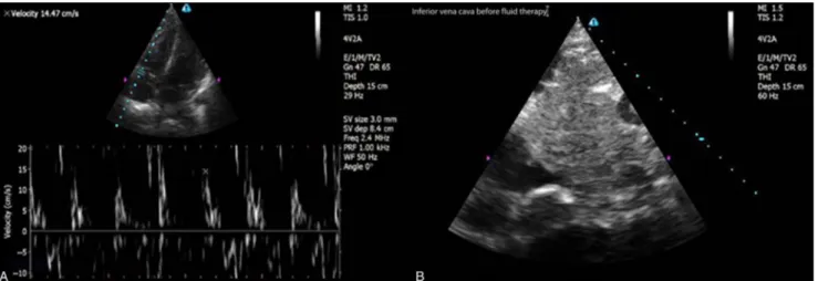
). The
velocity of the excursion of the tricuspid valve measured
at presentation was 14.47 cm/s and, together with
collapsed IVC, this
finding led to the decision to begin
fluid therapy immediately. Written informed consent
form was obtained from the patient prior to participation
in the study.
The patient underwent 2 L of
fluid therapy with
0.9% NaCl in a 2-h period. Control BUS after
fluid
therapy revealed decreased TDI velocity of tricuspid
annulus to 11.81 cm/s and dilated IVC not collapsing
suf
ficiently with respiration (Fig.
2
). Also, the blood
pressure and other
findings were normalized. The patient
received his maintenance therapy after admission to the
internal medicine department and was discharged from
the service after 3 days.
Discussion
Although the evidence is not strong enough, there is a
signi
ficant experience with the use of BUS to predict the
fluid responsiveness in critically ill patients [
1
]. Most
commonly, ED physicians prefer to use BUS since the
procedure is non-invasive and, moreover, physical
exami-nation
findings, hematocrit levels, and biochemical
mar-kers are not speci
fic indicators [
2
]. VTI is one of the BUS
measurements that have been shown to be highly
predic-tive of
fluid responsiveness if respirophasic changes of
VTI are greater than 20% [
3
]. The other method for
Fig. 1. (A) Bedside ultrasound (BUS) performed by the emergency department (ED) physician revealed increased TDI velocity of the right ventricle from the lateral annulus of the tricuspid valve. (B) BUS performed by the ED physician revealed a slit-like inferior vena cava under the left lobe of the liver
Fig. 2. (A) Control bedside ultrasound (BUS) afterfluid therapy revealed decreased TDI velocity of the right ventricle from the lateral annulus of the tricuspid valve. (B) Control BUS afterfluid therapy revealed that the inferior vena cava is dilated and not collapsing sufficiently with respiration
Fluid therapy monitoring by TDI
predicting
fluid response is identification of the
respira-tory-induced changes in peak aortic velocity [
3
]. This
same study showed that left ventricular end-diastolic area,
as a proxy of preload, did not discriminate between
responders and non-responders; this
finding was
con-firmed in a meta-analysis [
4
]. In some studies, it has been
shown that increasing respirophasic changes in the
diame-ter of IVC during positive pressure breathing, detect
fluid-responsive patients. These changes in diameter of the IVC
can effectively identify
fluid responsiveness in septic
patients, with a suggested cutoff point of 12% of the mean
diameter [
5, 6
]. In contrast to these dynamic
measure-ments of
fluid responsiveness with BUS, static ones, such as
IVC diameter, were not found to be clinically useful. In
addition, these methods do not depict the response of the
RV to
fluid challenge directly. We have shown that TDI
identi
fies RV function and pulmonary resistance directly.
TDI is superior to blood
flow Doppler as it directly reflects
myocardial function and is less subject to loading
condi-tions. Low values of systolic (Sm), diastolic early (Em), and
diastolic late (Am) tissue velocities of the right RV have
been proposed to re
flect systolic and diastolic RV
dysfunc-tion, respectively [
7
]. Only a limited number of researchers
have investigated the diagnostic accuracy of TDI on right
ventricular dysfunction in different clinical settings. In
mechanically ventilated patients, TDI velocities have been
shown to successfully discriminate patients with different
durations of weaning [
8
]. In a recent study of Harmankaya
et al. [
9
], RV-Sm was found lower in the non-surviving
septic shock patients compared with the surviving and the
control groups (11.8
± 4.2, 13.6 ± 3.3, and 15.1 ± 2.1
cm, respectively,
P = 0.002).
Conclusions
With the increased availability of portable ultrasound
devices with the TDI feature in EDs, monitoring
applica-tions with RV tissue Doppler in
fluid responsiveness may
find a clinical role, particularly if it is demonstrated to be
superior to the available alternatives in future strong
studies.
* * *
Funding sources: Nofinancial support was received for this study. Authors’ contribution: EEU and TE developed the concept and performed the drafting of the article; BK and SB performed the preparation and revision of the article; and EEU and TE performed the acquisition of data.
Conflict of interest: The authors declare no conflict of interest.
References
1. Wetterslev M, Haase N, Johansen RR, Perner A: Predicting fluid responsiveness with transthoracic echocardiography is not yet evidence based. Acta Anaesthesiol Scand 57, 692–697 (2013)
2. Dipti A, Soucy Z, Chandra S: Role of inferior vena cava diameter in assessment of volume status: A meta-analysis. Am J Emerg Med 30, 1414–1419.e1 (2012)
3. Feissel M, Michard F, Mangin I, Ruyer O, Faller JP, Teboul JL: Predicting volume responsiveness by using the end-expiratory occlu-sion in mechanically ventilated patients with septic shock. Chest 119, 867–873 (2001)
4. Marik PE, Cavallazzi R, Vasu T, Hirani A: Dynamic changes in arterial waveform derived variables and fluid responsiveness in mechanically ventilated patients: A systematic review of the literature. Crit Care Med 37, 2642–2647 (2009)
5. Feissel M, Michard F, Faller JP, Teboul JL: The respiratory variation in inferior vena cava diameter as a guide tofluid therapy. Intensive Care Med 30, 1834–1837 (2004)
6. Barbier C, Loubières Y, Schmit C, Hayon J, Ricôme J-L, Jardin F, Vieillard-Baron A: Respiratory changes in inferior vena cava diameter are helpful in predicting fluid responsiveness in ventilated septic patients. Intensive Care Med 30, 1740–1746 (2004)
7. Waggoner AD, Bierig SM: Tissue Doppler imaging: A useful echo-cardiographic method for the cardiac sonographers to assess systolic and diastolic ventricular function. J Am Soc Echocardiogr 14, 1143– 1152 (2001)
8. Papaioannou V, Stakos D, Dragoumanis C, Pneumaticos I: Rela-tion of tricuspid annular displacement and tissue Doppler imaging velocities with duration of weaning in mechanically ventilated patients with acute pulmonary edema. BMC Cardiovasc Disord 10, 20 (2010)
9. Harmankaya A, Akilli H, Gul M, Akilli NB, Ergin M, Aribas A, Cander B: Assessment of right ventricular functions in patients with sepsis, severe sepsis and septic shock and its prognostic importance: A tissue Doppler study. J Crit Care 28, 1111.e7– 1111.e11 (2003)