642
Rev Bras Cir Cardiovasc | Braz J Cardiovasc Surg
Rev Bras Cir Cardiovasc 2014;29(4):642-4 Ozyuksel A, et al. - Saccular aneurysm formation of the descending aorta
associated with aortic coarctation in an infant
RBCCV 44205-1599 DOI 10.5935/1678-9741.20140041
Saccular aneurysm formation of the descending
aorta associated with aortic coarctation in an infant
Formação de aneurismas saculares da aorta descendente associados com coarctação aórtica em criança
Arda Ozyuksel
1, MD; Emir Canturk
1, MD; Aygun Dindar
2, MD; Atif Akcevin
1, MD
1Istanbul Medipol University (Medipol UNV) and Department of
Cardio-vascular Surgery, Istanbul, Turkey.
2Istanbul University and Department of Pediatric Cardiology, Istanbul, Turkey.
This study was carried out at Istanbul Medipol University (Medipol UNV) and Department of Cardiovascular Surgery, Istanbul, Turkey.
No financial support.
Correspondence address: Arda Ozyuksel
Medipol University, Department of Cardiovascular Surgery TEM Otoyolu, Göztepe cikisi, No:1 Bagcilar, Istanbul, Turkey 34212 E-mail: [email protected]
Article received on August 26th, 2013
Article accepted on February 23th, 2014
BRIEF COMMUNICATION
Abstract
Aneurysm of the descending aorta associated with CoA is an extremely rare congenital abnormality. In this report, we present a 16 months old female patient in whom cardiac cath-eterization had been performed which had revealed a segment of coarctation and saccular aneurysm in the descending aorta. The patient was operated and a 3x2 centimeters aneurysm which embraces the coarcted segment in descending aorta was resect-ed. In summary, we present a case of saccular aortic aneurysm distal to aortic coarctation in an infant without any history of intervention or vascular inflammatory disease. Our case report seems to be the youngest patient in literature with this pathology.
Descriptors: Aortic Aneurysm, Thoracic. Aortic Coarctation.
Infant.
Resumo
Aneurisma da aorta descendente associada à coarctação aór-tica é uma anomalia congênita extremamente rara. Neste relato, apresentamos uma paciente de 16 meses de idade, nos quais o cateterismo cardíaco foi realizado, que havia revelado um seg-mento de coarctação e aneurisma sacular na aorta descendente. A paciente foi operada e um aneurisma de 3x2 centímetros, englo-bando o segmento coarctada na aorta descendente foi ressecado. Em resumo, apresentamos um caso de aneurisma sacular distal à coarctação da aorta em uma criança sem histórico de interven-ção ou doença inflamatória vascular. Nosso caso parece ser o do paciente mais jovem na literatura com esta afecção.
Descritores: Aneurisma da Aorta Torácica. Coarctação
Aórtica. Lactente
INTRODUCTION
Coarctation of the aorta (CoA) is defined as the hemody-namically significant narrowing of the descending thoracic aorta, usually just distal to the left subclavian artery where the ligamentum arteriosum originates[1]. CoA accounts for 6.5% of all congenital heart defects[2]. CoA is usually classi-fied into three categories: I.Isolated CoA, II. CoA with ven-tricular septal defect (VSD) and III. CoA with complex cardi-ac anomalies[3]. Aneurysm of the descending aorta associated with CoA is an extremely rare congenital abnormality. In
this report, we present the youngest patient per our literature search with CoA and saccular descending aortic aneurysm.
CASE PRESENTATION
A 16 months old female patient was admitted to our clin-ic with diagnosis of aortclin-ic coarctation and descending aor-tic aneurysm. She presented with poor feeding and failure to thrive (body weight: 8 kg). The prenatal and postnatal history was unremarkable; any umbilical vascular catheter-ization was not performed. Physical examination revealed
643
Rev Bras Cir Cardiovasc | Braz J Cardiovasc Surg
Rev Bras Cir Cardiovasc 2014;29(4):642-4 Ozyuksel A, et al. - Saccular aneurysm formation of the descending aorta
associated with aortic coarctation in an infant
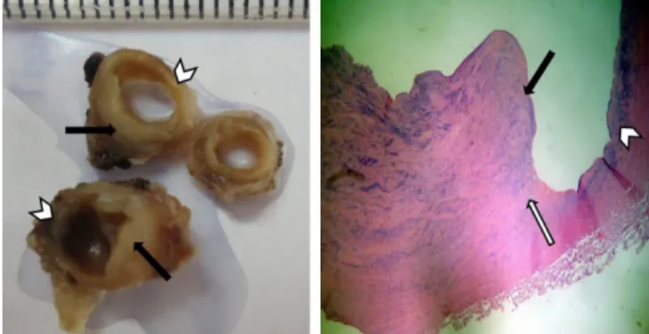
segments of both coarctation and aneurysm formation. Baso-philic degeneration and vacuole formation were remarkable (Figure 2). The postoperative period was uneventful and the patient was discharged at the sixth postoperative day without any complication. At the sixth postoperative month, a second intervention was performed because of recoarctation. The MR angiography revealed a recoarctation at the descending aorta, in which balloon dilatation was performed with a resid-ual gradient of 25 mmHg (Figure 3). The patient is followed up by echocardiographic evaluations with regular intervals.
Abbreviations, acronyms & symbols
CoA Coarctation of the aorta VSD Ventricular septal defect
80 mmHg systolic pressure difference between upper and lower extremities. Femoral pulses were weak but palpable. There was a systolic murmur (3/6) at the left sternal border which was also heard at the interscapular area. The leukocyte count, C-reactive protein level and erythrocyte sedimentation rate were all within normal limits. The patient had been ad-mitted to another clinic a few months ago and transthoracic echocardiography had demonstrated aortic coarctation with 60mmHg peak systolic gradient. Any intracardiac pathology had not been encountered. Cardiac catheterization had been performed in order to confirm the diagnosis and perform a balloon angioplasty if possible; however the catheterization had revealed a segment of coarctation and saccular aneurysm in the descending aorta (Figure 1). Therefore, the patient was referred to our clinic for surgical repair.
The patient was operated under general anesthesia and a left posterolateral thoracotomy was performed at the 4th inter-costal space. A 3x2 cm aneurysm which embraces the coarct-ed segment in descending aorta was encountercoarct-ed (Figure 1). Descending aorta was mobilized, resection and end-to-end anastomosis was performed with 6/0 polyprolene suture. The sutures were continuous at the posterior wall, whereas they were interrupted at the anterior wall of the anastomosis, in order to provide potential for growth. Residual gradient was less than 5 mmHg. The resected specimen was examined by the pathology department. Gross and microscopic cross sectional examination of the coarctation area revealed the
Fig. 1 - Coarctation and saccular aneurysm formation in the descending aorta in cardiac catheterization (left side - arrow). Saccular aneurysm embracing the segment of coarctation in the descending aorta, operative view (right side - arrow).
Fig. 2 - Gross cross sectional examination of the area of coarctation and aneurysm formation (left side) (black arrows indicate the area of coarctation and white arrow heads indicate the area of aneurysm formation). Hematoxylin eosin stained section revealing the zone of transition between the area of coarctation and aneurysm (right side) (x40 magnification) (black arrow indicates the area of coarctation and white arrow head indicates the area of aneurysm formation. Basophilic degeneration and vacuole formation were remarkable indicated with white arrow).
Fig. 3 - MR angiography revealing the recoarctation at the descending aorta (left) which was dilated with percutaneous intervention (right).
644
Rev Bras Cir Cardiovasc | Braz J Cardiovasc Surg
Rev Bras Cir Cardiovasc 2014;29(4):642-4 Ozyuksel A, et al. - Saccular aneurysm formation of the descending aorta
associated with aortic coarctation in an infant
Authors’ roles & responsibilities
AO Included in surgical team, preparation of the manuscript EC Included in surgical team, preparation of the manuscript AD Preoperative diagnostic workup of the patient, preparation
and final control of the paper
AA Head of surgical team, preparation and final control of the paper
DISCUSSION
Surgical repair of CoA mostly depends on the accom-panying cardiac anomalies in the patient. The main treat-ment target is providing a non stenotic aortic continuity with efforts to enhance the growth potential of the native vascular tissues, with or without the repositioning of the left subclavian artery[4]. Although rarely seen, aortic wall pathology such as aneurysm formation, aortic dissection and rupture are mainly the presenting symptoms of adult CoA. When pediatric age group is concerned, the saccu-lar aortic aneurysms distal to coarcted segment are very rare and data about these patients are only confined to limited case reports[5]. Our case report seems to be the youngest patient in literature with this pathology. In such cases, recoarctation may be encountered in the follow up which may be managed by percutaneous balloon dil-atation as presented in our case. Extensive mobilization of the aorta and its branches with a meticulous surgical technique is mandatory in order to reduce the tension at the anastomosis site.
In summary, we present a case of saccular aortic aneu-rysm distal to aortic coarctation in an infant without any history of intervention or vascular inflammatory disease. We speculate that localized weakness of the aortic wall may be responsible for aneurysm formation, since basophilic degen-eration and vacuole formation were remarkable at the transi-tion zone between the coarcted and aneurysmatic segments under microscope.
REFERENCES
1. Backer CL, Mavroudis C. Congenital Heart Surgery Nomenclature and Database Project: patent ductus arteriosus, coarctation of the aorta, interrupted aortic arch. Ann Thorac Surg. 2000;69(4 Suppl):S298-307.
2. Flyer DC. Report of the New England Regional Infant Cardiac Program. Pediatrics. 1980;64:432-36.
3. Backer CL, Paape K, Zales VR, Weigel TJ, Mavroudis C. Coarctation of the aorta. Repair with polytetrafluoroethylene patch aortoplasty. Circulation. 1995;92(9 Suppl):II132-6. 4. Croti UA, Braile DM, Marchi CH, Beani L. Aortic coarctation:
aortoplasty with interposition of the left subclavian artery (the Teles Mendonça technique). Rev Bras Cir Cardiovasc. 2007;22(2):255-6.
5. Celik T, Iyisoy A, Kursaklioglu H, Unlu M, Kose S, Ozmen N, et al. A large calcified saccular aneurysm in a patient with aortic coarctation. Int J Cardiovasc Imaging. 2006;22(1):93-5.