The relationship between fibrosis and nodule structure and
esophageal varices
Arif Mansur Coşar1 , Tolga Yakar2 , Ender Serin2 , Birol Özer2 , Fazilet Kayaselçuk3 1Department of Gastroenterology, Karadeniz Technical University School of Medicine, Trabzon, Turkey
2Departments of Internal Medicine and Gastroenterology, Başkent University School of Medicine, Adana, Turkey 3Departments of Pathology, Başkent University School of Medicine, Adana, Turkey
ABSTRACT
Background/Aims: The aim of the present study was to evaluate the histopathological findings of cirrhosis together with clinical and laboratory parameters, and to investigate their relationship with esophageal varices that are portal hypertension findings.
Materials and Methods: A total of 67 (42 male and 25 female) patients who were diagnosed with cirrhosis were included in the study. The mean age of the patients was 51.6±19.0 (1-81) years. The biopsy specimens of the patients were graded in terms of fibrosis, nod-ularity, loss of portal area, central venous loss, inflammation, and steatosis. The spleen sizes were graded ultrasonographically, and the esophageal varices were graded endoscopically.
Results: In the multivariate regression analysis, there was a correlation between the advanced disease stage (Child-Pugh score odds ratio (OR): 1.47, 95% confidence interval (CI): 1.018-2.121, p=0.040), presence of micronodularity (OR: 0.318, 95% CI: 0.120-0.842, p=0.021), grade of central venous loss (OR: 5.231, 95% CI: 1.132-24.176, p=0.034), and presence of esophageal varicose veins.
Conclusion: Although thrombocytopenia and splenomegaly may predict the presence of large esophageal varices, cirrhosis histopathol-ogy is the main factor in the presence of varices.
Keywords: Cirrhosis, histopathology, portal hypertension, esophageal varices
INTRODUCTION
Cirrhosis is histologically defined by the presence of re-generative nodules surrounded by fibrous tissue. This structural distortion leads to increased intrahepatic re-sistance and portal hypertension. Esophageal varices and ascites occur after portal pressure exceeds 10-12 mm Hg (1-3). The severity of the functional and structural impair-ment of the liver determines the course of the disease. Portal hypertension-related esophageal variceal bleeding, ascites, and hepatic encephalopathy generally determine the prognosis of patients with cirrhosis. In clinical prac-tice, some patients with cirrhosis live in an asymptomatic or compensated state, whereas others decompensate over time. Owing to the variability of the clinical course, it is beneficial to predict the clinical course using various laboratory or histological signs. The histological features of cirrhosis do not reflect the clinical severity or the stage of cirrhosis. As certain histological parameters can pre-dict clinical and biochemical decompensation, it is con-siderably important to be able to determine a relationship between specific histological parameters, such as fibrosis and nodule structure, on liver biopsies of patients with cirrhosis and clinical findings of portal hypertension, such
as esophageal varices. The aim of the present study was to evaluate the relationship between some histopatho-logical findings of cirrhosis with clinical, endoscopic, and laboratory parameters.
MATERIALS AND METHODS
The study was approved by the Başkent University Re-search and Ethics Committee. Data of 194 patients ad-mitted to our hospital between July 1999 and June 2006 who underwent liver biopsies for the evaluation of chronic liver diseases, liver masses, or liver function abnormalities were reviewed retrospectively. Patients with suitable liv-er biopsy specimens for histopathological reassessment were included in the study.
Histopathological assessment
All liver biopsies were fixed in 10% formalin and routine-ly stained with hematoxylin and eosin, reticulin, Masson trichrome, Periodic Acid-Schiff, and iron stain. The slides of each biopsy were reviewed by an experienced hepato-pathologist blinded to the patient’s clinical status. Liver biopsy samples >10 mm in length were accepted for his-topathological examination. In fragmented biopsies, the Cite this article as: Coşar AM, Yakar T, Serin E, Özer B, Kayaselçuk F. The relationship between fibrosis and nodule structure and
esophageal varices. Turk J Gastroenterol 2019; 30(7): 624-9.
Corresponding Author: Arif Mansur Coşar; arif@doctor.com
Received: September 14, 2018 Accepted: October 1, 2018 Available online date: April 8, 2019
© Copyright 2019 by The Turkish Society of Gastroenterology • Available online at www.turkjgastroenterol.org DOI: 10.5152/tjg.2019.18665
total length was estimated by combining the maximum dimensions of each individual fragment. Liver fibrosis was staged according to the Batts and Ludwig classification, a modification of the Scheuer classification, in which stage 0 corresponds to no fibrosis, stage 1 to portal fibrosis, stage 2 to periportal fibrosis, stage 3 to bridging fibrosis, and stage 4 to cirrhosis (4). Only biopsies categorized as stage 4 were included in the study.
The biopsies were re-evaluated for two parameters:
sinu-soidal fibrosis and nodule size. In the definition of
nod-ule size, the “mixed nodnod-ule” classification was used when there was at least one small nodule in addition to macro-nodules. For septal thickness, the thickness of the dom-inant type of septae in each specimen was scored. The histological parameters and scores assessed in the biopsy samples were as follows:
1. Sinusoidal fibrosis: 0, 1 (mild), 2 (moderate), and 3 (severe), 2. Septal thickness: 0, 1 (thin), 2 (medium), and 3 (thick),
3. Nodularity:
a. Small nodules: nodule size is comparable to the width
of the needle biopsy specimen,
b. Mixed nodules: presence of both small and large
nod-ules,
c. Large nodules: nodule size is larger than the needle
biopsy specimen. Endoscopic assessment
All patients underwent upper gastrointestinal endoscopy for the assessment of esophageal varices as an endoscopic finding of portal hypertension. The Pentax EPM-3500 and Olympus CV-70 endoscopy systems were used to deter-mine the presence of esophageal varices, and two experi-enced endoscopists performed the procedures. The esoph-ageal varices were graded as I-III according to the Japanese Classification of Esophageal Varices. Patients were classified dichotomously either as Group 1 (large, grade II-III esopha-geal varices) or Group 2 (none or small, grade I varices). Statistical analysis
All statistical analyses were performed using the SPSS software (version 14 for Windows; SPSS, Chicago, IL, USA). As most variables were not normally distribut-ed, data were presented as median and range (mini-mum-maximum) values. Owing to the heterogeneity of the distribution, the non-parametric Mann-Whitney U-test, the Kruskal-Wallis test, and the chi-square test were used. Logistic regression analysis was performed for multivariate analysis. A p value of <0.05 was considered statistically significant. A non-parametric statistical anal-ysis was applied to the data, such as esophageal varices and platelet count, which were the clinical and laborato-ry findings of portal hypertension, the histopathological findings, and the demographic data.
RESULTS
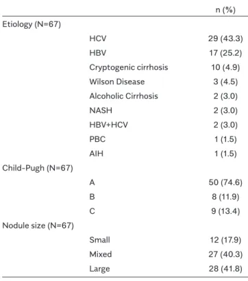
The study included 67 (25 (37.3%) female and 42 (62.7%) male) patients with histologically proven cirrhosis. The mean ages of the patients were 52.3±18.6 years for female and 51.2±19.5 years for male. Table 1 shows the etiology, Child-Pugh status, and nodule size of the patients. Most of the patients were hepatitis C virus or hepatitis B virus positive and Child-Pugh A, with large or mixed nodules. The relationship between the demographic parameters and the presence and group of esophageal varices with liver histopathology of the patients was examined for univariate analysis.
The results are as follows (Table 2):
1. A statistically significant relationship was detected between the nodule size and age (p<0.001), platelet
n (%) Etiology (N=67) HCV 29 (43.3) HBV 17 (25.2) Cryptogenic cirrhosis 10 (4.9) Wilson Disease 3 (4.5) Alcoholic Cirrhosis 2 (3.0) NASH 2 (3.0) HBV+HCV 2 (3.0) PBC 1 (1.5) AIH 1 (1.5) Child-Pugh (N=67) A 50 (74.6) B 8 (11.9) C 9 (13.4) Nodule size (N=67) Small 12 (17.9) Mixed 27 (40.3) Large 28 (41.8)
Table 1. Etiology, Child-Pugh status and nodule size distri-butions of patients
count (p=0.04), Child-Pugh score (p=0.001), esoph-ageal varices (p=0.001), and large esophesoph-ageal varices (p=0.03).
2. A statistically significant relationship was detected be-tween a high Child-Pugh score (p=0.047) and the thick-ness of septal fibrosis.
Table 3 presents the relationship between the presence of large esophageal varices (FII or FIII) and the other clin-ical and laboratory parameters of portal hypertension and demographic data. Table 4 presents the relationship
between the presence of esophageal varices and the similar parameters and data. A positive and statistical-ly significant relationship was detected between a high Child-Pugh score (p<0.001) and the presence of both large esophageal varices and esophageal varices. Multi-variate logistic regression analysis revealed a statistical-ly significant relationship between the Child-Pugh score (p 0.040), micronodularity (p 0.021), and presence of varices (Table 5).
DISCUSSION
The consequences of portal hypertension, such as var-iceal bleeding, are important factors in the morbid-ity and mortalmorbid-ity of patients with cirrhosis (5,6). At the
Presence of
Platelet Presence of large
number Child-Pugh oesophageal oesophageal
AST/ALT X103/mL Score varices varices Age Gender
ratio >1 (n/N) Med (Min-Max) Med (Min-Max) (n/N) (n/N) Med (Min-Max) (M/F)
NODULE SIZE Micronodules (n=12) 9/12 95.5 (27-443) 9.5 (5-12) 9/11 4/11 27.5 (1-55) 9/3 Mixed nodules (n=27) 15/27 139 (55-408) 6 (5-13) 11/27 4/27 54 (16-79) 16/11 Macro nodules (n=28) 14/28 150 (52-364) 5.5 (5-11) 5/28 1/28 57.5 (42-81) 17/11 p 0.34 0.04* 0.001* 0.001* 0.03* <0.001* 0.61 SEPTAL THICKNESS Thin (n=9) 5/9 146 (83-298) 5 (5-8) 2/9 0/9 66 (51-81) 7/2 Medium (n=31) 20/31 139 (27-408) 6 (5-13) 12/31 6/31 52 (1-79) 15/16 Thick (n=27) 13/27 138 (53-433) 6 (5-12) 11/26 3/26 53 (1-73) 20/7 p 0.45 0.92 0.047* 0.56 0.30 0.09 0.08
Med: Median, Min: Minimum, Max: Maximum
Table 2. Relationships between the demographic parameters and clinical findings of portal hypertension and the liver histo-pathology of the patients
Large oesophageal varices - + (n=57) (n=9) p n (%) n (%) Gender Male 34 (59.6) 7 (77.8) 0.300 Female 23 (40.4) 2 (22.2) Median Median (Min-Max) (Min-Max) Age 54 (1-81) 42.3 (3-72) 0.130 Platelet number X103/mL 146 (52-433) 80 (27-140) 0.004* Child-Pugh Score 6 (5-13) 9.2 (7-12) <0.001*
Table 3. Relationships between the presence of large oe-sophageal varices (FII or FIII) and other clinical and labora-tory parameters of portal hypertension and the demograph-ic data
Large oesophageal varices - + (n=41) (n=25) p n (%) n (%) Gender Male 23 (56.1) 18 (72.0) 0.196 Female 18 (43.9) 7 (28.0) Median Median (Min-Max) (Min-Max) Age 55 (36-81) 50 (1-78) 0.231 Platelet number X103/ml 154 (59-364) 120 (27-433) 0.038* Child-Pugh Score 5 (5-13) 7 (5-12) <0.001*
Table 4. Relationships between the presence of oesopha-geal varices and the other clinical and laboratory parameters of the portal hypertension and the demographic data
Third Baveno Conference on portal hypertension and its treatment, all patients with cirrhosis are suggested to be screened for the presence of esophageal varices re-gardless of predictor factors. Endoscopic scanning every 2 to 3 years for patients who do not show any presence of varices endoscopically and every 1 to 2 years for those who have ambiguous varices has been recommended (7,8). However, an endoscopy is an invasive and relatively expensive procedure that is not tolerated by some pa-tients. Moreover, the definition of variceal sizes depends on the endoscopist. Therefore, researchers have turned to alternative non-invasive methods for variceal screen-ing. Several clinical and laboratory parameters have been examined in terms of their ability to predict the presence of varices. In the literature, >10 studies have specifically investigated this issue (9-20). Chalasani et al. argued that the presence of thrombocytopenia and splenomegaly is
an independent predictor for the presence of esopha-geal varices (15). Madhotra showed that the presence of thrombocytopenia and splenomegaly is an independent predictor for the presence of large esophageal varices. However, in the present study, the presence of spleno-megaly could not be found in 32% of the cases where platelet counts were below the cut-off value (68,000/ mm3). The sensitivity and specificity of splenomegaly
and thrombocytopenia (<68,000/mm3) were 75% and
58% and 71% and 73%, respectively (16). Therefore, the presence of splenomegaly per se does not explain the thrombocytopenia seen in patients with cirrhosis. Other possible reasons may include “decreased thrombopoie-tin synthesis” and “altered immune mechanisms” (21). Other than the clinical and laboratory findings of portal hypertension, other remaining studies have focused on the histopathological characteristics of the liver and the development of esophageal varices.
For example, the Fibro Test is an alternative non-invasive method for analyzing liver fibrosis instead of histopathol-ogy. Its relationship with varices prediction was examined by Thabut et al. (14), who investigated the relationship of platelet count, prothrombin time, presence of asci-tes, Child-Pugh score, and Fibro Test results with large esophageal varices.
In their study, Fibro Test was found to be the parameter with the highest differentiation power. Nagula et al. (22) investigated portal vein pressure in patients with liver cir-rhosis and its relationship with histopathological parame-ters. A statistically significant relationship was found be-tween cirrhotic nodule size and septal thickness and the presence of portal hypertension. In the univariate analysis of data, Nagula et al. (22) found a statistically significant relationship between the presence of micronodules and platelet count, Child-Pugh score, presence of esophageal varices, presence of large varices, and age.
Common causes of micronodular cirrhosis include chron-ic alcohol usage, hemochromatosis, biliary tract obstruc-tion, chronic venous flow obstrucobstruc-tion, and childhood
Presence of large oesophageal varices Presence of oesophageal varices
p OR (C.I 95%) p OR (C.I 95%)
Thrombocytopenia 0.232 0.984 0.959-1.010 0.900 0.999 0.991-1.008
Child-Pugh Score 0.094 1.526 0.931-2.500 0.040 1.47 1.018-2.121
Nodularity 0.998 0.998 0.210-4.73 0.021 0.318 0.120-0.842
Table 5. The multivariate logistic regression analysis of the parameters related to the presence of oesophageal varices and large oesophageal varices in the univariate analysis
Nodule size
micro mix macro total
Viral etiology
HCV 2 10 17 29
HBV 1 8 8 17
HBV+HCV 0 1 1 2
Total. n(%) 3 (6.3) 19 (39.6) 26 (54.2) 48 (100)
In the entire study group ~ % 18 40 42 100
Table 6. Relationships between viral etiology and nodularity
Nodule size
micro mix macro total Child-Pugh score A 3 22 25 50 (74.6) B 3 3 2 8 (11.9) C 6 2 1 9 (13.4) Total. n(%) 12 (17.9) 27 (40.3) 28 (41.8) 67 (100) Child-Pugh score. Median (Min-Max) 9.5 (5-12) 6 (5-13) 5.5 (5-11) p=0.001 Table 7. Relationships between nodule size and Child-Pugh Score
metabolic diseases, and one of the rare causes is cirrho-sis secondary to chronic active hepatitis. In the current study group, the micronodular cirrhosis rate was almost 18%, whereas alcoholic cirrhosis was relatively infrequent at 3% (Table 1). The etiology of cirrhosis was hepatitis B or C infections or hepatitis B and C co-infection in 72% of the patients. The mixed nodularity rate in this group at almost 40% was classed as relatively high, in contrast with the conventional explanation that “the illnesses that cause macronodular cirrhosis from the beginning are chronically viral and autoimmune hepatitis” (23). In the present study, although the rate of alcoholic etiology in the patient population was very low, and most of the pa-tients showed viral etiology, micronodule formation in the pathology specimens was a very frequent finding when mixed nodularity was added (Tables 1 and 6). This can be attributed to the transformation of the cirrhosis mor-phology, which was initially micronodular and became macronodular during the course of the disease. Other possible explanations could include other etiological rea-sons that accompany the viral etiology but are not seen as dominant or sufficient enough to determine this mor-phological classification in most biopsy materials (23). In the current study, a statistically significant relationship was determined between micronodularity and the aver-age high level of the Child-Pugh score (Table 7). Accord-ingly, it may be advisable that patients with micronodular cirrhosis are closely monitored. The relationship between the presence of large varices and micronodularity was not statistically significant in the multivariate analysis, possi-bly due to the low number of patients with micronodular cirrhosis. The majority of patients with micronodular cir-rhosis in the current study group were viral hepatitis B, C, or D positive. The results of the study showed a sta-tistically significant relationship between the Child-Pugh score and the presence of varices in multivariate analysis. With respect to liver structure, compared with macro-nodules, micronodules can cause greater structural dam-age, leading to the development of portal hypertension and thereby contributing to the development of esopha-geal varices. Previous studies have shown the relationship between thrombocytopenia and liver fibrosis (14,24). However, as in alcoholic hepatitis without manifest fi-brosis, varices and other portal hypertension findings can occur in the early stage of chronic liver disease. In the cur-rent study, a statistically significant relationship was also found between the thickness of the fibrous septae and the Child-Pugh score (p=0.047). This relationship sup-ports a possible structural reason that negatively affects liver synthesis functions (Table 2). Micronodule formation was frequent in younger patients in the current study
group (p<0.001), as seen in the alcoholic cirrhosis and he-mochromatosis cases. This situation can be explained by an increase in the age of onset of cirrhosis or of discon-tinuing alcohol usage and by the transformative ability of cirrhosis, which was initially micronodular to macronod-ular cirrhosis following treatments, such as phlebotomy. A liver biopsy is still the gold standard in the diagnosis of liver diseases. When the cirrhosis diagnosis is prov-en by biopsy, reporting additional findings, such as nod-ule size and septal thickness, can serve as a guide for clinicians in predicting the development and course of portal hypertension. In the present study, which aimed to analyze the relationship between the findings of his-topathological cirrhosis and various parameters related to portal hypertension, some results that can be trans-ferred to clinical practice were obtained, although the study was retrospective, and the volume of data was relatively low. It can be recommended that all patients with cirrhosis are scanned in daily practice, in terms of the presence of esophageal varices, independently of the predicting factors. The most reliable method of var-iceal screening, which allows for possible intervention if needed, is endoscopic assessment. Endoscopic variceal screening is indicated and is cost-effective particularly in cases of compensated cirrhosis. Several alternative non-invasive methods have been defined. However, in case of negative results, it is important to keep in mind that the possibility of small varices cannot be excluded, in which case, the use of combined multiple parameters is recommended.
In conclusion, the presence of micronodularity can be ac-cepted as a predictor of varices. This is useful for patients who do not have clear clinical cirrhosis and who are diag-nosed after biopsy. It is possible to say that patients with micronodular cirrhosis have (1) a lower platelet count, (2) a higher Child-Pugh score, (3) a high probability of the presence of esophageal varices, (4) a high probability of the presence of large esophageal varices, and (5) lower age.
Ethics Committee Approval: Ethics committee approval was obtained from the local institutional ethical committee.
Informed Consent: N/A.
Peer-review: Externally peer-reviewed.
Author Contributions: Concept - A.M.C, E.S; Design - A.M.C, T.Y; Supervision - E.S, B.Ö; Resources - A.M.C, F.K; Materials - A.M.C,
F.K; Data Collectionand/or Processing - A.M.C, T.Y; Analysis and/ or Interpretation - A.M.C, E.S; Literature Search - A.M.C, S.E, T.Y; Writing Manuscript - A.M.C, T.Y; Critical Review - B.Ö.
Conflict of Interest: The authors have no conflict of interest to declare.
Financial Disclosure: The authors declared that this study has received no financial support.
REFERENCES
1. Garcia-Tsao G, Groszmann RJ, Fisher RL, Conn HO, Atterbury CE, Glickman M. Portal pressure, presence of gastroesophageal varices and variceal bleeding. Hepatology 1985; 5: 419-24. [CrossRef] 2. Morali GA, Sniderman KW, Deitel KM, et al. Is sinusoidal portal hy-pertension a necessary factor for the development of hepatic asci-tes? J Hepatol 1992; 16: 249-50. [CrossRef]
3. Casado M, Bosch J, Garcia-Pagan JC, et al. Clinical events after tran-sjugular intrahepatic portosystemic shunt: correlation with hemody-namic findings. Gastroenterology 1998; 114: 1296-303. [CrossRef] 4. Batts KP, Ludwig J. Chronic hepatitis. an update on terminology and reporting. Am J Surg Pathol 1995; 19: 1409-17. [CrossRef] 5. Arguedas MR, Heudebert GR, Eloubeidi MA, Abrams GA, Fallon MB. Cost-effectiveness of screening, surveillance, and primary prophy-laxis strategies for oesophageal varices. Am J Gastroenterol 2002; 97: 2441-52. [CrossRef]
6. Poynard T, Cales P, Pasta L, et al. Beta adrenergic-antagonist drugs in the prevention of gastrointestinal bleeding in patients with cirrhosis and oesophageal varices. An analysis of data and prognostic factors in 589 patients from four randomized clinical trials. Franco-Italian Multicenter Study Group. N Engl J Med 1991; 324: 1532-8. [CrossRef] 7. Thabut D, Trabut JB, Massard J, et al. Non-invasive diagnosis of large oesophageal varices with FibroTest in patients with cirrhosis: A preliminary retrospective study. Liver Int 2006; 26: 271-8. [CrossRef] 8. De Franchis R. Updating consensus in portal hypertension: Report of the Baveno III consensus workshop on definitions, methodology and therapeutic strategies in portal hypertension. J Hepatol 2000; 33: 846-52. [CrossRef]
9. Giannini E, Botta F, Borro P, Risso D, et al. Platelet count/spleen diameter ratio: proposal and validation of a non-invasive parameter to predict the presence of oesophageal varices in patients with liver cirrhosis. Gut 2003; 52: 1200-5. [CrossRef]
10. Giannini EG, Botta F, Borro P, et al. Application of the platelet count/spleen diameterratio to rule out the presence of oesophageal varices in patients with cirrhosis: A validation study based on fol-low-up. Dig Liver Dis 2005; 37: 779-85. [CrossRef]
11. Sharma SK, Aggarwal R. Prediction of large oesophageal varices in patients with cirrhosis of the liver using clinical, laboratory and imaging parameters. J Gastroenterol Hepatol 2007; 22: 1909-15. [CrossRef]
12. Schepis F, Camma C, Niceforo D, et al. Which patients with cir-rhosis should undergo endoscopic screening for oesophageal varices detection? Hepatology 2001; 33: 333-8. [CrossRef]
13. D’Amico G, Luca A. Natural history. Clinical-hemodyanamic cor-relations. Prediction of the risk of bleeding. Bailliere’s Clin. Gastroen-terology 1997; 11: 243-56. [CrossRef]
14. Thabut D, Trabut JB, Massard J, et al. Non-invasive diagnosis of large oesophageal varices with FibroTest in patients with cirrhosis: A preliminary retrospective study. Liver Int 2006; 26: 271-8. [CrossRef] 15. Chalasani N, Imperiale TF, Ismail A, et al. Predictors of large oesophageal varices in patients with cirrhosis. Am J Gastroenterol 1999; 94: 3285-91. [CrossRef]
16. Madhotra R, Mulcahy H E, Willner I, Reuben A. Prediction of oe-sophageal varices in patients with cirrhosis. J Clin Gastroenterol 2002; 34: 81-5. [CrossRef]
17. .Ng FH, Wong SY, Loo CK, et al. Prediction of oesophagogastric varices in patients with liver cirrhosis. J Gastroenterol Hepatol 1999; 14: 785-90. [CrossRef]
18. Pilette C, Oberti F, Aube C, et al. Non-invasive diagnosis of oe-sophageal varices in chronic liver diseases. J Hepatol 1999; 31: 867-73. [CrossRef]
19. Thomopoulos KC, Labropoulou-Karatza C, Mimidis KP, et al. Non-invasive predictors of the presence of large oesophageal var-ices in patients with cirrhosis. Digest Liver Dis 2003; 35: 473-8. [CrossRef]
20. Zaman A, BeckerT, Lapidus J, Benner K. Risk factors for the pres-ence of varices in cirrhotic patients without a history of variceal hemorrhage. Arch Intern Med 2001; 161: 2564-70. [CrossRef] 21. Giannini E, Botta F, Borro P, et al. Relationship between throm-bopoietin serum levels and liver function in patients with chronic liv-er disease related to hepatitis C virus infection. Am J Gastroentliv-erol 2003; 98: 2516-20. [CrossRef]
22. Nagula S, Jain D, Groszmann RJ, Garcia-Tsao G. Histological-he-modynamic correlation in cirrhosis-a histological classification of the severity of cirrhosis. J Hepatol 2006; 44: 111-7. [CrossRef] 23. Özbay G. Karaciğer Sirozunun Patolojisi. Hepato-Bilier Sistem ve Pankreas Hastalıkları (Göksoy E, Şentürk H, ed.) Sempozyum Dizisi No: 28 İstanbul, 97-100, 2002.
24. Taniguchi H, Iwasaki Y, Fujiwara A, et al. Long-term monitor-ing of platelet count, as a non-invasive marker of hepatic fibrosis progression and/or regression in patients with chronic hepatitis C after interferon therapy. J Gastroenterol Hepatol 2006; 21: 281-7. [CrossRef]