Left Ventricular Side Obstructive Pannus Formation
after Rheumatic Mitral Valve Replacement with
Preservation of the Subvalvular Apparatus
Macit Kalcßık, M.D.,* Mahmut Yesin, M.D.,† Sabahattin G€und€uz, M.D.,† Mustafa Ozan G€ursoy, M.D.,‡ Emrah Bayam, M.D.,† and Mehmet €Ozkan, M.D.†§
*Department of Cardiology, _Iskilip Atıf Hoca State Hospital, Cßorum, Turkey; †Department of Cardiology, Kosuyolu Kartal Heart Training and Research Hospital, _Istanbul, Turkey;‡Department of Cardiology, Gaziemir State Hospital, _Izmir, Turkey; and§Division of Health Sciences, Ardahan University, Ardahan, Turkey
(Echocardiography 2015;32:1887–1888)
Key words: prosthetic valve, transesophageal echocardiography Case Presentation:
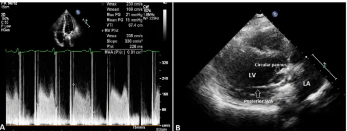
A 29-year-old woman was admitted to our out-patient clinic with progressive dyspnea. She had undergone mitral valve replacement (25 mm St. Jude Medical) due to rheumatic mitral stenosis 12 years earlier. Transthoracic (TTE), two-dimen-sional (2D), and real time three-dimentwo-dimen-sional (RT3D) transesophageal echocardiography (TEE) were performed for the evaluation of the mitral prosthesis. TTE revealed increased mean transprosthetic gradient (15 mmHg) and
decreased valve area (0.9 cm2) (Fig. 1A). In parasternal long-axis view, preservation of both anterior and posterior mitral subvalvular apparatus (SVA) was observed (Fig. 1B). 2D TEE showed a hyperechogenic circular mass on the left ventricular side of the prosthesis causing one leaflet stuck in closed position (Fig. 2A). RT3D TEE confirmed the diagnosis of left ventricular side obstructive pannus formation from both left atrial (Fig. 2B) and left ventricular views (Fig 2C). As the patient was symptomatic, she underwent
Figure 1. A. Transthoracic echocardiography (TTE) revealed mitral prosthetic valve obstruction with increased mean transpros-thetic gradient and decreased valve area. B. TTE parasternal long-axis view showed the preservation of both anterior and posterior mitral subvalvular apparatus (SVA). LA = left atrium; LV = left ventricle; SVA = subvalvular apparatus.
Data sharing: No additional data. Funding: No funding.
Address for correspondence and reprint requests: Macit Kalcßık, M.D., _Iskilip Atıf Hoca State Hospital, Meydan Mah. Toprak Sok. No:7/8 _Iskilip, Cßorum/Turkey. Fax: +903645113187;
E-mail: [email protected]
1887
© 2015, Wiley Periodicals, Inc.
redo valve surgery. Postoperative pannus speci-men was observed to be derived from chordal connections to the prosthetic annulus (Fig. 2D).
In nonrheumatic mitral valve replacement (MVR), preservation of the SVA is recommended to maintain annular–papillary continuity, which is known to be associated with improved left ven-tricular function in the early and late postopera-tive period. However, its applicability in rheumatic valves remains controversial.1 Preser-vation of SVA during MVR in rheumatic valve dis-ease may provoke pannus tissue formation on the left ventricular side of the mitral prostheses due to the presence of intensefibrosis and calcifi-cation in rheumatic valves. 2D and RT3DTEE have incremental value in the diagnosis of pannus for-mation on prosthetic heart valves.2–4
References
1. Coutinho GF, Bihun V, Correia PE, et al: Preservation of the subvalvular apparatus during mitral valve replace-ment of rheumatic valves does not affect long-term survival. Eur J Cardiothorac Surg 2015 Jan 18 [Epub ahead of print].
2. Ozkan M, G€und€uz S, Yildiz M, et al: Diagnosis of the pros-thetic heart valve pannus formation with real-time three-dimensional transoesophageal echocardiography. Eur J Echocardiogr 2010;11:E17.
3. Kalcßık M, Toprak C, Gursoy MO, et al: An unusual cause of left ventricular outflow tract obstruction: obstructive pannus on the left ventricular side of a mechanical mitral prosthesis. J Echocardiogr 2014;12:120–122. 4. G€ursoy MO, Kalcßık M, Karakoyun S, et al: The current
sta-tus offluoroscopy and echocardiography in the diagnosis of prosthetic valve thrombosis– a review article. Echocar-diography 2015;32:156–164.
Figure 2. A. Two-dimensional transesophageal echocardiography (TEE) revealed a hyperechogenic circular mass on the left ven-tricular side of the prosthesis causing one leaflet stuck in closed position. Real time three-dimensional TEE B. left atrial and C. left ventricular views showed left ventricular side obstructive pannus formation and one stuck leaflet in closed position. D. Postopera-tive specimen of mitral prosthetic valve with obstrucPostopera-tive pannus formation on the left ventricular side of the prosthesis. The pannus formation was observed to be derived from chordal connections to the prosthetic annulus. LA = left atrium; LV = left ventricle; SVA = subvalvular apparatus.
1888 Kalcßık, et al