39 (3): 414-423, 1992
SKIN IN THE KIDS OF THE IMPROVED BALKAN CAPRlC BREED: !TS
HISTOMORPHOLOGlCAL CHARACTERISTlCS IN DORSAL, VENTRAL
AND LEG AREAS
Mihelie, D.I, A. Hraste2, K. Babic3, Vesna Gjurcevic Kantura4, Z. JanickiS, Snjezana Curkovic6
Islah edilmiş Balkan keçisi oğlaklarında deri: Sırt, karınaltı ve bacak bölgelerinde derinin histomorfolojik özellik4eri.
Suınmary: Skin in tlze kids of tlze improı'ed Balkan capric breed: ;ts 'Izistomorplzologic clzaracteristics in donal, verıtral and leg areas
Skin in tlze kids of tlze improved Balkan capric breed is the tlzickest in dorsal area being the most tender in ventral area. The epidermal surfaee is veıy homy, especially in dorsal and leg skin. VIiithin the stratum retieulare of donal area the skin callagen fibres are l'Oughforming a dense network while the elastic fibres are more tender and more dence within the stratum papillare and near the hair follides, the skin glands and tlze walls of blood vessels. The stratum papillare in tlze skin of leg area as to the density of fibrillar elements can be divided in two parts: a part of a crisp constitution below the epidermis, and the other more dense part toward the stratum retiıulare.
Özet: Islah edilmiş Balkan keçisi oğlaklarında deri, sırt bölgesinde enkalın durumdadır; en ince olduğu bölge ise kannaltıdır. Epidermis yüzeyi, özellikle sırt ve bacaklarda fazıa kornifiye olmuştur. Sırt bölgesinde derinin stratum reticulare'si kaba kollagen iplikler içerir ve bunlar sıkı bir keçe örgüsü oluşturur-lar. Buna karşılık, çok daha ince olan elaslik iplikler ise stratum papillare' de
i M. V. Sei., Veterinary Faeulty, Department of anatomy, hisıology and embryology. Zagreb University, Croatia.
2 Prof. Dr. Vcterinary Faeulty, Department of anatomy, histology and embryology. Zagreb University, Croatia.
3 Prof: Dr. Vcterinary Faeulıy, Department of anatomy, histology and embryology. Zagreb University, Croatia.
4 Prof. Dr. Veterinary Facıılty, Department of anatomy, histology and embryology. Zagreb University, Croatia.
5 Ass. D.V.:\1. Veterinary Faeulıy, Department of biology and pathology of wild animals. Zagreb University, Croatia
6 D.V.M. Veterinary Faeulty, Department of anatomy, histology and embryology. Zagreb University, Croatia.
SKIN IN THE KIDS OF THE IMPROVED BALKAN... 415
ve bunun içindeki kıl Jolliküllerine, deri bez/erine ve kan damarlarına yakın olan bölgelerde yoğunlaşmışlardır. Bacak bölgesi derisinde stratum papillare, ipliksel oluşumların yoğunluğu bakımından iki kısma ayrılabilir: Bu oluşumlar epidermisin altında seyrek ve incedir,. stmtum reticulara'ye doğru ise hem sıklaşır, hem de kalınlaşırlar.
Introduction
In previous papers some authors such as Karasek and Dehlcrt (6, 7) compared the histochemical constitution of porcine and human skin. Monteiro-Riviers (16) compared young porcine and human skin. Mc Even Jankimon and Lloyd (9) made a histochemical comparison of boyine and sheep skin deseribing their similarities and differences. The skin in swine, bovines and ~heep was investigated by Marcarian and Calhoun (ıo), Lloyd et aL. (8), Meyer et aL. (ll), Warren et aL. (19) and Galatik et al .
rı).
Rusan (16) investigated histomorphologic and chemical composition of skin in domestic Simmental bovines kept intensively and extensively. He stated ıhat the re were some differences as to the thickness of skin, of collagen and elastic fibres as well as of each skin stratum. Analogue findings in bovine skins from the extensive and intensive breeding were described by Rusan and Hraste (17). Tanyolaç et aL.(18) investigated the changes occurred durini~ year period within the skin of the Turkish Angora goats aged from 1 to 9 years. They noted that the thickness of the skin was not the same whole year round. It was rather identical in the age ranging from 2 to 9 years depending on the quantity of fat tissue within the subcutis although it was always thinner within the lateral parts of the body. The authors did not define the discerniblc boundary between cutis and subcutis.Hraste et aL. (4) described histomorphologic, stereologic and physiocemical properties of porcine skins proceeding from the ex-tensiye and intensive breeding . In the investigated samples wme minor differences in hitomorphologic constitution were observed. The in-vestigations of physicochemical and stereologic properties showed much more differences. On the basis of the obtained results the authors stated that the skins proceeding from pigs kept extensively were more profitable in lcather industry.
Hraste et aL. (3) investigated histomorphologic and stereologic constitution of raw and tanned skins of crossbred Simmental bovines kept intensively and extensively. The authors stated that there were
:;ome differences in skin eonstitution of the investigated animals de-pending on the way of their keeping, thidmess of the skin and presence of callagen and clastic fibres within the skin before and after tanning. Mihelie et aL. (I 2) described the histomorpologie constitution of kid skin in the Sana and Togemburg eaprie breeds. They stated that between the papillary and the reticular leyers and between the eutis and the subeutis therc was a visible bOLlndary formed by a more dense net of ela"tic fibres nınning paralldly with the skin surfaee. Thcre were also somc differcnees in the constitution of skin in various body regions. In his histomorphologie deseription of skins in goats of various breeds Ropae( i5) diseussed the cxistcnce of bounelaries between the subeutis and the eutis and between the strata papillare and retieulare. This author also noted a different thiekness of goat skins in various breeds and in differcnt body regions.
Matedal and Methods
The 2 cm samples of skin from dorsal, ventral and leg areas were taken from LO kids of the improved Balkan eaprie brecd. Immediately af ter the saerification the animal samplcs were put into the LO
%
for-malin solution being dehydrated in the akohols of increasing grada-tion s and being mounted in paraffin. Then the 8 um thick paraffin slices were made and divided in two groups. One group was stained with hematoxylin and eosin after Majer (Svob, i974) and the second group was stained by differential staining proeedure for the appearance of eonnective tissue fibres by means of acid oreein and by Giemsa's method af ter Pineus (Svob, 1974).Results
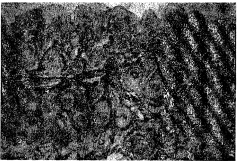
Within the semplcs of skin from dorsal area one could see rather a thick epidermis, its surfaee being very homy. The stratum papillare was medium thiek and well developed without manifesting eonneetive tissue papillae. Within the papillary layer there were same hair foI-lides along which, abit deeper, weıı developed scbaeeous glands were ranged (Fig. I). Mm. arreetores pilorum werc ranged along the hair failides. Elastic fibres were tender forming a network bclow the epi-dermis paralleııy witlı thc skin surfal'e, although somc of them were vertil'al to the basal epidermal layer to which they were adhering. The distribution of dastic fibres was more dense around the hair follides
SKIN IN THE KIDS OF THE IMPROVED BALKAN... 417
Figure ]., Skinfrom dorsa] area stained with hematoxylin and eosİn. x 40. Şekil ]. Sırt bölgesinde deri, H-E., x 40.
and where the papillary layer of the corium passed into the reticular one forming thus a boundary between these two layers (Fig. 2). Colla-gen fibres ,vere distributed within a dense net of clastic fibres. Some rough collagen fibres forming a dense network were dominant within the retiClılar layer of the corium (Fig. 3). Within a net of callagen fib-res there wcre some tender clastic fibres generaııy around the hair bulb together with sebaceous glands, blood vessels and sweat glands. The nuc1ei of fibrocytes and fibroblasts could be seen among the fib-res (Fig. 4). Where eutis passed İnto subcutis the elastic fibfib-res were extending paraııeııy with skin sur'ace and they were more dense.
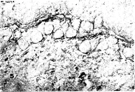
The sectİons of the skin from ventral area showcd a rclatively thick epidermis; its surface being very horny. The papillary layer of the corium was wide and of crisp constituion with visible sebaceous glands, hair foııides and mm. arrectores piııorum. Elastic fibres were tender, paraııcl with the skin surface and more dense around the hair foIlides (Fig. 5). Within a fine net of elastic fibres some rough collagen fibres appeared. Some rougher collagen fibres dominated within the reticular layer of the corİum and among them therc were some tender clactic fibres espeeİaııy around haİr bulbs, sebaceous and sweat glands.
Figure 2. Skin from dorsal area stained after Pinkus. x 40. Şekil 2. Sırt bölgesinde deri. Pinkus'a göre boyanmış. x 40.
SKIN IN THE KIDS OF THE IMPROVED BALKAN ... 419
:':f5
Figure 4. Reticular layer of the skin from dorsal area with visible fibrocyte and fibroblast nucIei stained with hematoxylin and eosin. x 100.
Şekil 4. Sırt bölgesinde derinin retiküler katmanı. Fibrosit ve fibroblast'ların çekirdekleri görülmekte. x 100.
Figure 5. Skin from ventral area with the boundary between the papiIlary and the reticular layers stained af ter Pinkus. x 40.
Şekil 5. Karın bölgesinde derinin papillar katmanından retikıiler katmanına geçiş alanı. Pinkus'a göre boyanmış. x 40.
Within a net of callagen and elastic fibres some hair bulbs as well as some fibrocyte and fibrablast nuclei could be seen. The boun-dary between subcutis and cutis was rather c1ear and it was formed by elastic fibres ranged parallelly with the skin surface.
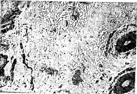
In the samples of skin taken from leg area a thick epithelium with horny surface was noted. The stratum papillare was wide and according to density offibre e1ements it could be divided in two parts. The part below the epidermis was crİsp while the other part toward the reticular layer was more dense; its fibres being thicker (Fig. 6). In the papillary layer there were some sebaceous glands along the haİr foIlicIes connected by mm. arrectores pilorum. The reticular layer was thick and formed by bundles of rough collagen fibres and by few dastic ones. Some fibrocyte and fibroblast nucIeİ as well as the hair bulbs and sweat glands could be secn among the bundles of fibres.
Figurc 6. Stratum papillare of the skin from leg area stained with hematoxylin and eosin. x 120.
Şekil 6. Bacak bölgesinde derinin stratum papiııare katmanı. H.-E.) x 120.
Discussion and Conclusions
Marcarİan and Calhoun (10), Warren et aL. (19), Galatik et aL. (1), Rusan (16), Ropac (15), Mihelİc et aL. (12) in their papers
sta-5KIN IN THE KIDS OF THE IMPROVED BALKAN... 421
ted the differences as to the thickness of skin in different animals and in various parts of the body. Our findings agreed with those of previous authors. In dorsal area the skin epidermis was relatively thin and rat-her horny on its surface while the papillae were not manifested pre-cisely because of thinnes of the epidermis. The papillary layer was well developed and wide . Elastic fibres were more dense around the hair follides and sebaceous glands forming a network within which few collagen fibres could be seen. A boundary toward the reticular la-yer was not sharp although elastic fibres were more dense in this arca. Collagen fibres were rough, strong and they formed a dense network in the retieular layer. In this layer there were few dastic fibres, espe-cially around the glands, the hair bulbs as well as in the boundary toward the subcutis where they were more dense and paralle! with the skin surfaee. A thinner epidermis, horny on the surface, could be noted in the skin taken from ventral area. No connective tissue papil-lae could be noted whatsoever. Elastic fibres in the papillary layer for-med a network thinner than those within dorsal skin. Collagen fibres were rough and strong in the reticular layer forming a thin ııetwork and being more tender than the ones found in dorsal area.
In the skin of kg area the epidermi~ was thin and very horny without connective tissue papillae. Below the epidermis the stratum papillare was crisp becoming more dense toward the reticular layer. Collagen fibres in the retieular layer were rougher and more dense than those in the ventral area skin, although they were tender forming a more crisp net than the one in the same layer of dorsal skin. Sebaceous glands in the skin of this body area were more dense than those in ventral and dorsal skin.
On the basis of the [acts expounded it appears that the skin in dorsal area is the most thick one and that collagen fibres of the reti-cular layer of the corium are the roughest and the most densdy dist-ributed ones. Theyare most tendcr in verntral skin where theyare forming the most crisp net. Elastic fibres are more dense and more numerous in ventral skin than in other two body arcas. Elastic fibres constituted the boundaries between subcutis and cutis as well as between the papillary and the reticular layers. In these areas they are mor e dence and theyare running parallelly with the skin surface. Such location of clastic fibres in the boundary between the reticular and the papillary layers as well as between subcutis and cutis corres-ponds to the findings of Hraste ct aL.(4), Rusan and Hraste (17) in
boyine skins, and of Ropac (15) and Mihclic et aL. (12) in kid's skin. In the kids of the improved Balkan capric breed the skin is thicker; collagen fibres being rougher and morc densc while dastic fibres are more scarce but stronger than in skins of the kids of the Sana and the Togcmburg capric brecds( i2). Such differences they most probably depand on keeping conditions because the kids were kept o:tensivcIy being exposed to constant changes of embiental temperaturc, humi-dity, sunlight and to other atmospheric contingencies. Such a conclu-sion corresponds to the findings of Galatik et aLp), Rusan (16), Hraste et aL. (3, 4), Mihelic (13), Ropac (I:)) in skins of pigs, bovines, goats and kids. Sebaceous and sweat glands are more numerous in the skin from leg area than in other two body arcas. The epidermal surface is mor e horny in those parts that were more exposed to mechanic influences and the corium too is thicker. Our investigations of kids' skin ~howed an essential influence of the bre ed and thc way of keeping on the histologic constitution and on the thickness of the layers in raw skins, which should be taken in consideration while processing skins into final proclucts.
References
I. Galatik, A., A. Blazaj, J.Z. Vaculik, Z. Krul (1986). Moıphologie uııd Hislologie der
Schweinhaul. Das Leder 37: 213-220.
2. Heidemann, E., S. Allam (1974). Slruklurelle Untersuchuııg der Sch!.Veinhaut im Vergleü;h zur Rindshaut. Das Leder 25 (10): 190-197.
3. Hraste, A., Z. Rusan, Vesna Gjurcevic-Kantura, K. Babic, Vera Jukic-Bres-tovec (I 988). Stereoloska islra;;:ivanja ko::e goveda kod radicitog ızaciıla drzaıı}a i ishrane.
Vet. arlıiv., 58: 67-73.
4. Hraste, A., Vesna Gjurcevic-Kantura, K. Babic, Z. Rusan, D. Mihelic, Vera Jukic-Brestovec (I 989). Fi::ikalnokcmijska i s!ereoloska islraziuan}a lIeirıje71eko"e suin}a
kod razlicitog naciııa drzaııja. Vet. arhiv., 59: 339-345.
5. Karasek, J., W. Dehlert (I%S al. Die Ullraslruklur der l;;Pidermis des Sclıweirıes. i.
Stratum basale und spinosum. Z. mikrosk. anat. Forsch., 7S: 133-144.
6. Karasek, J., W. Dehlert (I96S b). Die Ullraslruklur der Schwei71upidermis. II. Stratum
granulosum und corneum. Z. mikrosk. anat. Forsch., 79: 157-169.
7. Kozlowski, G.P., M.L. Calhoun (I 969). Mieroscopic analom)' of Ihe Inlegumenl of Sheep.
Am. J.Vet. Res., 30: (IS) 1267-1279.
8. Lloyd, D.H., S.F. Amakiri, D Mc Evan Jenkinson (1979). Slrl/clur of Ihe sheep
SKIN IN THE KIDS OF THE IMPROVED BALKAN ... 423
9. Me Evan Jenldnson, D., D.H. Uoyd (I 979). The topography of the skin surftue of Cattle
and sheep.Brit. Vet. J., 135: 376-379.
LO. Mareamn, G.H., L.M. Calhoun (1966). Microscopit: Anatamy of the integument of adult
swine. Am. J. Vet. Res., 17: (118) 765-772.
ll. Meyer, W., K. Neurand, B. Radke, (1980). Aspect of fibre arrangements in thf skin of
the pig. 13. Kongres der Europaeischen Vereinigung der Veterinaeranatomen. Liverpool
2.- 5. September.
12. Mihe1ie, D., K. Babie, A. Hraste, Vesna Gjurcevie-Kantura, Z. Jameki (1992).
Histomorfowske osobitosti koze jarica sanskeitogemburske pasmine. Vet. arhiv.
13. Mihe1ie, D. (1991). HistomorfoloskeiIıistocnzimske osobitostiijarecih koza. Magistarski rad Veterinarski fakultet, Zagreb.
14. Monteiro-Riviere, Naney A. (1986). Vltrastructurai eoaiuation of the pareine inltgument.
Swine Biomedical Res., 7: 641-655.
15. Ropae, M. (1991). Vtjacaj pasmine iııacina drzanja ııo morfowske ihistoencimske esobi/osti koze koza. Disertacija Veterinarski fakultet, Zagreb.
16. Rusan, Z. (1986). Parametrijizikaino-mehanickih soojstaoa kao rezultat strukture ko;:e. Diser-tacija Tehnoloski fakultet Zagreb.
17. Rusan, Z., A. Hraste (1989). Histomorphologische Eigenschaften oon Rindsclıauten aus
iıı-tensiucr und ebteıısioer Zudıt, Das Leder 40: 67-72.
18. Tanyo1aç,A., W. Meyer, M. Sağlam, A. Ozer, Z. Özean (1989). Mikhroscopisclıe
Un-/ersuchungm an tkr Haut tkr T,irkischen Angoraziege. Dtsch. Tieraerztl. Wscht., 96 (10):
374-512.
19. Warren, G.H., P.J. James, A.M. NevUle (1983). A morphonıetricanarysis of the changes
with age in the skin surface wax and sebaceous area of Merino sheep.Aust. Vet. J.,60 (8):