• Sonuç bulunamadı

Are perioperative near-infrared spectroscopy values correlated with clinical and biochemical parameters in cyanotic and acyanotic infants following corrective cardiac surgery?

N/A
N/A
Protected

Academic year: 2021

Share "Are perioperative near-infrared spectroscopy values correlated with clinical and biochemical parameters in cyanotic and acyanotic infants following corrective cardiac surgery?"

Copied!
6
0
0

Yükleniyor.... (view fulltext now)

Tam metin

(1)

Perfusion 1 –6 © The Author(s) 2015 Reprints and permissions: sagepub.co.uk/journalsPermissions.nav DOI: 10.1177/0267659115588632 prf.sagepub.com

Introduction

Near-infrared spectroscopy (NIRS) is a non - invasive method for the assessment of regional oxygen supply and demand in different tissue beds. The different measurements obtained from different parts of the body provide the determination of adequacy of local organ perfusion.1 Acute kidney injury is a consequence

of systemic inflammation, ischemia, emboli and low cardiac output states.2 The newborn period has some

unique properties when the immaturity of the renal system is considered, especially, the vascular resistance is high in the newborn and infantile periods.3 Although

the estimation of renal function is possible by the determination of several parameters in children and adolescents with cyanotic and acyanotic congenital heart diseases, data about the infancy period are lim-ited.4 Cerebral blood flow and oxygen supply may be

preserved in low cardiac output states due to the effective

autoregulation mechanisms in the brain and, there-fore, different measurements from different body parts is important when using NIRS monitoring.5 Although

Are perioperative near-infrared spectroscopy

values correlated with clinical and biochemical

parameters in cyanotic and acyanotic infants

following corrective cardiac surgery?

Cihangir Ersoy,

1

Arda Özyüksel,

1

Tijen Alkan Bozkaya,

1

Pelin Karaaslan,

2

Tuğrul Örmeci,

3

Akif Ündar,

4

Atıf Akçevin

1

and Halil Türkoğlu

1

Abstract

Background: Near-infrared spectroscopy (NIRS) is a useful non-invasive tool for monitoring infants undergoing cardiac

surgery. In this study, we aimed to determine the NIRS values in cyanotic and acyanotic patients who underwent corrective cardiac surgery for congenital heart diseases.

Methods: Thirty consecutive infants who were operated on with the diagnosis of ventricular septal defect (n=15) and

tetralogy of Fallot (n=15) were evaluated retrospectively. A definitive repair of the underlying cardiac pathology was achieved in all cases. A total of six measurements of cerebral and renal NIRS were performed at different stages of the perioperative period. The laboratory data, mean urine output and serum lactate levels were evaluated along with NIRS values in each group.

Results: The NIRS values differ in both groups, even after the corrective surgical procedure is performed. The recovery

of renal NIRS values is delayed in the cyanotic patients.

Conclusion: Even though definitive surgical repair is performed in cyanotic infants, recovery of the renal vasculature may

be delayed by up to two days, which is suggestive of a vulnerable period for renal dysfunction. Keywords

near-infrared spectroscopy; tetralogy of Fallot; ventricular septal defect; pediatric cardiac surgery; renal circulation

1 Department of Cardiovascular Surgery, Medipol University, Istanbul,

Turkey

2Department of Anesthesiology, Medipol University, Istanbul, Turkey 3Department of Radiology, Medipol University, Istanbul, Turkey 4 Penn State Hershey College of Medicine, Department of Pediatrics,

Surgery & Bioengineering, Hershey, PA, USA

Corresponding author:

Arda Özyüksel, Department of Cardiovascular Surgery, Medipol University, Medipol Mega Hospital, TEM Goztepe Cikisi No: 1, Bagcilar, Istanbul 34214, Turkey.

Email: ozyukselarda@yahoo.com

Presented at the 64th International Congress of the European Society

for Cardiovascular and Endovascular Surgery (ESCVS), March 26-29, 2015, Istanbul, Turkey.

(2)

cardiopulmonary bypass (CPB) is not considered as a major determinant of neurological impairment follow-ing cardiac surgery, perfusion is a modifiable factor that enhances the quality of cerebral protection.6,7

In this study, we aimed to determine the relation-ship between the perioperative NIRS values (cerebral, renal) and the clinical and biochemical parameters of renal function in two groups of patients with the diagnosis of cyanotic and acyanotic congenital heart diseases. The question was whether the clinical effects of preoperative renal and cerebral perfusion with deoxygenated blood in the cyanotic patients were promptly relieved and NIRS values, as well as clinical parameters of renal dysfunction, became equivalent in both groups following corrective cardiac surgery in infants.

Patients and Methods

A retrospective analysis of the perioperative NIRS data of 30 consecutive infants who were operated on for iso-lated ventricular septal defect (VSD) or tetralogy of Fallot (TOF) were evaluated following the approval of the institutional ethical committee. All of the patients were operated on in the infancy period (range 2 to 12 months). Group I were acyanotic infants with isolated VSD (n=15) whereas Group II were cyanotic patients with TOF (n=15). Patients with a palliative surgical treatment instead of a definitive repair were excluded. Preoperative renal dysfunction and emergency surgical interventions were other criteria for exclusion. The common target for surgical intervention was deter-mined as an unobstructed biventricular outflow without any residual shunts at atrial and ventricular levels. All the patients received low to moderate doses of positive inotropic support.

The perioperative regional cerebral and renal oxygen saturation were continuously monitored by a NIRS sen-sor (INVOSTM-5100 C, Medtronic Inc., Minneapolis,

MI, USA). A total of six measurements for both cerebral NIRS (c-NIRS) and renal NIRS (r-NIRS) were per-formed at different stages of the perioperative period; I: before induction of the anesthesia, II: at the time of cross-clamping, III: following the termination of cardio-pulmonary bypass, IV: at the 4th, V: 24th and VI: 48th

postoperative hours. The laboratory data which were evaluated together with the total urine output at the 24th

postoperative hour included serum creatinine and lac-tate levels.

Surgical technique and cardiopulmonary

bypass

Following the induction of anesthesia and endotracheal intubation, a midline sternotomy was performed in all

cases. Intravenous heparin was administered in order to achieve an activated clotting time >480 seconds. The ascending aorta and the superior and inferior vena cavae were cannulated with an 8-10 Fr aortic cannula and 12-14 Fr venous cannulae (DLP, Medtronic Inc.,), respectively. All the cases were operated under CPB and aortic cross-clamping at moderate to low hypothermia. The CPB circuit was primed with heparinized fresh blood and the hematocrit level was maintained between 28% and 35% during CPB. Pulsatile perfusion was implemented with a pulsatile roller pump (Maquet HL20, Hirrlingen, Germany) and a Capiox Baby RX05 hollow-fiber membrane oxygenator (Terumo Corp., Tokyo, Japan) in both groups. Pulsatile flow was used in all cases in which the base flow rate, start time and stop time were 10%, 20% and 80%, respectively. The mean arterial pressure was maintained at 40 mmHg during CPB. Myocardial management was achieved with inter-mittent cold (4°C) blood cardioplegia. Weaning from CPB was commenced at a rectal temperature of 36.7°C. Ultrafiltration (Hemofilter 6S, Hospal, Lyon, France) was implemented in all cases at the rewarming phase until the end of the termination of CPB, with a blood flow through the arterial line to the filter at a rate of 10-15 mL/kg/min.8

The surgical technique included complete correction of the underlying cardiac pathology in both groups. In Group I, the VSDs were closed using interrupted poly-prolene sutures, with the autologous pericardial patch treated with glutaraldehyde. All the patients had isolated VSDs, but six cases had patent foramen ovale that were concomitantly closed. In Group II, the patients with TOF underwent total correction with VSD closure and enlargement of the right ventricular outflow tract (RVOT) stenosis. Transannular patch enlargement was implemented in nine of the cases, whereas a subannular patch with preservation of the pulmonary valve was implemented in six cases. We did not encounter any sig-nificant residual shunts or impairments in ventricle function in our patients in the postoperative echocardi-ographic evaluation.

Statistical analysis

Results were analyzed with the Statistical Package for the Social Sciences (SPSS) version 17.0 (SPSS Inc., Chicago, IL, USA). Comparisons between the groups were performed with the ‘Mann-Whitney U Test’ whereas the ‘Wilcoxon Signed Rank Test’ was used for the statistical analysis in the groups for comparison of preoperative and postoperative values. The lactate val-ues (I to VI) in the groups were compared with the ‘Friedman test’ and the determination of the group that was leading to the statistically significant difference was performed with ‘Bonferoni-adjusted Wilcoxon Signed

(3)

Rank Test’ (p<0.01). Statistically significant difference was determined with a p-value lower than 0.05.

Results

The patients in both groups were statistically identical when age, gender and bodyweight were considered. The mean age of the patients in Groups I and II were 6.3 ± 3.7 and 6.8 ± 3.1 months, respectively. The mean body-weight of the patients in Groups I and II were 6.0 ± 2.1 and 6.0 ± 2.2 kg, respectively.

The preoperative oxygen saturation values were 94.6 ± 14.3% and 81.9 ± 5% for Groups I and II, respectively. The preoperative blood hematocrit level was 35.9 ± 2.3% in Group I whereas it was 38.6 ± 2.1% in Group II. When the postoperative hematocrit levels were consid-ered, Group I had a hematocrit level of 31.4 ± 2.3% whereas Group II had a hematocrit level of 29.9 ± 2.3%. There was a statistically significant difference between preoperative and postoperative hematocrit levels when the two groups were compared. The hematocrit changes (ΔHct), defined as the difference between the preopera-tive and postoperapreopera-tive hematocrit values in Group I and Group II, were 4.5 ± 3.1% and 8.7 ± 3.8%, respectively (p=0.026). Table 1 presents the preoperative demo-graphic properties and hematocrit changes of the patients as well as the CPB and aortic cross-clamp times.

Both the preoperative renal and cerebral NIRS values were statistically different between Groups I and II. Beginning at the induction of the anesthesia, there were significant differences between the two groups, accord-ing to the cerebral and renal NIRS values, continuaccord-ing until the postoperative 48th hour. When the renal NIRS

values were evaluated, the renal NIRS values were at the lowest level at stage II, which is the time of aortic cross-clamping. In both of the groups, the NIRS values began to rise at stage III, reaching a steady state at stage VI; they were statistically higher than at stage I (p<0.001). When the cerebral NIRS values were evaluated, the lowest value was recorded in stage II in both groups. However, in Group I, there is a steep rise in stages III and IV; moreo-ver, the basal level is reached at stage V, which is earlier than Group II (p<0.001). In Group II, the rise in cerebral NIRS values began at the third stage and reached a level more than the basal values in stage VI (p<0.001). The six

stage (I to VI) measurements for c-NIRS and r-NIRS are presented Figure 1 and Figure 2, respectively.

Serum lactate levels (mmol/L) were similar in both groups at stages I and II. After stage III, serum lactate levels were significantly higher in Group II until the postoperative 48th hour (stage VI) when the difference Table 1. The demographic properties, hematocrit changes, cardiopulmonary bypass and aortic cross-clamp times of the patients are presented.

Groups Age (months) Weight (kg) Δ Hct X-clamp time (min) CPB time (min)

Group I 6.3 ± 3.7 6 ± 2.1 4.5 ± 3.6 48.6 ± 10.4 77.3 ± 21.1

Group II 6.8 ± 3.1 6 ± 2.2 8.7 ± 3.8 86.9 ± 23.8 126.3 ± 23.7

p ns ns 0.026 0.0001 0.0001

Δ Hct: hematocrit change, X-clamp: aortic cross-clamp, CPB: cardiopulmonary bypass, ns: non-significant.

0 10 20 30 40 50 60 70 80 90 100 I II III IV V VI %

Renal NIRS levels

I II * * * * * *

* p<0.01, Acyanoc (Group I) vs. Cyanoc (Group II)

Figure 1. The renal NIRS levels in both groups at six different stages of measurement are demonstrated.

Stage I: before the induction of anesthesia, Stage II: after cross-clamping of the aorta, Stage III: after the termination of cardiopulmonary bypass, Stage IV: 4h post-CPB, Stage V: 24h post-CPB, Stage VI: 48h post-CPB.

0 10 20 30 40 50 60 70 80 90 100 I II III IV V VI %

Cerebral NIRS levels

I II

* * * *

* p<0.01, Acyanoc (Group I) vs. Cyanoc (Group II) † p<0.05, Acyanoc (Group I) vs. Cyanoc (Group II)

Figure 2. The cerebral NIRS levels in both groups at six different stages of measurement are demonstrated.

Stage I: before the induction of anesthesia, Stage II: after cross-clamping of the aorta, Stage III: after the termination of cardiopulmonary bypass, Stage IV: 4h post-CPB, Stage V: 24h post-CPB, Stage VI: 48h post-CPB.

(4)

became insignificant between the two groups again. The six stage (I to VI) measurements for lactate levels are presented in Table 2.

All the patients had normal serum creatinine levels at the preoperative evaluation. The mean urine output (MUO) at the postoperative 24th hour was 93.3 ± 50.4

mL/kg/day and 108.6 ± 50.5 mL/kg/day (p = 0.18) for Groups I and II, respectively. Preoperative and postop-erative 24th hour serum creatinine levels (mg/dL) were

similar for both of the groups (p>0.05). Peritoneal dialy-sis was performed in three patients in Group I (2.66 days/patient) and in nine patients in Group II (4.33 days/patient). (Table 3). Any permanent renal dysfunc-tion was not encountered in either group at the time of discharge.

Discussion

Intraoperative and postoperative management of the adverse events related to low cardiac output in infants undergoing congenital heart surgery is an essential component of successful treatment. Conventional monitoring for the detection of tissue hypoxia is pro-vided with mixed venous oxygen saturation and serum lactate levels.9 However, mixed venous oxygen

satura-tion may not yield accurate results in patients with cyanotic congenital heart defects. Although serum lac-tate is a reliable marker of tissue oxygen delivery and cellular perfusion, it does not provide real-time moni-toring of the patient.

Near-infrared spectroscopy measures the equilibrium of oxyhemoglobin and deoxyhemoglobin in a mixture of veins, arteries and capillaries, with the application of the ‘Beer-Lambert’ law for the measurement of the con-centration of a substance according to its absorption of

light.10 However, there are some limitations with NIRS

monitoring, such as a lack of precise contribution of various vascular beds, the size of the area explored and a lack of a direct reference against which to correlate val-ues.11,12

In our study, we primarily aimed to evaluate the NIRS values following a corrective cardiac repair, which, theoretically, provides oxygenated blood supply to renal and cerebral beds, both in cyanotic and acy-anotic patients. Our first observation was the lack of appearance of the effects of oxygenated blood perfu-sion to renal and cerebral beds, where cerebral and renal measurements were concerned. After complete surgical repair in the cyanotic group, the renal and cer-ebral NIRS measurements were still lower than the acy-anotic group, which was statistically significant until the postoperative 48th hour. The serum lactate levels

were significantly lower in the acyanotic group in the first postoperative 24 hours; however, the levels on the second day were statistically insignificant when both groups were compared. The postoperative 24th hour

serum creatinine levels and mean urine output were statistically insignificant, as well. In our experience, early prophylactic peritoneal dialysis provides an effec-tive electrolyte balance and input-output volume bal-ance in patients undergoing cardiac surgery.13

Therefore, we performed peritoneal dialysis in both groups, 20% in Group I and 60% in Group II. Although a statistically significant difference is demonstrated between the two groups with regard to the implementa-tion of peritoneal dialysis, we perform peritoneal dialysis more liberally in cyanotic patients and we do not attrib-ute this difference to any NIRS measurement.

During cardiac surgery, the effect of hemoglobin lev-els on NIRS monitoring is inevitable.14 The hematocrit Table 2. Six-stage serum lactate levels (mmol/L) in both groups.

Groups I II III IV V VI

I 0.6 ± 0.7 0.8 ± 0.9 5.8 ± 4.8 5.7 ± 3.6 1.6 ± 0.3 1.0 ± 0.6

II 1 ± 0.7 1.2 ± 0.9 9.4 ± 4.8 9.0 ± 3.8 6.0 ± 0.5 1.6 ± 0.5

p ns ns 0.026 0.021 0.0018 ns

Stage I: before the induction of anesthesia, Stage II: after cross-clamping of the aorta, Stage III: after the termination of cardiopulmonary bypass, Stage IV: 4h post-CPB, Stage V: 24h post-CPB, Stage VI: 48h post-CPB.

Table 3. Peroperative and postoperative serum creatinine levels, mean urine output and peritoneal dialysis in patients in both groups.

Groups Preop. Cr Postop. Cr MUO mL/kg/day PD (n/total) day(s)

I 0.1 ± 0.06 0.5 ± 0.1 93.3 ± 50.4 3/15 (2.66 days)

II 0.1 ± 0.05 0.7 ± 0.1 108.6 ± 50.5 9/15 (4.33 days)

p ns ns ns 0.0012

(5)

changes in Groups I and II were statistically significant; however, its effect on the postoperative NIRS values of the cyanotic patients is multifactorial. We did not encounter high hematocrit levels in order to use blood-free prime in cyanotic patients, which was probably related to low ages at the time of the corrective cardiac surgery. Although the kidneys and brains of the patients in both groups, theoretically, should begin to receive oxygenated blood at the termination of CPB, there was a statistically significant difference in the cerebral and renal NIRS values in both cyanotic and acyanotic patients. The renal NIRS surge of the cyanotic and acy-anotic patients suggests that the renal perfusion of these patients becomes similar at least 2 days after surgery. Therefore, this postoperative period should be opti-mally managed in terms of renal perfusion and urine output in cyanotic infants.

Our previous experience correlated the renal resis-tive index values with the cerebral and renal NIRS meas-urements as well as clinical outcomes in infants with an age of less than 3 months, all of whom were in the high-risk group according to the ‘Aristotle Basic Complexity Risk Stratification’ score. A renal resistive index value greater than 0.8 was found to be associated with lower postoperative mean urine output and significantly higher lactate levels as well as a significantly higher postoperative intensive care unit and hospital stay of the patients.15

Herein, we would like to clarify one issue about our study design. We did not aim to directly relate the NIRS values with renal or cerebral malperfusion in two pathophysiologically different patient popula-tions. To the best of our knowledge, the NIRS surges in cyanotic and acyanotic infants have not been reported in the literature with regard to the hypothesis that both patients should have similar hemoglobin-oxygenation dynamics following the definitive repair of the underlying congenital heart disease without further ongoing intracardiac shunts. The major limi-tation of our study is the different CPB and aortic cross-clamping times, as well as the preoperative hemoglobin values which may directly affect the renal perfusion in the postoperative period. However, mean urine output and postoperative serum creatinine lev-els were similar in both groups, which may suggest effective renal protection in both groups. Our find-ings suggest that serum lactate levels become statisti-cally similar in the 48th postoperative hour; however,

cerebral and renal NIRS values still remain low in cyanotic patients. In our opinion, since these infants are candidates for early definitive surgical treatment of right to left shunts, persistently high levels of hema-tocrit and its adverse effects on microvasculature is not the only culprit for delayed recovery of the vascu-lar bed in kidneys.

Our study suggests that the postoperative period, including the first 48 hours, is a vulnerable period for renal function with regard to NIRS measurements in infants undergoing cardiac surgery. Although serum lactate levels and cerebral NIRS values become normal at the end of the second day, renal NIRS values still remain low. However, if an effective mean urine output is provided, the renal NIRS values demonstrate signs of recovery two days after the operation.

Declaration of Conflicting Interest

The authors declare that there is no conflict of interest.

Funding

This research received no specific grant from any funding agency in the public, commercial or not-for-profit sectors.

References

1. Zulueta JL, Vida VL, Perisinotto E, et al. Role of intra-operative regional oxygen saturation using near infrared spectroscopy in the prediction of low output syndrome after pediatric heart surgery. J Card Surg 2013; 28: 446–452.

2. Ghatanatti R, Teli A, Tirkey SS, et al. Role of renal bio-markers as predictors of acute kidney injury in car-diac surgery. Asian Cardiovasc Thorac Ann 2014; 22: 234–241.

3. Bude RO, DiPietro MA, Platt JF, Rubin JM, Miesowicz S, Lundquist C. Age dependency of the renal resistive index in healthy children. Radiology 1992; 184: 469–473.

4. Amornchaicharoensuk Y, Werawatganon T,

Tohsukhowong P, et al. Comparison of renal function between cyanotic and acyanotic congenital heart disease in children and adolescent. J Med Assoc Thai 2012; 95: 1501–1508.

5. Ortmann LA, Fontenot EE, Seib PM, et al. Use of near infrared spectroscopy for estimation of renal oxygena-tion in children with heart disease. Pediatr Cardiol 2011; 32: 748–753.

6. Durandy Y, Rubatti M, Couturier R. Near infrared spec-troscopy during pediatric cardiac surgery: errors and pit-falls. Perfusion 2011; 26: 441–446.

7. Hogue CW Jr, Palin CA, Arrowsmith JE. Cardiopulmonary bypass management and neurologic outcomes: an evi-dence-based appraisal of current practices. Anesth Analg 2006; 103: 21–37.

8. Ziyaeifard M, Alizadehasl A, Massoumi G. Modified ultrafiltration during cardiopulmonary bypass and postoperative course of pediatric cardiac surgery. Res

Cardiovasc Med 2014; 3: e17830. doi:

10.5812/cardiovas-cmed.17830.

9. Tweddell JS, Ghanayem NS, Hoffman GM. Pro: NIRS is “standard of care” for postoperative management. Semin

Thorac Cardiovasc Surg Pediatr Card Surg Annu 2010; 13:

44–50. doi: 10.1053/j.pcsu.2010.02.008.

10. Li J, Van Arsdell GS, Zhang G, et al. Assessment of the relationship between cerebral and splanchnic oxygen

(6)

saturations measured by near-infrared spectroscopy and direct measurements of systemic haemodynamic variables and oxygen transport after the Norwood pro-cedure. Heart 2006; 92: 1678–1685.

11. Andropoulos DB, Stayer SA, Diaz LK, et al. Neurological monitoring for congenital heart surgery. Anesth Analg 2004; 99: 1365–1375.

12. Watzman HM, Kurth CD, Montenegro LM, et al. Arterial and venous contributions to near-infrared cerebral oxi-metry. Anesthesiology 2000; 93: 947–953.

13. Alkan T, Akçevin A, Türkoglu H, et al. Postoperative prophy-lactic peritoneal dialysis in neonates and infants after com-plex congenital cardiac surgery. ASAIO J 2006; 52: 693–697. 14. Owen-Reece H, Smith M, Elwell CE, et al. Near infrared

spectroscopy. Br J Anaesth 1999; 82: 418–426.

15. Örmeci T, Alkan-Bozkaya T, Özyüksel A, et al. Correlation between cerebral-renal NIRS and ipsilateral renal perfu-sion parameters as clinical outcome predictors after open heart surgery in neonates and infants. Artificial Organs 2015; 39: 53–58.

Şekil

Figure 2.  The cerebral NIRS levels in both groups at six  different stages of measurement are demonstrated.
Table 3.  Peroperative and postoperative serum creatinine levels, mean urine output and peritoneal dialysis in patients in both  groups.

Referanslar

Benzer Belgeler

Systemic right ventricle in elderly patients with congenitally corrected transposition of the great arteries: Clinical profile, cardiac.. biomarkers, and

This study examined the relationship between congenital heart disease and oxidative stress by measuring the total antioxidant status (TAS), total oxidant status (TOS), and

Whi- le analysing the rheograms, the following values had been assessed: A (in ohm) – reflecting blood supply to cerebral vascular basin dependent on cardiac out- put and tension
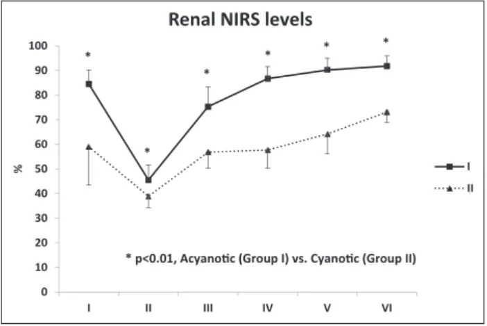
Background:­ This study aims to investigate cerebral oxygenation differences between cyanotic and non-cyanotic pediatric cardiac surgery patients using near-infrared

Prior to attempting surgery, it is important to consider the presence of preoperative pneumonia, acidosis, and poor postoperative forced expiratory volume in 1 second and

Hint-İran geleneğin- den doğarak Türk ve İslami gelenekle harmanlanmış daire-i adalete içkin yö- netim bilgisi Osmanlı Devleti’ne aktarılmış; Osmanlı Devleti’nin

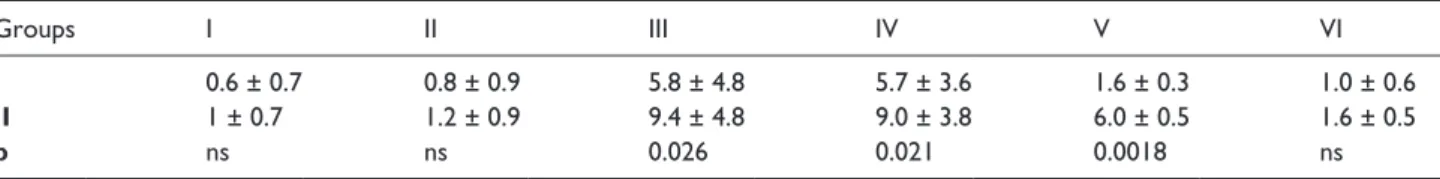
Abbreviations: RScO 2 , right cerebral oxygen saturation; LScO 2 , left cerebral oxygen saturation; NS, not significant.. determine the baseline NIRS values of patients who

高血壓自我保健 返回 醫療衛教 發表醫師 李彩鳳醫師 發佈日期 2010/09/07 高血壓自我保健 中醫科李彩鳳主治醫師