Successful laparoscopic removal of mesenteric and omental
cysts in toddlers: 3 cases with a literature review
Arzu Pampal
a,⁎
, Aydin Yagmurlu
ba
Ufuk University, Faculty of Medicine, Department of Pediatric Surgery, Ankara, Turkey
b
Ankara University, Faculty of Medicine, Department of Pediatric Surgery, Ankara, Turkey Received 11 January 2012; revised 24 March 2012; accepted 25 March 2012
Key words: Mesenteric cyst; Omental cyst; Laparoscopy; Minimally invasive surgery; Children
Abstract Mesenteric and omental cysts are rare benign intraabdominal anomalies with uncertain etiologies. Surgical removal is the preferred treatment owing to complications related to cyst enlargement. A 1-year-old boy with an intrauterine diagnosis of a cystic mass adjacent to his stomach and liver, a 3-year-old girl, and a 3-year-old boy with an incidental diagnosis of intraabdominal cysts were scheduled for laparoscopic surgery. The mass of the 1-year-old boy was a multiloculated cyst originating from the lesser omentum, the incidental mass in the girl was a multiseptated cyst located in the jejunoileal mesentery, and the incidental mass of the 3-year-old boy was a uniloculated cyst originating from the ileal mesentery. All the cysts were excised either laparoscopically or in a laparoscopy-assisted manner. The laparoscopic or laparoscopy-assisted excision of the mesenteric and omental cysts seems to be a feasible, safe, and cost-effective surgical procedure with shorter operative times, even in toddlers.
© 2012 Elsevier Inc. All rights reserved.
Mesenteric and omental cysts are uncommon intraabdom-inal masses, generally of a benign nature. The incidence of mesenteric cysts is nearly 1 of 105,000 hospital admissions, and the incidence of omental cysts is even more rare. These cysts are more common in the adults, with only 25% of cases diagnosed before the age of 10 years[1]. Despite the recent literature setting cystic intraabdominal lymphatic malforma-tions apart from the mesenteric and omental cysts with regard to their pathologicfindings and clinical behavior, all omental, mesenteric, and retroperitoneal cysts are actually the exten-sions of what were originally retroperitoneal anlage during embryogenesis and complete excision of the intraabdominal cysts of any nature is the treatment of choice.
Despite the earlier findings of limited intraabdominal working space for laparoscopic procedures in infants and young children leading to more difficult and time-consuming
operations, with time and operator experience, minimally invasive surgery in pediatric-aged individuals has become more widely used. This report describes 3 toddlers with intraabdominal mesenteric or omental cysts successfully excised laparoscopically or with laparoscopy-assisted opera-tions and reviews the pediatric cases treated by minimally invasive surgery in the literature.
1. Case reports
1.1. Case 1
A 13-month-old boy with an intraabdominal cystic mass that had been discovered at the 36th week of gestation was scheduled for a laparoscopic excision. The boy was a term baby with no signs or symptoms of gastrointestinal obstruction. The prenatal ultrasound revealed a 2-cm cystic ⁎ Corresponding author. Tel.: +90 312 2044269; fax: +90 312 2026213.
E-mail address:[email protected](A. Pampal).
www.elsevier.com/locate/jpedsurg
0022-3468/$– see front matter © 2012 Elsevier Inc. All rights reserved. doi:10.1016/j.jpedsurg.2012.03.080
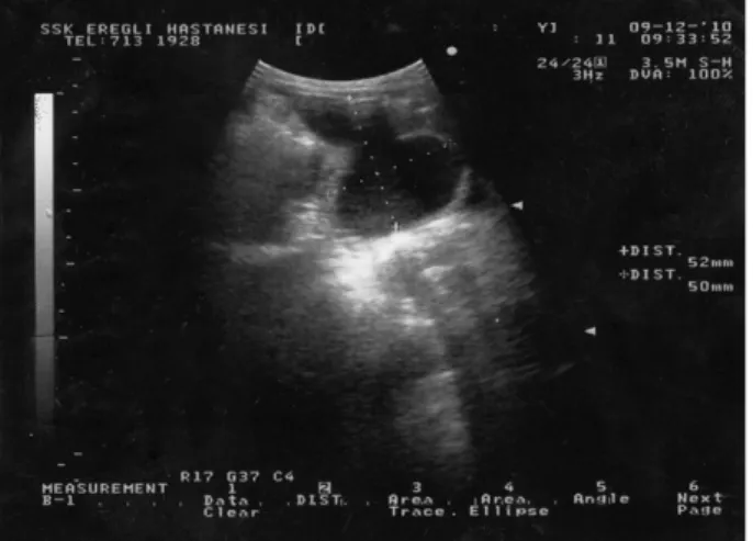
mass adjacent to the stomach, and the boy received consecutive ultrasounds after birth. The ultrasound at the age of 1 year revealed a 67 × 39 mm multiseptated cyst with dense contents that was located in the prepancreatic area, adjacent to the stomach and left lobe of liver, expanding down to the right pelvic area (Fig. 1). Consecutive ultrasounds revealed a growing mass and a suspicion of a lymphatic malformation or omental cyst, so surgical removal was required.
The boy was taken to the operating room after premedication, intubated endotracheally, and ventilated. A 10-mm trocar was inserted via umbilicus using an open technique, and the exploration of the abdominal cavity revealed a multiseptated cystic mass originated from the lesser omentum, adjacent to the liver, adherent to the stomach and transverse colon, and extending down to right lower quadrant. Two additional 5-mm trocars were inserted. Tight adhesions to the stomach and transverse colon were divided with a harmonic scalpel. The serosanguineous content of the cyst was aspirated, the cystic wall was totally removed from the lesser omentum using the harmonic scalpel, and the mass was extracted through the trocar. The procedure took 30 minutes overall. The boy was fed during the postoperative period as soon as he wanted to eat, and he was discharged on thefirst postoperative day. The pathologic evaluation of the cystic mass revealed a multiloculated cyst with a columnar mesothelial lining and no cellular atypia. The cystic wall showed vascular dilation and a minimal infiltration of lymphocytes but no significant lymphatic vessel formation. The postoperative course was uneventful, and there has been no recurrence.
1.2. Case 2
A previously healthy two-and-a-half-year-old girl was treated in the emergency department for bladder distention.
After the insertion of a urinary catheter, she was evaluated by a pelvic ultrasound study. A 4 × 2 cm cystic mass adjacent to the bladder was observed. Her urine culture was sterile, she was diagnosed as having cystitis and medicated. Because of the cystic mass, her parents requested that she receive consecutive follow-up ultrasound scans. At the sixth month follow-up period, the ultrasound revealed a 57 × 45 × 29 mm multiseptated cystic mass located cranial to bladder and an additional 32 × 21 × 10 mm cyst located in the mesentery. Because of the enlargement of the cystic mass, she was scheduled for a laparoscopic excision.
The girl was taken to the operating room after premedica-tion. She was ventilated using a laryngeal mask. A 10-mm trocar was inserted via the umbilicus, and the exploration of the abdominal cavity revealed a multiseptated cystic massfilling the right lower quadrant of her abdomen (Fig. 2). After placing 2 additional 5-mm trocars, the cyst was evaluated and was noted to arise from the jejunoileal mesentery. The serosangui-neous content of the cyst was aspirated, the cystic wall was totally removed from the mesentery with a harmonic scalpel, and the mass was extracted through the trocar. The umbilical opening was widened, and the jejunoileal segment was exteriorized. After the exploration of the residual tissue and the establishment of hemostasis, the mesenteric defect was sutured with interrupted stitches. The procedure took 35 minutes overall. The girl was fed during the postoperative period as soon as she wanted to eat and was discharged on the first postoperative day. The pathologic evaluation of the cystic mass revealed a multiloculated cyst with a columnar mesothelial lining and no cellular atypia, compatible with a mesenteric cyst. The postoperative course was uneventful with no recurrence of the mass.
1.3. Case 3
A previously healthy 3-year-old boy was evaluated because of chronic abdominal pain and constipation. On
Fig. 1 The ultrasound scan of the multiloculated cystic mass of the first case located at the prepancreatic area adjacent to the stomach and liver.
Fig. 2 The laparoscopic view of the mesenteric cyst (*) of the second case located in the jejunoileal mesentery.
ultrasound study, it was noted that he had a 5.5 × 5.5 mm cystic mass adjacent to his bladder. The child was observed, and at 2 months of follow-up, the cystic mass enlarged to 75 × 36 ×50 mm, so he was scheduled for laparoscopic excision (Fig. 3).
The boy was taken to the operating room after premedication. He was ventilated using a laryngeal mask. A 10-mm trocar was inserted via umbilicus with an open technique, and the exploration of the abdominal cavity revealed an segment of ileum lying on a uniloculated cystic mass. After placing 2 additional 5-mm trocars, the cyst was evaluated, revealing intimate contact with the bowel. The umbilical opening was widened, and the jejunoileal segment was exteriorized. The serosanguineous content of the cyst was aspirated, and the cystic wall was totally removed from the mesentery using electrocautery preserving the bowel. The mesenteric defect was sutured with interrupted stitches.
The procedure took 45 minutes. The boy was fed during the postoperative period as soon as he wanted to eat and was discharged on the first postoperative day. The pathologic evaluation of the cystic mass revealed a uniloculated cyst with a cuboidal mesothelial lining and no cellular atypia, compatible with a mesenteric cyst. The postoperative course was uneventful with no recurrence.
2. Discussion
Mesenteric, omental, and retroperitoneal cysts are unusual causes of intraabdominal masses in childhood. Different developmental theories for these cysts are discussed, including the benign proliferation of the ectopic mesenteric lymphatics that do not communicate with the rest of the lymphatic system, the failure to join the embryonic lymphatic spaces to the venous system, the failure of the fusion of mesenteric leaves, the deficiency of the normal lymphaticovenous shunts, trauma, neoplasm, and the localized degeneration of lymph nodes[2]. These cysts are present either incidentally, with nonspecific abdominal complaints or with acute abdominal pain. The spectrum of presentation depends primarily on the location and the size of the cyst. Any emerging complications, including enlargement, intracystic hemorrhage, torsion, infection, or rupture, are common indications of a surgical excision[2].
Ultrasound is a very sensitive and specific radiologic imaging modality used not only for the diagnosis but also for the follow-up of these cysts, even in the prenatal period
[3]. Despite providing additional information about the location, size, and invasion of the cysts into the surrounding tissues, computed tomography is not generally necessary while evaluating mesenteric cysts in childhood because of the high radiation exposure. Magnetic resonance Fig. 3 The ultrasound scan of the uniloculated cystic mass of the
third case adjacent to the bladder.
Table Pediatric cases of mesenteric, omental, and retroperitoneal cysts treated by MIS
Age Sex Diagnosis Treatment
Esposito et al[4] 15 d M Intraperitoneal cystic lymphangioma Laparoscopy-assisted excision
Kuga et al[5] 47 d F Omental cyst Laparoscopy-assisted excision
51 d F Omental cyst Laparoscopy-assisted excision
Al-Zaiem[6] 11 mo M Intraperitoneal cystic lymphangioma Laparoscopy-assisted excision Wildhaber et al[7] 18 mo F Retroperitoneal cystic lymphangioma Transperitoneoscopic excision 4 y M Retroperitoneal cystic lymphangioma Transperitoneoscopic excision
Singh et al[8] 15 y F Retroperitoneal cystic lymphangioma Retroperitoneoscopic + laparoscopic excision Kenney et al[9] 16 y F Intraperitoneal cystic lymphangioma Laparoscopic excision + gastropexy
Yao et al[10] 15 y F Omental cyst Laparoscopic excision
Raghupathy et al[11] M Mesenteric cyst Laparoscopic excision
de Lagauise et al[12] 0.9 mo F Intraperitoneal lymphangioma Laparoscopic excision
3 y M Intraperitoneal lymphangioma Laparoscopic omentectomy
4 y M Intraperitoneal lymphangioma Laparoscopic excision
8 y M Intraperitoneal lymphangioma Laparoscopic excision
9 y F Intraperitoneal lymphangioma Laparoscopic excision
14 y F Intraperitoneal lymphangioma Laparoscopic excision
M indicates male; F, female.
E7 Laparoscopic removal of mesenteric and omental cysts
imaging could be an alternative for evaluation, especially in prenatal cystic lesions. The treatment includes the complete excision of the cystic mass. Sclerosing agent therapies either alone or followed by marsupialization can be used with a moderate risk of recurrence in the case of complete excision failure.
Minimally invasive surgery (MIS) is a surgical treatment option. These cysts can be excised either laparoscopically or in a laparoscopy-assisted manner. The literature review revealed a moderate number of presentations in adulthood, but pediatric cases are limited. Only 16 pediatric cases were operated using MIS (Table). No matter where the cysts are located, the surgical teams were able to complete the procedures with shorter operation times and better postoperative cosmesis. Infants with intraabdominal cysts were generally operated on in a laparoscopy-assisted manner[4-6]. Minimally invasive surgery was presented as an advantageous procedure for this age group. The other cases mentioned in the case reports were operated either laparoscopically or retroperitoneoscopically without the need for laparotomy[7-11].
In a series by de Lagausie et al [12], of the 9 children undergoing a laparoscopic excision of abdominal cystic lymphatic malformations, 6 cases were completed laparosco-pically. The reasons noted for laparotomy in the remaining 3 cases included the need for bowel resection and adhesion of the cyst to the inferior vena cava. We did not require conversion during our surgical procedures. Instead, we enlarged the umbilical openings to repair the defect of the mesentery in the second case and to excise the uniloculated cyst that had intimate contact with small bowel in the third case. If the necessity for bowel resection arose, we believe that the same extended umbilical opening would also be suitable to complete the procedure in a laparoscopy-assisted manner.
Sakurai et al[13] described the successful laparoscopic excision of an omental cyst located at the lesser omentum in a 50-year-old patient. The authors stated that the procedure was feasible and advantageous. Despite being a safe alternative to laparotomy, the authors stated that there were a limited number laparoscopic cases for adult omental or mesenteric cysts reported in the literature. They linked the limited use of laparoscopy to the difficulties in preoperative diagnosis, the possibility of malignancy, and the probable need for a bowel resection during cyst excision. Because a neoplasm was a possible etiologic factor in the cysts in elderly patients and the need for a bowel resection and anastomosis can be handled in a laparoscopy-assisted
manner in young children, the laparoscopic excision of mesenteric and omental cysts, even those located at the lesser omentum in children, seems more probable. We excised an omental cyst from the lesser omentum in a 1-year-old infant, and we think the procedure is very advantageous.
Unless specific contraindications exist for laparoscopic excision, we think that either laparoscopy or laparoscopy-assisted surgery, even in toddlers, is an appropriate surgical option, offering shorter operation times, shorter hospital stays, reduced postoperative ileus, decreased risk of intestinal adhesions, and better postoperative cosmesis.
References
[1] Ricketts RR. Mesenteric and omental cysts. In: Grosfeld JL, O'Neill JAJ, Coran AG, Fonkalsrud EW, editors. Pediatric surgery. Philadel-phia (Pa): Mosby Elsevier; 2006. p. 1399-406.
[2] Lin JI, Fisher J, Caty MG. Newborn intraabdominal cystic lymphatic malformations. Semin Pediatr Surg 2000;9:141-5.
[3] Konen O, Rathaus V, Dlugy E, et al. Childhood abdominal cystic lymphangioma. Pediatr Radiol 2002;32:88-94.
[4] Esposito C, Alicchio F, Savanelli A, et al. One-trocar ileo-colic resection in a newborn infant with a cystic lymphangioma of the small-bowel mesentery. J Laparoendosc Adv Surg Tech A 2009;19:447-9.
[5] Kuga T, Inoue T, Taniguchi S, et al. Laparoscopic surgery in infants with intra-abdominal cysts: two case reports. JSLS 2000;4:243-6. [6] Al-Zaiem MM. Assisted laparoscopic excision of huge abdominal
cysts in newborns and infants using the umbilical laparoscopic port incision. J Pediatr Surg 2011;46:1459-63.
[7] Wildhaber BE, Chardot C, Le Coultre C, et al. Total laparoscopic excision of retroperitoneal cystic lymphangioma. J Laparoendosc Adv Surg Tech A 2006;16:530-3.
[8] Singh RR, Govindarajan KK, Bowen C, et al. Retroperitoneal cystic lymphangioma: a rare presentation in childhood, treated laparoscopi-cally. J Laparoendosc Adv Surg Tech A 2009;19:249-50.
[9] Kenney B, Smith B, Bensoussan AL. Laparoscopic excision of a cystic lymphangioma. J Laparoendosc Surg 1996;6(Suppl 1):S99-101. [10] Yao CC, Wu TL, Wong HH, et al. Laparoscopic resection of an
omental cyst with pedicle torsion. Surg Laparosc Endosc Percutan Tech 1999;9:372-4.
[11] Raghupathy RK, Krishnamurthy P, Rajamani G, et al. Intraabdominal cystic swelling in children—laparoscopic approach, our experience. J Indian Assoc Pediatr Surg 2003;8:213-7.
[12] de Lagausie P, Bonnard A, Berrebi D, et al. Abdominal lymphangio-mas in children: interest of the laparoscopic approach. Surg Endosc 2007;21:1153-7.
[13] Sakurai Y, Taniguchi K, Uyama I, et al. Laparoscopic excision of the cystic lymphangioma occurred in the lesser omentum: report of a case and review of literature. Surg Laparosc Endosc Percutan Tech 2009;19:e11-4.