KADIR HAS UNIVERSITY
GRADUATE SCHOOL OF SCIENCE AND ENGINEERING
IN SILICO IDENTIFICATION OF PHYSIOLOGICAL SUBSTRATES AND INHIBITORS OF SERUM PARAOXONASE 1 ENZYME
TALHA KARABIYIK
January, 2014
T alha K ara bı yı k M .S c. T he sis 2014
IN SILICO IDENTIFICATION OF PHYSIOLOGICAL SUBSTRATESAND
INHIBITORS OF SERUMPARAOXONASE 1 ENZYME
TALHA KARABIYIK
Submitted to the Graduate School of Science and Engineering in partial fulfillment of the requirements for the degree of
Master of Science in
Computational Biology and Bioinformatics
KADIR HAS UNIVERSITY
January, 2014 APPENDIX B
KADIR HAS UNIVERSITY GRADUATE SCHOOL OF SCIENCE AND ENGINEERING
IN SILICO IDENTIFICATION OF PHYSIOLOGICAL SUBSTRATESAND
INHIBITORS OF SERUMPARAOXONASE 1 ENZYME
TALHA KARABIYIK
APPROVED BY:
Prof. Dr. Kemal Yelekçi (Advisor) KHAS _____________________
Prof. Dr. Safiye Sağ Erdem MÜ _____________________
Asst. Prof. Dr. Şebnem Eşsiz Gökhan KHAS _____________________
APPROVAL DATE: 10/Jan/2014 APPENDIX B
i
“I, Talha Karabıyık, confirm that the work presented in this thesis is my own. Where information has been derived from other sources, I confirm that this has been indicated in the thesis.”
© TALHA KARABIYIK İstanbul, 2014
All Rights Reserved
i
Abstract
IN SILICO IDENTIFICATION OF PHYSIOLOGICAL SUBSTRATESAND
INHIBITORS OF SERUMPARAOXONASE 1 ENZYME
TALHA KARABIYIK
Master of Science in Computational Biology and Bioinformatics Advisor: Prof. Dr. Kemal Yelekçi
January, 2014
Paraoxonase 1 (PON1), as an important antioxidant enzyme against oxidative stress, has been implicated in the pathogenesis of a number of disorders including cancer, cardiovascular and several other diseases. Although there has been considerable progress in understanding the PON1 enzyme, its precise physiological substrate and function still remain inconclusive. Discovery of new PON1 substrates or inhibitors will provide better understanding of PON1’s cardiovascular protective and
antioxidant effects.
PON1 is known to show lactonase, aryl esterase and phosphotriesterase
(paraoxonase) activity. PON1, having two calcium ions within its central tunnel, shows six-bladed β-propeller, with each arm comprising of four β-sheets. The structural Ca2+ is buried, whereas the catalytic Ca2+ lies at the bottom of the active-site cavity.
In this study, metabolites of Human Metabolome Database (HMDB) version 3.0 containing over 40,000 metabolites were docked against the PON1 structure (PDB ID: 3SRE) determined by Ben-David et al. in a virtual screening scenario using AutoDock 4.2 and metabolites were evaluated in terms of docking energy and docking pose.
ii
The best 2000 metabolites in terms of docking energy were inspected one by one and 97 of them were selected due to their chemical groups that the PON1 may work on. These 97 metabolites were further evaluated in terms of their docking poses, and this further evaluation revealed that 10 out of 97 had the correct docking pose.
These aforementioned metabolites are sorgolactone, indoxyl sulfate, 5-methoxyhinokinin, enterolactone, (-)-arctigenin, epoxybergamottin,
pandamarilactone 32, (-)-matairesinol, alectrol, isoalantolactone. Except indoxyl sulfate, all of them are of plant origin.
It is known that PON1 activity is negatively correlated with high intake of vegetables. With this data and docking energies of metabolites of plant origin mentioned above in hand, it is suggested that these metabolites of plant origin may be PON1 inhibitors.
Indoxyl sulfate plays roles in mechanisms of various diseases and causes oxidative stress. This metabolite also has considerably low docking energy and may also be a PON1 inhibitor.
To be certain about the PON1 inhibitory potential of these mentioned 10 metabolites laboratory assays should be carried out.
iii
Özet
SERUM PARAOKSONAZ 1 ENZİMİNİN FİZYOLOJİK SÜBSTRATLARININ VE İNHİBİTÖRLERİNİN İN SİLİCO TANIMLANMASI
TALHA KARABIYIK
Hesaplamalı Biyoloji ve Biyoinformatik, Yüksek Lisans Danışman: Prof.Dr. Kemal Yelekçi
Ocak, 2014
Oksidatif strese karşı önemli bir antioksidan olarak görev yapan paraoksonaz 1 (PON1), kanser ve kardiyovasküler hastalıklar başta olmak üzere birçok hastalığın patogeneziyle ilişkilendirilmektedir. PON1’in fizyolojik rolünün anlaşılmasında çok yol alınmış olsa da bu enzimin fizyolojik rolü ve sübstratları hala tam olarak
bilinmemektedir. Yeni PON1 sübstrat veya inhibitörlerinin keşfi, PON1’in kardiyovasküler koruyucu ve antioksidan etkilerinin daha iyi anlaşılmasını sağlayacaktır.
PON1’in laktonaz, aril esteraz ve fosfotriesteraz aktivitesi gösterdiği bilinmektedir. PON1, merkez tünelinde iki tane kalsiyum iyonu bulunan, her bir kanadında 4 tane β-tabakası olan 6 kanatlı β pervane katlanması gösterir. Yapısal Ca2+ gömülü durumda iken katalitik Ca2+ aktif bölge kavitesinin dibinde yer alır.
Bu araştırmada 40.000’in üzerinde metabolit içeren İnsan Metabolom Veritabanı (HMDB) versiyon 3’teki metabolitler Ben-David ve arkadaşlarınca tanımlanan PON1 (PDB ID: 3SRE) yapısına karşı sanal tarama metodolojisiyle AutoDock 4.2 kullanılarak taranmıştır ve metabolitler bağlanma (docking) enerjilerine ve bağlanma pozlarına göre değerlendirilmiştir.
iv
Docking enerjilerine göre en iyi 2000 metabolit tek tek incelenmiştir ve PON1’in etki edebileceği düşünülen kimyasal gruplara sahip olan 97 metabolit ayıklanmıştır. Bu 97 tane metabolit daha sonra bağlanma (docking) pozlarına göre
değerlendirilmiştir ve bu değerlendirme sonucunda 10’unun kataliz için doğru pozda olduğu görülmüştür.
Bu on metabolit şunlardır: Sorgolakton, indoksil sülfat, 5-metoksihinokinin, enterolakton, arktigenin, epoksibergamottin, pandamarilakton 32,
(-)-matairesinol, alektrol, izoalantolakton. Bunlardan indoksil sülfat hariç hepsi bitkisel kökenlidir.
Yüksek sebze tüketimi ile PON1 aktivitesinin ters korelasyon gösterdiği
bilinmektedir. Üstte bahsedilen bitkisel kökenli bileşiklerin bağlanma enerjileri de dikkate alındığında bu bitkisel kökenli bileşiklerin PON1 inhibitörü olduğu
düşünülmektedir.
İndoksil sülfatın çeşitli hastalık mekanizmalarında etkin olduğu ve oksidatif strese yol açtığı bilinmektedir. İndoksil sülfatın da bağlanma enerjisi oldukça düşükür ve bu indoksil sülfatın da bir PON1 inhibitörü olduğunu düşündürmektedir.
Üstte bahsedilen metabolitlerin PON1 inhibitörleri olduklarının kesin tespiti laboratuvar deneyleriyle ortaya koyulmalıdır.
v
Acknowledgements
Foremost, I would like to thank my advisor Prof. Kemal Yelekçi for the continuous support of my M.S. study and research, for his patience, encouragement, jokes, and immense knowledge. His guidance helped me in all the time of research and writing of this thesis. I could not have imagined having a better advisor and mentor for my M.S. study.
Besides my advisor, I would like to thank the rest of my thesis committee: Prof. Safiye Erdem and Asst. Prof. Şebnem Eşsiz Gökhan for their encouragement, insightful comments, and hard questions.
A special thank goes to Serkan Altuntaş, for letting me use his program (YaVST) and his passion for the dissemination of his knowledge.
Last but not the least, I would like to thank my parents for their genes and unwavering support throughout my life.
vi
Table of Contents
Abstract ... i Özet ... ii Acknowledgements ... vii Table of Contents ... viList of Figures ... viiii
List of Tables ... viii
1. Chapter 1: Objective ... 1
2. Chapter 2: Introduction ... 3
2.1 Paraoxonase ... 3
2.2 Metabolomics ... 17
2.2 Docking ... 19
3. Chapter 3: Materials and Methods ... 1
3.1 Preparation of PON1 Structure for Docking ... 23
3.2 Preparation of Human Metabolome for Docking ... 24
3.3 Preparation of Known Ligands of PON1 for Docking ... 25
3.4 Docking Parameters ... 25
3.5 Generation of the Necessary Files for Virtual Screening ... 26
3.6 Presentation and Compilation of Virtual Screening Results ... 26
4. Chapter 4: Results ... 30
5. Chapter 5: Discussion ... 33
Curriculum Vitae ... 37
Refererences ... 38
vii
List of Figures
Figure 2.1: PON1 structure. ... 5
Figure 2.2: Hydrolysis mechanisms ... 6
Figure 2.3: 2HQ in the liganded structure ... 7
Figure 2.4: Interactions of 2HQ with the active site residues ... 8
Figure 2.5: Proposed mechanism for the lactone hydrolysis of PON1. ... 9
Figure 2.6: A small molecule “docks” to the protein target ... 19
Figure 3.1: Prepared PON1 for docking. ... 24
Figure 3.2: The top view of the protein with the cubic grid ... 26
Figure 3.3: A sample table with HMDB IDs ... 27
Figure 3.4: A sample HTML table ... 28
Figure 4.1: Paraoxon and phosphate (yellow) are superimposed. ... 30
Figure 4.2: 2-hydroxyquinoline (2HQ) superimposed with phosphate (yellow) ... 31
Figure 5.1: The binding poses of 50., 100., 150., 200., and 250. metabolites in experiments with 70 X 70 X 70 grid sizes ... 33
Figure 5.2: Indoxyl sulfate and phosphate are superimposed. ... 35
viii
List of Tables
Table 2.1: The basic features of paraoxonase family ... 4 Table 4.1: Docking energies of known ligands ... 29 Table 4.2: Selected metabolites having the “correct pose” ... 32
1
1. Chapter 1: Objective
Serving as an important antioxidant against oxidative stress paraoxonase 1 (PON1), is associated with the pathogenesis of many diseases mainly cancer, and
cardiovascular disease. Even though understanding the physiological role of PON1 taken a long way, the physiological role and the substrate of this enzyme are still not fully understood. This unknown status is the motivation source of this study.
PON1 has been identified in the early 1950s after Abraham Mazur's note about an animal enzyme capable of hydrolyzing organophosphates. Due to this enzyme’s capability of hydrolysing paroxon, a toxic metabolite of pesticide parathion, this enzyme was named as paraoxonase. At the same time PON1 hydrolyzes some other organophosphate compounds (for example, chlorpyrifos and diazoxon) also nerve gases like soman and sarin. PON1 is known to exhibit lactonase, arylesterase and non-selective (promiscuous) phosphotriesterase activity.
The first crystal structure of paraoxonase (PDB ID: 1V04) belongs to PON1, and it was obtained and analyzed by Harel and his friends. This crystal structure shows propeller folding that has two calcium ions in the center tunnel, 4 units of β- layer at each wing with 6 wings of β propeller. Structural Ca2+ is buried and catalytic Ca2 + resides at the bottom of the active side.
In this study, a new technique and / or algorithm will not be used. What makes this study different is the approach. Namely: to investigate the physiological substrates or inhibitors of PON1, whole human metabolome is docked against PON1 with
2
structure-based virtual screening method. Virtual screening in drug research is a computational technique often used to uncover inhibitors.
In this study, metabolites in the Human Metabolome Database version 3 (HMDB) containing over 40,000 metabolites will be scanned using AutoDock 4.2 against structure of PON1 (PDB ID: 3SRE), that is defined by Ben-David and his friends, with the virtual screening methodology and metabolites will be evaluated according to the binding energy and the binding pose.
The goal of this study is to reveal physiological substrates and / or inhibitors of PON1 in silico. The discovery of new PON1 substrates or inhibitors will be beneficial to a better understanding of cardiovascular protective and antioxidant effects of PON1.
3
2. Chapter 2: Introduction
2.1. Paraoxonase
2.1.1. Paraoxonase Family
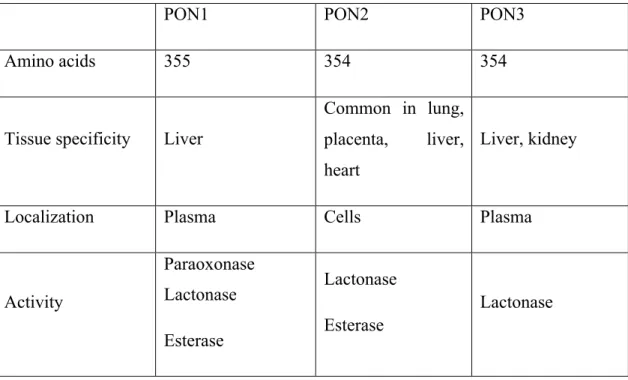
Located side by side on the long arm of chromosome 7 PON1, PON2 and PON3 constitutes paraoxonase family in human (1). Adjacent status and important
structural homology between these three genes indicates that they take origin through duplication from a common genetic precursor; these genes approximately show similarity in 65% amino acid level and in 70% the nucleotide level. PON2 is the oldest member of the family evolutionarily and although having limited substrate spectrum only shows lactonase activity (2, 3). PON3 is expressed both in the liver and kidneys, but the expression of PON1 only can be in the liver (4,5). These two enzymes are released into the bloodstream, where they merged with HDL. Extra codon (lysine) at position 106 of PON1 distinguishes this enzyme from the other two PON(4,5). Unlike the other two enzymes PON2 cannot be found in the blood, but can widely be expressed in various tissues which includes heart, kidney, liver, lung, placenta, small intestine, spleen, stomach and testes(6). In all three enzymes there are lactonase activity (Table 2.1). Even though in these days most attention is directed to figure out PON2 and PON3, PON1 is still the most researched and understood paraoxonase.
4
Table 2.1: The basic features of paraoxonase family (4-6)
PON1 PON2 PON3
Amino acids 355 354 354
Tissue specificity Liver
Common in lung, placenta, liver, heart
Liver, kidney
Localization Plasma Cells Plasma
Activity Paraoxonase Lactonase Esterase Lactonase Esterase Lactonase
2.1.2. The Discovery of Paraoxonase 1
PON1 has been identified in the early 1950s after Abraham Mazur's note about an animal enzyme capable of hydrolyzing organophosphates. Due to this enzyme’s capability of hydrolysing paroxon, a toxic metabolite of pesticide parathion, this enzyme was named as paraoxonase (7, 8). At the same time PON1 hydrolyzes some other organophosphate compounds (for example, chlorpyrifos and diazoxon) also nerve gas like soman, sarin because of that in mammals this, plays a protective role in chronic exposure to toxic compounds (9). Enzyme, due to these effects attracted the attention of toxicologists for a long time. This situation have changed after the reports in 1991. These reports states that this enzyme can prevent the accumulation of lipid peroxides in LDL and thus it might have potential antiatherogenic properties (10). However, the exact physiological role of the enzyme remains unclear.
5 2.1.3. Structure of Paraoxonase 1
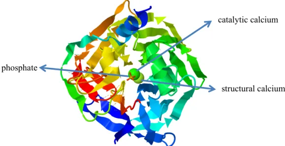
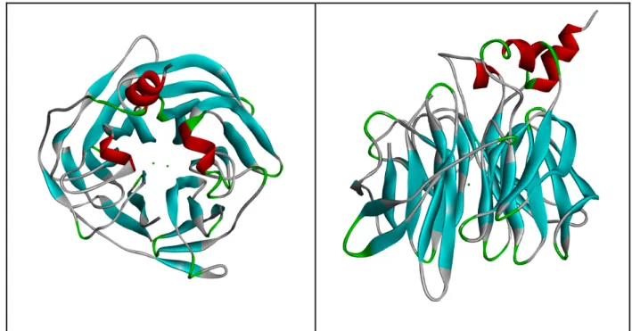
From this protein which is a gene product of PON1, weighs 43 kDa, contains 355 amino acids, at the stage of maturation and secretion terminal methionine is removed and the leader sequence that ensures HDL particles to bind, are protected (11). The first crystal structure of paraoxonase (PDB ID: 1V04) belongs to PON1, and it was obtained and analyzed by Harel and his friends (12). This crystal structure shows propeller folding that has two calcium ions in the center tunnel, 4 units of β- layer at each wing with 6 wings of β propeller (Figure 2.1).
Figure 2.1: Top view of the 6-blade propeller of the structure. At the center from top to bottom respectively phosphate ion catalytic calcium ion and structural calcium.
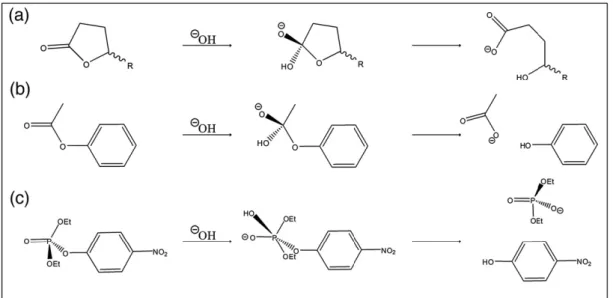
The structural Ca2+ is burried, whereas the catalytic Ca2+ resides at the bottom of the active site. In the hydrolysis of lactones and aryl esters (C-O bond cleavages via tetrahedral intermediates; figure 2. 2) His115-His134 dyad deprotonates a water molecule to produce attacking hydroxide and catalytic calcium stablizes the resulting
phosphate
structural calcium catalytic calcium
6
tetrahedral oxyanionic intermediate (13). Unlike the hydrolysis of the aryl esters and lactones enzyme shows promiscuous hydrolysis activity to phosphotriesters (P-O bond cleavage via a pentacovalent intermediate; figure 2.2). His115 mutations, reducing lactonase and aryl esterase activity, increases the rate of hydrolysis of phosphotriesters. This situation arises from the promiscuous activity of paraoxonase against the phosphotriesters (13,14).
Figure 2.2: General mechanisms of hydrolysis of three substrate types: lactones (a), esters (b) and phosphotriesters (c) (15)
First published crystal structure showed folding of PON1 and key features of the active region but this structure was obtained at pH 4.5, at this pH level enzyme is almost inactive (12). Not having ligand at this structure and also the absence of active region loop (72-79 residues covering the longest loop in the active region) made it impossible to have a more detailed description(15) . Ben-David and his friends was obtained two crystal structures at pH 6.5 which is close to the optimal pH in 2012. In one of these novel crystals, there is a lactone analogue
7
IDs of these novel crystals with a ligand and without a ligand are 3SRG and 3SRE respectively. As described earlier Ben-David and his friends also have used
recombinant rePON1-G2E6 that is in sequence ~ %90 similar with both rabbit and human PON1 and showing the same enzymatic specificity (16).
Ben-David and his friends mentioned the different conformations of the enzyme in their article. The structure (3SRG) with the ligand shows closed conformation. Apo structures (1V04 and 3SRE) shows open conformation (15). These different
conformations were shown previously with molecular dynamics studies (17) but Ben-David et al.’s experimental work is intrinsically more valuable.
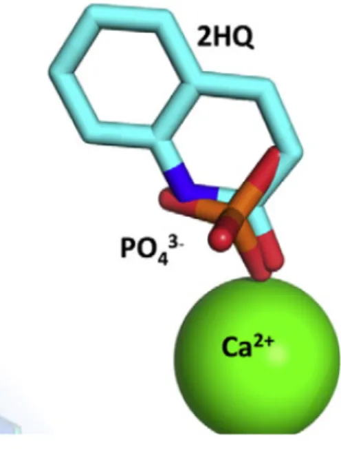
When 3SRG with ligand and 3SRE are superimposed NH and carbonyl oxygen of 2HQ coincides with the phosphate oxygens in the apo structure (figure 2.3). This overlap supports the idea that binding poses of both 2HQ and phosphate resembles the binding poses of substrates and/or reaction intermediates (15).
Figure 2.3: 2HQ in the structure with ligand and phosphate in the apo structure are superimposed (15).
8
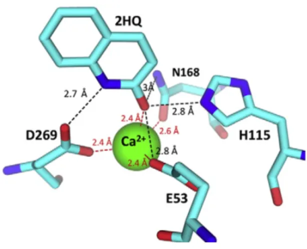
In addition to the interaction with catalytic calcium 2HQ interacts with the side chains of H115, D269, E53, and N168 (figure 2.4). Apart from the immobilization of 71-81 loop, no change in the backbone of the enzyme is observed as a result of 2HQ binding (15).
Figure 2.4: Interactions of 2HQ with the active site residues (15)
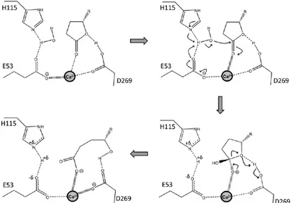
Proposed mechanism for lactone hydrolysis of PON1 (the same mechanism applies also for aryl esterase activity) demonstrated at figure 2.5 (15). In figure 2.5 , charge relay between the reaction intermediate, the calcium atom, and its ligating residues are shown with the wide and narrow broken lines. The deprotonation of water and the accompanying protonation of E53 weakens the interaction of E53 with calcium and thus the negatively charged oxyanion intermediate compound may be further stabilized. The reverse charge flow may apply to the leaving-group protonation and D269, although the evidence for the latter's role as general acid is indirect (15).
9
Figure 2.5: Proposed mechanism for the lactone hydrolysis of PON1.
2.1.4. PON1 Polymorphisms
PON1 gene coding region, comprises two polymorphic region positions;in position 55 leucine (L) and methionine transition (M) (55 L> M) and in position 192
glutamine (Q) and arginine transition (R) (192 Q> R). In addition to this polymorphism in the coding region, especially in the positions of 107/108,
significant changes in the promoter region have also been reported (18). Because of the connection of polymorphism in the PON1 promoter region, 55 L> M
polymorphism affects the enzyme concentration. 55 L> M polymorphism is within the N-terminal region of PON1 responsible for binding to HDL (19, 20). 192 Q> R polymorphism is responsible for the differences in the substrate preferences of the enzyme. In 192 Q / R polymorphism, lower PON1 enzyme activity observed among individuals who have the Q allele compared to those with the R allele. As for in 55 L / M polymorphism; MM homozygote individuals has lower PON1 enzyme activity compared to LL homozygotes against paraoxonase (18, 20, 21) .
Isoform 192 R hydrolyzes paraoxon whereas isoform 192 Q hydrolyes diazoxon, soman and sarin (22). Hydrolysis of paraoxon capacity in the blood (PON activity) is frequently used as a marker of PON1 enzyme activity. This enzyme activity reflects 192 Q> R polymorphism and the change in the concentration of PON1 enzyme.
10
Amino acid settlements at position of 192 (QR) causes to two alloenzymes.
Alloenzyme Q prevents the accumulation of lipid peroxides on LDL more, compared to R alloenzyme (23). Second exonic polymorphism of the PON1 gene, occurs in the 55 (L / M) position. This polymorphism affects PON1 activity less compared to 192 polymorphism. PON1 gene polymorphisms, by preventing the protective function of the enzyme increases the risk of coronary artery disease (24, 25).
2.1.5. Paraoxonase 1 and Coronary Artery Disease
Studies in recent years have showed that calcium-dependent paraoxonase which is found on HDL have roles in the metabolism of oxidized lipids and in the protection from atherosclerosis. Relationship between PON1 and atherosclerosis is connected to the antiatherogenic properties of HDLs. The PON1 hydrolyzing biologically active LDL, plays a strong role in preventing the fatty streak formation by significantly reducing the lipid peroxides formation. On HDL PON1 is associated with apo A-I with its N-terminal and may reduce the risk of vascular disease by breaking down proinflammatory molecules formed with LDL oxidation (19,26). For the unification of PON with HDL PON’s N-terminal hydrophobic signal peptide was found to be necessary. In case of deficiency of apolipoproteins the N-terminal hydrophobic peptide is connected directly to phospholipids and it is observed that PON in circulation is affiliated to phospholipids on HDL (27). Serum PON1 activity in patients with myocardial infarction, familial hypercholesterolemia, and in patients with diabetes, were lower than in healthy group (28).
Purified PON has a concentration-dependent inhibitory effect on the oxidation of HDL and it was observed that increased levels of PON in serum increased oxidation resistance of HDL. It was observed that PON inhibitors reduce serum
11
PON activity and increase of HDL oxidation. Effects of HDL-mediated PON or purified PON in the initiation, propagation and termination phases of LDL oxidation process were studied and HDL on LDL oxidation inhibitory effect was found to be from metal ion chelation or peroxidase-like activity (29). In the studies of knockout and transgenic mice the powerful role of PON1 in vascular diseases have been proposed ; on mice who have low levels of serum PON1 predisposition to atherosclerosis and the rate of stenosis has been shown to increase (30).
In studies examining the relationship between PON1 Q192R polymorphism and coronary heart disease the conflicting results were obtained. In some studies PON1 192 RR genotypes present in a higher incidence of coronary artery disease has been shown , PON1 Q192R polymorphism is thought to be a risk factor in atherosclerosis (31, 32). Even though some studies were performed in Turkish society and
supported this idea (33), such a relationship could not be found in some studies (34, 35).
2.1.6. Paraoxonase 1; Obesity, Metabolic Syndrome and Type 2 Diabetes Mellitus
Hyperglycaemia causes oxidative stress and atherosclerosis. It is clear that paraoxonase has an important role in diabetic patients. In diabetic patients, hyperglycemia, hyperinsulinemia, elevated free fatty acids and dyslipidemia may stem from increased reactive oxygen species. Diabetic patients with diabetic retinopathy and/or hypertension show reduced serum PON1 activity. These complications in diabetic patients may be due to increased predisposition to lipid
12
peroxidation (36). Environmental and genetic factors affecting serum PON1 levels affect the HDL’s protective role in LDL oxidadion (atherosclerosis) (37).
PON1-192RR and PON1-55LL genotypes are more frequently observed than other PON1 genotypes in type 2 diabetes cases. A variety of studies reported that in dieases such as DM, hypercholesterolemia and kidney failure which are associated with coronary heart disease, low serum PON1 activity is independent of the genotype present (38, 39). Asssociation of PON1 with HDL is impaired in diabetic patients and in various studies PON1 activity have been found to be reduced (40).
Low PON1 enzyme activity is attributed to glycation of the enzyme rather than decreased synthesis (41) and it is concluded that there exist reduced PON1 activity in diabetic patients with complications such as neuropathy, nephropathy and
retinopathy (42).
In metabolic syndrome, a risk factor for cardiovascular diseases, oxidative stress causes insulin resistance and it is proposed that this situation lowers the PON1 activity (43). Senti et al. showed decreased activity of PON1 in metabolic syndrome (44). In another study PON LL genotype was shown to be related with the severity of insulin resistance (45).
Obesity is an independent risk factor for cardiovascular disease and diabetes. Ferreti et al. found lower HDL-PON activity in obese patients than in control group in the study investiganting lipoproteins, oxidative stress and HDL-PON activity in healthy and obese individuals and they suggested that changes in the content of HDL reduce the enzyme activity by affecting the binding of the enzyme to the surface of the HDL particle (46 ). 5% – 10% of women of reproductive age is affected by the polycystic ovary syndrome (PCOS), being the most common reproductive endocrinopathy and
13
associated with the increased risk for the development diabetes, hypertension and atherosclerotic heart disease (48). There are studies suggesting that decreased PON1 activity contributes carbohydrate metabolism impairment, insulin resistance and atherosclerotic heart disease in women with PCOS (49, 50). Fortunato et al. reported that paraoxonase gene polymorphism in middle-aged women with PCOS is an indepedendent risk factor increasing carotid intima-media thickness (51). Fenkci et al. found that total antioxidant levels in PCOS patients is significantly lower than that of control group (52). Again in patients with PCOS it is suggested that increased oxidative condition contributes to the increase in cardiovascular risk. HDL antioxidant effect is attributed to HDL-associated enzymes up to a point and particularly PON1 and platelet activating factor acetyl hydrolase (PAF-AH) enzymes are emphasized (53 , 54).
2.1.7. Paraoxonase 1 and Liver Diseases
Chronic HBV infection is an important disease with high morbidity and mortality due to being a risk factor for liver failure, liver cirrhosis, and liver cancer (55). Süleyman et al. found lower PON1 activity in patients with chronic hepatitis than in healhty control group and they attributed this condition in chronic hepatitis patients to either decreased PON expression in damaged liver cells or changes in HDL dynamics (56). NASH, not associated with excessive alcohol intake, is a form of chronic hepatitis and shows the histological properties of alcoholic liver disease (57). NASH pathogenesis is not yet fully clear, but the two mechanisms are pointed out: First, increased oxidative stress and lipid peroxidation are related with increased fat accumulation in liver, second the tumor necrosis factor-mediated damage (58).
14
Bafikol et al. reported low levels of PON1 in NASH patients and they suggested that inflammation may be responsible for this condition and coversion of HDL from antiinflammatory/antioxodant complex to proinflammatory/prooxidant complex (59). In chronic liver disease, changes in the level of and in the structure of HDL is related with hepatic LCAT activity (60). In a study involving 49 patients with
hepatosteatosis and 25 healthy people, it was found that PON1 activity and NO levels were significantly lower in the patient group than in the control group and MDA levels were found higher in the patients. It was concluded that in patients with hepatosteatosis oxidative stress represses PON1 significantly (61). In patients with cirrhosis PON1 activity is much lower than in patients with other hepatitis forms and albumin and bilirubin levels correlates with the PON1 activity (62). PON1 activity was found to be related with the severity of alcoholic liver disease(63).
2.1.8. Paraoxonase1 and Neurological Diseases
Genetic and environmental factors play roles in the complex etiopathogenesis of neurological diseases. Alzheimer's disease is neurodegenerative disorder and is the most common cause of dementia over 65 years. Oxidative stress is an important mechanism in the development of degenerative diseases such as atherosclerosis, diabetes and Alzheimer's disease (64).
In Alzheimer's patients PON1 activity is significantly lower and patients carrying R allele shows more serum PON1 activity than those carrying Q allele. Dantoine et al. suggested PON1 192 polymorphism as a reliable marker to distinguish Alzheimer's patients from vascular dementia and healthy people (65).
15
Epidemiological studies suggested a link between the pesticide exposure and Parkinson's disease (66). Environmental neurotoxins that can be metabolized by PON1 may be held responsible for the age related neurodegeneration in sporadic idiopathic cases. The B allele was suggested as a genetic predisposition to Parkinson's disease (67). Human PON1 catalyzes the hydrolysis of
organophosphates, aromatic carboxylic acid esters and carbamates. Differences in PON1 activity in individuals with a risk of exposure increase the risk of
organophosphate intoxication by affecting organophosphate metabolism (18). Recent studies showed that in patients with schizophrenia and stroke PON1 activity is lower than in healthy subjects (68, 69). Investigations probing the PON1 activity in mixed connective tissue disease, Behçet’s disease, SLE, rheumatoid arthritis,
osteoarthritis and fibromyalgia revealed reduced PON1 activities (70).
2.1.9. Paraoxonase 1 and Cancer
Oxidative stress is one of the major etiological factors in carcinogenesis. Since PON1 is a member of endogenous free radical scavenging system, its roles in the etiology and prevention of cancers are of interest. Elkıran et al. showed in a study that the serum PON1 activity in lung cancer patients was found to be significantly lower than in healthy people (71). In a similar case-control study by Lee et al showed that in the 177 patients carrying PON1 gene Q/Q genotype risk of developing lung cancer significantly increased (72). Patients having gastric or pancreatic cancer showed lower paraoxonase levels than healthy control group (73.74).
16
SNP L55M is responsible for an amino acid change in exon 3 of PON1, and this SNP reduces the PON activity by decreasing the amount of the enzyme (75). PON1
activity was found to be reduced in gastroesophageal cancer, increased inflammation and cancer related anemia. Drops in PON1 activity were associated with lymph node metastasis(76). The study investigating the distribution of PON 192 polymorphism among 42 high-grade glioma and 42 menengioma patients and 50 healthy controls showed significantly lower serum PON1 activities in both tumor groups than in control group. Relationships of antioxidants with serum PON1 in the development of brain tumor are being investigated (77). During successive ovulations oxidative stress can damage DNA in the ovarian epithelial cells and this DNA damage increases the potential for malignant transformation(78). It was suggested that there exists significant links between PON1 SNPs and ovarian cancer risk. Serum PON and arylesterase activities were measured in epithelial ovarian cancer patients and both of them were found to be lower in the patient group than in the control. There was a inverse correlation between the PON activity and CA-125 level and disease stage(79).
Stevens et al. reported that in women with MM genotype there exist 57% increased breast cancer and 85% increased invasive breast cancer incidence for L55M
polymorphim (80).
2.2. Metabolomics 2.2.1. Description
The Human Genome Project which was completed in 2003 revealed that in human body there are 30,000-40,000 genes. 99.9% of the human genome is same in all
17
humans. This 0.1% difference will be instrumental in understanding the differences in responses to drugs, disease severities and disease predispositions among humans. The nucleotide sequence of the human genome alone is not very helpul in
understanding human nature. To investigate the function of these genes proteomics and transcriptomics studies were done. However, the data gathered from the
aforementioned researches is not enough to explain clinical phenotypes. Because the information that determine the clinical phenotype is hidden in metabolites occured within the cell (81). During more than 100 years, biochemical studies were carried to uncover enzymes, substrates, products, intermediates, kinetic features of enzymes and the factors affecting the cell metabolism. Today most of the metabolic pathways, used to understand disease and normal states, are known. This accumulated
biochemical data is used to increase the human life quality and longevity and today with new technologies and data evaluation methods this biochemical data is brought to new dimensions (82).
Metabol means “change” in Greek and “ome” means set. All metabolism in a cell or a living system is metabolome and metabolome study is called metabolomics. A more detailed description of metabolimics is the detection, quantition, and identification of small molecules within the metabolome with high throughput technologies. These small molecules, having molecular weight below 1500 Da, are metabolites such as peptides, oligonucleotides, sugars, nucleosides, organic acids, ketones, aldehydes, amines, amino acids, lipids, steroids, alkaloids and drugs, and human-bacterium products. Body fluids such as serum, urine, cerebrospinal fluid, plasma and saliva may be used for metabolomics studies. These analyses are used in field of clinical chemistry, pharmacology, preclinical drug research, toxicology, transplant monitoring, cancer metabolism and newborn screening.
18
Fundamentally in metabolomics studies two technologies are used and these are NMR and different mass spectrometries. NMR can identify all metabolites in a sample and measure the concentrations of the metabolites very quickly. Mass spectrometry complements the role of the NMR by uncovering the profile of thousands of metabolites more sensitively, reevaluating and calculating. Metabolite profiles are stored in computers and evaluated with software (82).
2.2.2. Human Metabolome Database
The Human Metabolome Database (HMDB) is a freely available electronic database containing detailed information about small molecule metabolites found in the human body. It is intended to be used for applications in metabolomics, clinical chemistry, biomarker discovery and general education. The database is designed to contain or link three kinds of data: 1) chemical data, 2) clinical data, and 3)
molecular biology/biochemistry data. The database (version 3.0) contains 40,260 metabolite entries including both water-soluble and lipid soluble metabolites as well as metabolites that would be regarded as either abundant (> 1 uM) or relatively rare (< 1 nM) (93 - 95).
2.3. Docking 2.3.1. Description
Docking, a molecular modeling method, predicts the proper orientation of a molecule to the another molecule when bound to each other to make a stable comlex (96). Binding affinity or the strength of association between bound molecules may be
19
estimated from knowledge of the preferred orientation using for example scoring functions.
The binding orientation of drug candidates to their protein receptors is frequently predicted by docking in order to estimate the affinity and activity of the drug candidate. Hence docking is heavily used in the rational drug design (97). A lot of effort has been directed towards improving the docking methods.
Figure 2.6: A small molecule “docks” to the protein target (Wikipedia). 2.3.2. Description of the Docking Problem
Molecular docking can be seen as a problem of lock-and-key. Here, the protein can be seen as the lock and the ligand can be seen as the key. Defining molecular docking as a optimization problem may be appropriate. This would describe the
20
“best-fit” orientation of a ligand binding to the protein of interest. However, a hand-in-glove analogy is more appropriate than lock-and-key due to the flexibility of both the ligand and the protein (98). During the binding process, changes in the
conformations of both the protein and the ligand occur to get an overall best-fit and this kind of conformational adjustments culminating in the overall binding is referred to as indiuced-fit (99).
In the molecular docking, the molecular recognition process is computationally simulated. Docking tries to find the lowest possible free energy of the overall system by achieving an optimized conformation for both the protein and ligand and relative orientation between protein and ligand.
2.3.3. Docking Methods
There are especially two approaches popular within the docking community. One of them utilizes a matching method that describes the protein and the ligand as
complementary surfaces (100 – 102). The second approach simulates the actual binding process by calculating ligand-pairwise interaction energies (103). Both approaches have significant advantages as well as some limitations. These are mentioned below.
Geometric matching/shape complementarity techniques define the receptor and small molecule as a set of characteristics that make them dockable (104). These
characteristics may include complementary or molecular surface descriptors. In this case, the molecular surface of the receptor is defined in terms of its solvent
accessible surface area and the molecular surface of the ligand is defined in terms of its matching surface description. The complementarity between the two surfaces
21
purports the shape matching description that may help finding the complementary pose of docking the receptor and the ligand molecules. Another approach is to describe the hydrophobic properties of the receptor using turns in the main chain atoms. Using Fourier shape descriptor technique is an yet another approach (105-107). Whereas the shape complementarity techniques are typically robust and fast, they cannot model the dynamic changes occurring in the ligands and proteins accurately, although recent improvements allow these methods to take account the ligand flexibility. Shape complementarity are very fast and can quickly scan through several thousand ligands in seconds and actually figure out whether they can bind to the active site of the protein, and usually scalable to even protein-protein
interactions. These methods are more suitable to pharmacophore based approaches, since they use geometric descriptions.
The simulation of the binding process as such is a much more complicated process. The receptor and the ligand are separated by some physical distance, and the ligand is steered into the protein’s active site after a certain number of moves in its
conformational space. The moves incorporate internal changes to the ligand’s
structure including torsion angle rotations as well as rigid body transformations such as rotations, translations. Each of these moves in the conformation space of the ligand changes the total energy of the system. The apparent advantage of the system is suitability to incorporate ligand flexibility whereas in shape complementarity techniques inclusion of the ligand flexibility is harder due to their nature. Another advantage is that the process is physically closer to what happens in reality. Since this technique has to explore a rather large energy landscape, this technique takes longer time to evaluate the optimal pose of binding. However grid-based techniques as well as fast optimization methods have significantly fixed these problems.
22 2.3.4. Virtual Screening
Virtual screening (VS) is a computational technique used to screen small molecule libraries in order to identify hits which are most likely to bind to a drug target,
typically protein receptor or ezyme (108, 109). Virtual screening has been defined as the ”automatically evaluating very large libraries of compound” using computer programs (110). There are two broad categories of virtual screening techniques. Ligand-based screening and structure-based screening are those two categories (111).
23
3. Chapter 3: Materials and Methods
In this study, metabolites of human metabolome are docked against paraoxonase 1 protein structure by using structure based virtual screening strategy. This study consists of several steps, and these steps are:
• preparation of PON1 structure for docking
• preparation of ligands and metabolites for docking
• adjusting the docking parameters and the generation of files required for virtual screening
• after the screening process evaluation of results. 3.1. Preparation of PON1 Structure for Docking
The enzyme structure with PDB ID 3SRE (15) was downloaded from Protein Data Bank (114). Water molecules and pollutants were removed from the protein structure by using the Accelrys Discovery Studio 3.1. Using the same program hydrogens were added to the structure and “clean geometry” was done on the structure. In the resulting structure, there remained polypeptide chain, 2 calcium ions and and a phosphate ion. This structure was kept for comparison and from this structure phosphate ion was removed to get the structure for docking, and again removing the phosphate ions from the lattice structure of the protein to be used in docking were obtained. As a result, the structure that was used in docking was containing the polypeptide chain and 2 calcium ions. Figure 3.1 shows the top and side views of the protein structure used in docking.
24
Figure 3.1: Prepared PON1 for docking. Top view on the left, side view on the right.
3.2. Preparation of Human Metabolome for Docking
A single SDF file which contains all of the metabolites was downloaded from
Human Metabolome Database (HMDB) (93-95). The metabolites in this single SDF were “washed” to remove ions and subjected to energy minimization with MMFF94x force field by using Molecular Operating Environment (MOE) 2009.10 software. Open Babel (115) was used to cleave this single SDF into single metabolite files totalling 40,209 files. Again using Open Babel each generated SDF file was converted to a respective PDB file. AutoGrid produces grids for docking and supports 15 atom types described in its default atomic parameter file. Supported atoms are: H, C, N, O, F, Mg, P, S, Cl, Ca, Mn, Fe, Zn, Br, and I. To filter out PDB files containing the unsupported atom type(s), a Python script (Appendix A:
atomTypeChecker.py) coded by me was used. 147 of the PDB files were excluded from the study due to the existence of unsupported atom type(s).
25
3.3. Preparation of Known Ligands of PON1 for Docking
Known substrates and inhibitors of PON1 compiled from various articles (15, 17, 117, 118, 119) were docked along with metabolites and their docking scores were used as references. These molecules were either downloaded from PubChem (120) or manually drawn using Accelrys Draw 4.1. These molecules are listed below.
N-acyl homoserine lactone (AHL) molecules used by various bacteria in “quorum sensing” and hydrolyzed by PON: 3-hydroxy-7-cis-C14-HSL, 3-hydroxy-C4-HSL, 3-oxo-C10-HSL, 3-oxo-C12 -HSL, 3-oxo-C6-HSL, 7-cis-C14-HSL, C4-HSL, and C6-HSL
Substrates: 2-oxo-clopidogrel, gamma-butyrolactone, (+/-)-4-HDoHE, 5-Hete, coumarin delta-undecalactone, delta-valerolactone, gamma-undecalactone, homocysteine thiolactone, homogentisic lactone, paraoxon, phenyl acetate, 4-thiobutyl-gamma-butyrolactone (TBBL).
Inhibitors: 2-hydroxyquinoline (2HK), ethylmaleimide.
3.4. Docking Parameters
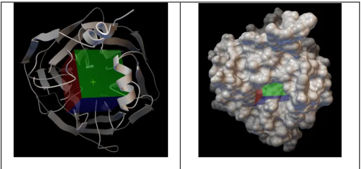
The protein’s active site was represented with a 0.375 Å spaced 40 X 40 X 40 grid. The center of mass coordinates of the phosphate was used to adjust the center of the grid. Figure 3.2 shows the grid. Lamarckian Genetic Algorithm (LGA) and the maximum number of energy evaluation of 2,000,000 were seleceted for docking. 100 runs for known ligands and 10 runs for metabolites were chosen. The charge of calciums was set to +2. The software used for docking was AutoDock 4.2 (116).
26
Figure 3.2: The top view of the protein with the cubic grid. On the left ribbon representation of the protein, on the right molecular surface representation
3.5. Generation of the Necessary Files for Virtual Screening
YaVST (Yet Another Virtual Screening Tool) is a program developed by Serkan Altuntaş. Basically this program produces the files necessary for the operation of AutoDock and AutoGrid and makes batch docking of ligands possible. In the in silico experiments metabolites were divided into groups containg 3000 metabolites for batch docking.
3.6. Presentation and Compilation of Virtual Screening Results
DLG files produced during docking process contains both energies and docking poses of molecules when bound to their receptors. A Python script (Appendix A: autodock-results.py) by Serkan Altuntaş extracts the best docking energy of the molecule in the DLG and forms a list of energies from all DLGs within the same directory. The lists from different directories were combined using "cat" Unix
27
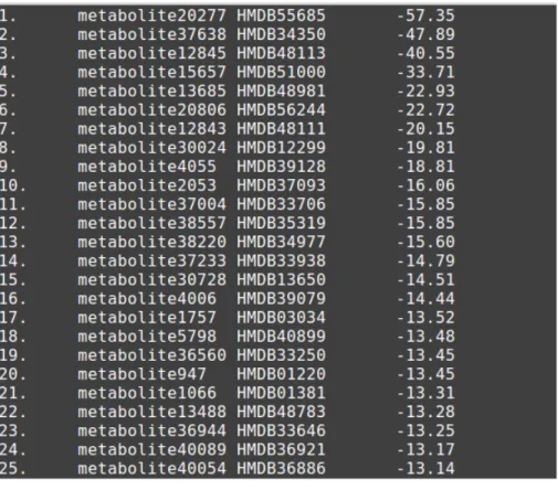
command and sorted using "sort" Unix command according to the second column numerically. IDextractor.py (Appendix A) by me formed a tab separated list (Figure 3.3) containing energy values as well as HMDB IDs. This list with HMDB IDs is in plain ASCII and can be opened with Excel for data analysis. This list was used to create a HTML table (figure 3.3) that eases the visual inspection of the metabolites by using HTMLGen.py script by me. After the visual inspection the best docking poses of selected metabolites were extracted using extract_first_structure.py script (Appendix A) by Serkan Altuntaş for 3D analysis together with the PON1 structure using AutoDockTools and/or Accelrys Discovery Studio 3.5 Visualizer.
Figure 3.3: From left to right columns, respectively: order, file name, HMDB ID, docking energy in kcal/mol
28 Figure 3.4: HTML table
29
4. Chapter 4: Results
Table 4.1: Docking energies of known ligands
Compound Docking Energy (kcal/mol)
Paraoxon -10.06 (+/-)-4-HDoHE -9.24 5-Hete -9.07 7-cis-C14-HSL -7.68 3-oxo-C10-HSL -7.56 3-oxo-C12-HSL -7.37 3-hydroxy-7-cis-C14-HSL -7.23 Gamma-undecalactone -6.62 Delta-undecalactone -6.59 3-oxo-C6-HSL -6.51 C6-HSL -6.44 2-oxo-clopidogrel -6.41 Coumarin -6.06 C4-HSL -6.00 TBBL -5.93 3-hydroxy-C4-HSL -5.86 2-hydroxyquinoline -5.84 Homogentisic lactone -5.68 Phenyl acetate -5.11 Delta-valerolactone -4.76 Gamma-butyrolactone -4.39 Homocysteine thiolactone -4.26 Ethylmaleimide -4.14
Substrates or inhibitors used in the research are listed in table 4.1. Energies are sorted in ascending order. Paraoxon had the best docking energy value, but its docking pose was incorrect. Paraoxon was not in a position suitable for the reaction to take place (figure 4.1). In the same manner, binding poses of 3-oxo-C10-HSL and C12-HSL were unfavorable. Except these three mentioned molecules (paraoxon, 3-oxo-C10-HSL and 3-oxo-C12-HSL) all of the ligands had correct poses. What I mean by the “correct pose” is the positioning of the chemical groups, that may be acted by
30
PON1, above the catalytic calcium in a suitable distance. An excellent example of the “correct pose” can be seen in Figure 2.3 in page 7. 2HQ in the structure (PDB ID: 3SRG) with ligand and phosphate in the apo structure (PDB ID: 3SRE) are
superimposed for comparison (15). The docking pose of 2-hydroxyquinoline having the correct pose is shown in figure 4.2.
Figure 4.1: Paraoxon and phosphate (yellow) are superimposed. Nitro group of paraoxon is above the catalytic calcium.
31
Figure 4.2: 2-hydroxyquinoline (2HQ) superimposed with phosphate (yellow) has the “correct pose”
The best 2000 metabolites in terms of docking energy were analyzed. The metabolites having the chemical groups suitable for the action of PON1 were selected. It was found that 10 out of these selected metabolites had the “correct pose”. Table 4.2 shows these 10 metabolites.
Table 4.2: Selected metabolites having the “correct pose”
Order HMDB ID D. E. (kcal/mol) Compound Name
235. HMDB39607 -9.90 Sorgolactone 395. HMDB00682 -9.52 Indoxyl sulfate 475. HMDB38950 -9.39 5-methoxyhinokinin 584. HMDB06101 -9.25 Enterolactone 589. HMDB30087 -9.25 (-)-Arctigenin 885. HMDB39067 -8.94 Epoxybergamottin 935. HMDB39767 -8.91 Pandamarilactone 32 1351. HMDB35698 -8.57 (-)-Matairesinol 1575. HMDB41372 -8.41 Alectrol 1863. HMDB35934 -8.23 Isoalantolactone
32
5. Chapter 5: Discussion
First of all, docking the human metabolome against an enzyme, even if it does not include anything new technically, as an approach it is new. There are two things which make this approach valuable and meaningful. The first one of these: although there were taken long ways in the understanding of the physiological role of
paraoxonase 1, the physiological role of this enzyme and substrates of it are still not fully understood (121). The second one: the PON1 enzyme shows lactonase, aryl esterase and phosphotriesterase activity and it shows effect against more than one substrate type. The discovery of PON1 substrates or inhibitors will be useful in understanding the cardiovascular protective and antioxidant effects much better. It is way more common to come across enzyme specificity rather than enzyme promiscuity. Promiscuous enzymes such as PON1 are very few. In nature there exist three lactonase enzyme families each of which belongs to a different superfamily. Surprisingly, key active site architectures of members of these three lactonase family converged as a result of evolutionary processes. Interestingly, in all three enzyme families “promiscuous” organophosphate hydrolase activity was observed (122). There are two cases in which it is known that bacterial metallo-lactonases gained paraoxonase activity. After the introduction of parathion, these bacterial metallo-lactonases evolved to become very efficient paraoxonases in a few decades (123-125). The active site of PON1 is very evolvable and by protein engineering more efficient PON1s can be created. Engineered PON1 can be used against various toxins during chemical warfare.
33
The grid size was taken as 70 X 70 X 70 in initial experiments. It was observed that the best 1000 metabolites in terms of docking energy were almost all triglyceries bound to the exterior of the PON1 structure (figure 5.1). In expreriments involving a very heterogenous chemical space, the protein’s active site should be represented in smallest volume possible. During the experiments no physical/chemical filtering (except due to technical difficulties) was performed, but it is reasonable to do filtering according to some criteria. Filtering may reduce the labor required for the anlysis of the results.
In this study, the protein structure with PDB ID 3SRE was used. This selection was not gratuitous. There were 3 PON1 crystal structure at the time the study began. The structure 3SRE having open conformation was obtained at pH 6.5. Using this open conformation larger molecules can be docked.
Figure 5.1: The binding poses of 50., 100., 150., 200., and 250. metabolites in experiments with 70 X 70 X 70 grid sizes
34
The best 2000 metabolites in terms of docking energy were analyzed. The metabolites having the chemical groups suitable for the action of PON1 were selected. It was found that 10 out of these selected metabolites had the “correct pose”. Table 4.2 shows these 10 metabolites. Except indoxyl sulfate all of these compounds are of plant origin. Being 90% of the metabolites of plant origin is very interesting. High vegetable consumption considered as a good habit reduces the risk of coronary heart disease, but shows a negative correlation with the PON1 activity (129,130). With these data at hand, these 9 metabolites of plant origin may be considered as PON1 inhibitors due to their low docking energies. Laboratory assays should be carried to reveal the possible inhibitory potential of these metabolites on PON1.
Indoxyl sulfate is a dietary protein metabolite, and also the metabolite of the common amino acid tryptophan. Indoxyl sulfate is a circulating uremic toxin
stimulating glomerular sclerosis and interstitial fibrosis. Indoxyl sulfate is one of the well known substances of a group of protein-bound uremic retention solutes. Indoxyl sulfate increases the rate of progression of renal failure. In plasma, indoxyl sulfate is a protein-bound uremic solute that induces endothelial dysfunction by inhibiting endothelial proliferation and migration in vitro. Some studies suggest that indoxyl sulfate is also involved in oxidative stress. In hemodialyzed patients, serum levels of indoxyl sulfate are associated with levels of pentosidine, a marker of carbonyl and oxidative stress; in vitro, indoxyl sulfate increases reactive oxygen species (ROS) production in tubular cells, and increases NAD(P)H oxidase activity in endothelial cells. Indoxyl sulfate impairs osteoblast function and induces abnormalities of bone turnover. Indoxyl sulfate strongly decreases the levels of glutathione, one of the most active antioxidant systems of the cell (131-134). Indoxyl sulfate has a low docking
35
energy. This situation may indicate the possible inhibitory effect on PON1.
Laboratory studies should be done to understand the role of this metabolite on PON1.
Figure 5.2: Indoxyl sulfate and phosphate are superimposed. Phosphate is shown in yellow.
36
Curriculum Vitae
Talha Karabıyık was born in 15 April 1982 in Bandırma, Balikesir. His B.S. degree in biology was awarded in 2004 from Fatih University, İstanbul. He started his medical residency in clinical biochemistry in 2009 in Şişli Hamidiye Etfal Training and Research Hospital, İstanbul. After the completion of his medical residency, he was appointed to Haseki Training and Research Hospital as a clinical (bio)chemistry specialist in 2013. His main research interests are structural bioinformatics and computational biology.
37
References
1. Primo-parmo SL, Sorenson RC, Teiber J, La du BN. The human serum paraoxonase/arylesterase gene (PON1) is one member of a multigene family. Genomics. 1996;33(3):498-507.
2. Draganov DI, La du BN. Pharmacogenetics of paraoxonases: a brief review. Naunyn Schmiedebergs Arch Pharmacol. 2004;369(1):78-88.
3. La du BN, Aviram M, Billecke S, et al. On the physiological role(s) of the paraoxonases. Chem Biol Interact. 1999;119-120:379-88.
4. Mochizuki H, Scherer SW, Xi T, et al. Human PON2 gene at 7q21.3: cloning, multiple mRNA forms, and missense polymorphisms in the coding sequence. Gene. 1998;213(1-2):149-57.
5. La du BN, Aviram M, Billecke S, et al. On the physiological role(s) of the paraoxonases. Chem Biol Interact. 1999;119-120:379-88.
6. Ng CJ, Shih DM, Hama SY, Villa N, Navab M, Reddy ST. The paraoxonase gene family and atherosclerosis. Free Radic Biol Med. 2005;38(2):153-63. 7. Mazur A. An enzyme in animal tissues capable of hydrolysing the
phosphorus-fluorine bond of alkyl fluorophosphates. J Biol Chem. 1946;164:271-89.
8. Aldridge WN. Serum esterases. II. An enzyme hydrolysing diethyl p-nitrophenyl phosphate (E600) and its identity with the A-esterase of mammalian sera. Biochem J. 1953;53(1):117-24.
9. Costa LG, Cole TB, Vitalone A, Furlong CE. Measurement of paraoxonase (PON1) status as a potential biomarker of susceptibility to organophosphate toxicity. Clin Chim Acta. 2005;352(1-2):37-47.
38
10. Mackness MI, Arrol S, Durrington PN. Paraoxonase prevents accumulation of lipoperoxides in low-density lipoprotein. FEBS Lett. 1991;286(1-2):152-4. 11. Sorenson RC, Bisgaier CL, Aviram M, Hsu C, Billecke S, La du BN. Human
serum Paraoxonase/Arylesterase's retained hydrophobic N-terminal leader sequence associates with HDLs by binding phospholipids : apolipoprotein A-I stabilizes activity. Arterioscler Thromb Vasc Biol. 1999;19(9):2214-25. 12. Harel M, Aharoni A, Gaidukov L, et al. Structure and evolution of the serum
paraoxonase family of detoxifying and anti-atherosclerotic enzymes. Nat Struct Mol Biol. 2004;11(5):412-9.
13. Khersonsky O, Tawfik DS. The histidine 115-histidine 134 dyad mediates the lactonase activity of mammalian serum paraoxonases. J Biol Chem. 2006;281(11):7649-56.
14. Yeung DT, Lenz DE, Cerasoli DM. Analysis of active-site amino-acid residues of human serum paraoxonase using competitive substrates. FEBS J. 2005;272(9):2225-30.
15. Ben-david M, Elias M, Filippi JJ, et al. Catalytic versatility and backups in enzyme active sites: the case of serum paraoxonase 1. J Mol Biol. 2012;418(3-4):181-96.
16. Aharoni A, Gaidukov L, Yagur S, Toker L, Silman I, Tawfik DS. Directed evolution of mammalian paraoxonases PON1 and PON3 for bacterial expression and catalytic specialization. Proc Natl Acad Sci USA. 2004;101(2):482-7.
17. Hu X, Jiang X, Lenz DE, Cerasoli DM, Wallqvist A. In silico analyses of substrate interactions with human serum paraoxonase 1. Proteins. 2009;75(2):486-98.
39
18. Gupta N, Gill K, Singh S. Paraoxonases: structure, gene polymorphism & role in coronary artery disease. Indian J Med Res. 2009;130(4):361-8.
19. Humbert R, Adler DA, Disteche CM, Hassett C, Omiecinski CJ, Furlong CE. The molecular basis of the human serum paraoxonase activity polymorphism. Nat Genet. 1993;3(1):73-6.
20. Adkins S, Gan KN, Mody M, La du BN. Molecular basis for the polymorphic forms of human serum paraoxonase/arylesterase: glutamine or arginine at position 191, for the respective A or B allozymes. Am J Hum Genet. 1993;52(3):598-608.
21. Leviev I, Deakin S, James RW. Decreased stability of the M54 isoform of paraoxonase as a contributory factor to variations in human serum paraoxonase concentrations. J Lipid Res. 2001;42(4):528-35.
22. Davies HG, Richter RJ, Keifer M, Broomfield CA, Sowalla J, Furlong CE. The effect of the human serum paraoxonase polymorphism is reversed with diazoxon, soman and sarin. Nat Genet. 1996;14(3):334-6.
23. Mackness MI, Arrol S, Mackness B, Durrington PN. Alloenzymes of paraoxonase and effectiveness of high-density lipoproteins in protecting low-density lipoprotein against lipid peroxidation. Lancet. 1997;349(9055):851-2. 24. Garin MC, James RW, Dussoix P, et al. Paraoxonase polymorphism Met-Leu54 is associated with modified serum concentrations of the enzyme. A possible link between the paraoxonase gene and increased risk of cardiovascular disease in diabetes. J Clin Invest. 1997;99(1):62-6.
25. Mackness MI, Mackness B, Durrington PN, et al. Paraoxonase and coronary heart disease. Curr Opin Lipidol. 1998;9(4):319-24.
40
26. Tuncel P, Örmen M, Şişman AR. Paraoksonazın biyolojik varyasyonu ve HDL-Kolesterol ile ilişkisi. Türk Klinik Biyokimya Derg 2009;7(1):17-22. 27. Blatter MC, James RW, Messmer S, Barja F, Pometta D. Identification of a
distinct human high-density lipoprotein subspecies defined by a lipoprotein-associated protein, K-45. Identity of K-45 with paraoxonase. Eur J Biochem. 1993;211(3):871-9.
28. Mackness MI, Harty D, Bhatnagar D, et al. Serum paraoxonase activity in familial hypercholesterolaemia and insulin-dependent diabetes mellitus. Atherosclerosis. 1991;86(2-3):193-9.
29. Hegele RA, Brunt JH, Connelly PW. A polymorphism of the paraoxonase gene associated with variation in plasma lipoproteins in a genetic isolate. Arterioscler Thromb Vasc Biol. 1995;15(1):89-95.
30. Rozenberg O, Rosenblat M, Coleman R, Shih DM, Aviram M. Paraoxonase (PON1) deficiency is associated with increased macrophage oxidative stress: studies in PON1-knockout mice. Free Radic Biol Med. 2003;34(6):774-84. 31. Odawara M, Tachi Y, Yamashita K. Paraoxonase polymorphism
(Gln192-Arg) is associated with coronary heart disease in Japanese noninsulin-dependent diabetes mellitus. J Clin Endocrinol Metab. 1997;82(7):2257-60. 32. Imai Y, Morita H, Kurihara H, et al. Evidence for association between
paraoxonase gene polymorphisms and atherosclerotic diseases. Atherosclerosis. 2000;149(2):435-42.
33. Taşkiran P, Cam SF, Sekuri C, et al. [The relationship between paraoxanase gene Leu-Met (55) and Gln-Arg (192) polymorphisms and coronary artery disease]. Turk Kardiyol Dern Ars. 2009;37(7):473-8.
41
34. Ombres D, Pannitteri G, Montali A, et al. The gln-Arg192 polymorphism of human paraoxonase gene is not associated with coronary artery disease in italian patients. Arterioscler Thromb Vasc Biol. 1998;18(10):1611-6.
35. Ko YL, Ko YS, Wang SM, et al. The Gln-Arg 191 polymorphism of the human paraoxonase gene is not associated with the risk of coronary artery disease among Chinese in Taiwan. Atherosclerosis. 1998;141(2):259-64. 36. Maritim AC, Sanders RA, Watkins JB. Diabetes, oxidative stress, and
antioxidants: a review. J Biochem Mol Toxicol. 2003;17(1):24-38.
37. Deakin SP, James RW. Genetic and environmental factors modulating serum concentrations and activities of the antioxidant enzyme paraoxonase-1. Clin Sci. 2004;107(5):435-47.
38. Mackness M, Durrington P, Mackness B. Paraoxonase 1 activity, concentration and genotype in cardiovascular disease. Curr Opin Lipidol. 2004;15(4):399-404.
39. Mackness B, Mackness MI, Arrol S, et al. Serum paraoxonase (PON1) 55 and 192 polymorphism and paraoxonase activity and concentration in non-insulin dependent diabetes mellitus. Atherosclerosis. 1998;139(2):341-9. 40. Ikeda Y, Suehiro T, Inoue M, et al. Serum paraoxonase activity and its
relationship to diabetic complications in patients with non-insulin-dependent diabetes mellitus. Metab Clin Exp. 1998;47(5):598-602.
41. Hedrick CC, Thorpe SR, Fu MX, et al. Glycation impairs high-density lipoprotein function. Diabetologia. 2000;43(3):312-20.
42. Flekac M, Skrha J, Zídková K, Lacinová Z, Hilgertová J. Paraoxonase 1 gene polymorphisms and enzyme activities in diabetes mellitus. Physiol Res. 2008;57(5):717-26.
42
43. Paolisso G, Tagliamonte MR, Rizzo MR, Giugliano D. Advancing age and insulin resistance: new facts about an ancient history. Eur J Clin Invest. 1999;29(9):758-69.
44. Sentí M, Tomás M, Fitó M, et al. Antioxidant paraoxonase 1 activity in the metabolic syndrome. J Clin Endocrinol Metab. 2003;88(11):5422-6.
45. Barbieri M, Bonafè M, Marfella R, et al. LL-paraoxonase genotype is associated with a more severe degree of homeostasis model assessment IR in healthy subjects. J Clin Endocrinol Metab. 2002;87(1):222-5.
46. Ferretti G, Bacchetti T, Moroni C, et al. Paraoxonase activity in high-density lipoproteins: a comparison between healthy and obese females. J Clin Endocrinol Metab. 2005;90(3):1728-33.
47. Knochenhauer ES, Key TJ, Kahsar-miller M, Waggoner W, Boots LR, Azziz R. Prevalence of the polycystic ovary syndrome in unselected black and white women of the southeastern United States: a prospective study. J Clin Endocrinol Metab. 1998;83(9):3078-82.
48. ACOG Practice Bulletin No. 108: Polycystic ovary syndrome. Obstet Gynecol. 2009;114(4):936-49.
49. Dursun P, Demirtaş E, Bayrak A, Yarali H. Decreased serum paraoxonase 1 (PON1) activity: an additional risk factor for atherosclerotic heart disease in patients with PCOS?. Hum Reprod. 2006;21(1):104-8.
50. Lenarcik A, Bidzińska-speichert B, Tworowska-bardzińska U. The role of chronic inflammation and Leu55Met PON1 polymorphism in the pathogenesis of polycystic ovary syndrome. Gynecol Endocrinol. 2010;26(9):673-83.
43
51. Fortunato G, Rubba P, Panico S, et al. A paraoxonase gene polymorphism, PON 1 (55), as an independent risk factor for increased carotid intima-media thickness in middle-aged women. Atherosclerosis. 2003;167(1):141-8.
52. Fenkci V, Fenkci S, Yilmazer M, Serteser M. Decreased total antioxidant status and increased oxidative stress in women with polycystic ovary syndrome may contribute to the risk of cardiovascular disease. Fertil Steril. 2003;80(1):123-7.
53. Mackness MI, Durrington PN, Mackness B. The role of paraoxonase 1 activity in cardiovascular disease: potential for therapeutic intervention. Am J Cardiovasc Drugs. 2004;4(4):211-7.
54. Chait A, Han CY, Oram JF, Heinecke JW. Thematic review series: The immune system and atherogenesis. Lipoprotein-associated inflammatory proteins: markers or mediators of cardiovascular disease?. J Lipid Res. 2005;46(3):389-403.
55. Wang FS. Current status and prospects of studies on human genetic alleles associated with hepatitis B virus infection. World J Gastroenterol. 2003;9(4):641-4.
56. Kilic SS, Aydin S, Kilic N, Erman F, Aydin S, Celik I. Serum arylesterase and paraoxonase activity in patients with chronic hepatitis. World J Gastroenterol. 2005;11(46):7351-4.
57. Alba LM, Lindor K. Review article: Non-alcoholic fatty liver disease. Aliment Pharmacol Ther. 2003;17(8):977-86.
58. Angulo P. Nonalcoholic fatty liver disease. N Engl J Med. 2002;346(16):1221-31.
44
59. Başkol M, Başkol G, Deniz K, Ozbakir O, Yücesoy M. A new marker for lipid peroxidation: serum paraoxonase activity in non-alcoholic steatohepatitis. Turk J Gastroenterol. 2005;16(3):119-23.
60. Sabesin SM, Hawkins HL, Kuiken L, Ragland JB. Abnormal plasma lipoproteins and lecithin-cholesterol acyltransferase deficiency in alcoholic liver disease. Gastroenterology. 1977;72(3):510-8.
61. Atamer A, Bilici A, Yenice N, Selek S, Ilhan N, Atamer Y. The importance of paraoxonase 1 activity, nitric oxide and lipid peroxidation in hepatosteatosis. J Int Med Res. 36(4):771-6.
62. Ferré N, Camps J, Prats E, et al. Serum paraoxonase activity: a new additional test for the improved evaluation of chronic liver damage. Clin Chem. 2002;48(2):261-8.
63. Marsillach J, Ferré N, Vila MC, et al. Serum paraoxonase-1 in chronic alcoholics: relationship with liver disease. Clin Biochem. 2007;40(9-10):645-50.
64. Campion D, Dumanchin C, Hannequin D, et al. Early-onset autosomal dominant Alzheimer disease: prevalence, genetic heterogeneity, and mutation spectrum. Am J Hum Genet. 1999;65(3):664-70.
65. Dantoine TF, Drouet M, Debord J, Merle L, Cogne M, Charmes JP. Paraoxonase 1 192/55 gene polymorphisms in Alzheimer's disease. Ann N Y Acad Sci. 2002;977:239-44.
66. Le couteur DG, Mclean AJ, Taylor MC, Woodham BL, Board PG. Pesticides and Parkinson's disease. Biomed Pharmacother. 1999;53(3):122-30.
45
67. Clarimon J, Eerola J, Hellström O, Tienari PJ, Singleton A. Paraoxonase 1 (PON1) gene polymorphisms and Parkinson's disease in a Finnish population. Neurosci Lett. 2004;367(2):168-70.
68. Sarandol A, Kirli S, Akkaya C, Ocak N, Eroz E, Sarandol E. Coronary artery disease risk factors in patients with schizophrenia: effects of short term antipsychotic treatment. J Psychopharmacol (Oxford). 2007;21(8):857-63. 69. Kim NS, Kang K, Cha MH, et al. Decreased paraoxonase-1 activity is a risk
factor for ischemic stroke in Koreans. Biochem Biophys Res Commun. 2007;364(1):157-62.
70. Goswami B, Tayal D, Gupta N, Mallika V. Paraoxonase: a multifaceted biomolecule. Clin Chim Acta. 2009;410(1-2):1-12.
71. Elkiran ET, Mar N, Aygen B, Gursu F, Karaoglu A, Koca S. Serum paraoxonase and arylesterase activities in patients with lung cancer in a Turkish population. BMC Cancer. 2007;7:48.
72. Lee CH, Lee KY, Choe KH, et al. [Effects of oxidative DNA damage induced by polycyclic aromatic hydrocarbons and genetic polymorphism of the paraoxonase-1 (PON1) gene on lung cancer]. J Prev Med Public Health. 2005;38(3):345-50.
73. Akçay MN, Yilmaz I, Polat MF, Akçay G. Serum paraoxonase levels in gastric cancer. Hepatogastroenterology. 2003;50 Suppl 2:cclxxiii-cclxxv. 74. Akçay MN, Polat MF, Yilmaz I, Akçay G. Serum paraoxonase levels in
pancreatic cancer. Hepatogastroenterology. 2003;50 Suppl 2:ccxxv-ccxxvii. 75. Leviev I, Deakin S, James RW. Decreased stability of the M54 isoform of
paraoxonase as a contributory factor to variations in human serum paraoxonase concentrations. J Lipid Res. 2001;42(4):528-35.