R.T.
UNIVERSITY OF DICLE
INSTITUTE OF NATURAL AND APPLIED SCIENCES
SCREENING OF ANGUCYCLINE ANTIBIOTICS AS POTENTIAL
DRUG CANDIDATES AGAINST MRSA BY DOCKING ANALYSIS
Hazem Abbas Tofiq AL-BUSTANY
M.Sc. THESIS
DEPARTMENT OF BIOLOGY
DIYARBAKIR
January - 2016
T.C.
DİCLE ÜNİVERSİTESİ
FEN BİLİMLERİ ENSTİTÜSÜ
ANGUSİKLİN ANTİBİYOTİKLERİN DOKİNG ANALİZLERİ
YARDIMIYLA MRSA’YA KARŞI POTANSİYEL İLAÇ ADAYI
OLARAK TARANMASI
Hazem Abbas Tofiq AL-BUSTANY
YÜKSEK LİSANS TEZİ
BİYOLOJİ ANABİLİM DALI
DİYARBAKIR
Ocak – 2016
APPROVAL OF THE THESIS
UNIVERSITY OF DICLE
INSTITUTE OF NATURAL AND APPLIED SCIENCES
DIYARBAKIR
Screening of Angucycline Antibiotics as Potential Drug Candidates Against MRSA
by Docking Analysis, Submitted by Hazem Abbas Tofiq AL-BUSTANY in partial
fulfillment of the requirements for the degree of Master of Science in Biology/Molecular
Biology.
Examination Committee:
Title
Name & Surname
Signature
Chairman
: Prof. Dr. Hasan Ç. ÖZEN
Member (Supervisor) : Prof. Dr. Ebru İNCE
Member
: Yrd. Doç. Dr. Selami ERCAN
Date of Thesis Defense: 15/01/2016
I approve accuracy of the above information.
Assoc. Prof. Dr. Mehmet YILDIRIM
MANAGER OF THE INSTITUTE
I
ACKNOWLEDGEMENT
I want to express my gratitude to my supervisor Prof. Dr. Ebru ĠNCE and
co-supervisor Prof. Dr. Necmettin PĠRĠNÇÇĠOĞLU, for their enthusiasm, inspiration and
great efforts to explain things clearly and simply, in addition to their scientific advice
throughout my study courses. For me this is completely new and challenge topic
‘Computer Aided Drug Design’ that my co-supervisor gave a step-by-step guidance and
brought me most bioinformatics knowledge.
I am especially grateful to Prof. Dr. Murat KIZIL for scientific lectures in
‘Medicinal Chemistry’ throughout my study courses.
I am grateful for University of Dicle and the head of Biology Department Prof.
Dr. A. Selçuk ERTEKĠN that gives me an opportunity to continue my higher education
here and such a chance to contribute a little improvement in related area.
Special thanks are extended to all friends specially Dr. Süleyman ÖZAKIN,
Ekrem KUM & Hayrettin DĠNÇ, for their help and support.
My deepest gratitude goes to my family for their assistance and encouragement
throughout the period of study.
Finally my great thanks to all which they helped me in anyway.
II
CONTENTS
Page
Acknowledgement ……….………... I
Contents ……….………... II
Abstract in Turkish (OZET) ……….……… IV
Abstract ……….…….……….….. V
List of Tables ……….……….….. VI
List of Figures ……….………. VII
List of Appendix ……….……….……… IX
Abbreviations ……….. X
1. INTRODUCTION ….………. 1
2. LITERATURE REVIEW ………. 5
2.1. Current Status of MRSA ……….. 5
2.2. Treatment approaches for MRSA ………. 7
2.3. Targeted pathway ………. 9
2.4. Secondary Metabolites ………. 11
2.4.1. Function and Importance of Secondary Metabolites ………. 12
2.4.2. Genetic of Secondary Metabolites ………. 14
2.5. Angucycline Compounds ………. 15
2.6. Actinomycetes, especially genus streptomyces (Angucyline producers) ……… 17
2.7. Protein (Receptor) Structure & Function ……….. 19
2.7.1. Introduction to Proteins ………. 19
2.7.2. Protein Structure ……… 21
2.7.3. Protein Data Bank ……….. 22
2.8. Ligands ………. 23
2.9. Protein-Ligand Interaction ……… 23
2.9.1. Non-covalent binding ……….……… 24
2.9.2. Electrostatic Interaction ………. 24
2.9.3. Hydrophobic Interaction ……… 25
2.9.4. Binding Affinity and Binding Site ……….… 26
III
2.10. Molecular Docking ……….……….. 27
2.10.1. AutoDock Vina ………. 28
3. MATERIALS AND METHODS ……….…………... 31
4. RESULTS AND DISCUSSION ……….………. 39
5. CONCLUSION ……….……….. 65 6. REFERENCES ……… 67 APPENDIX I ……….…………... 75 APPENDIX II ………... 90 APPENDIX III ………. 105 APPENDIX IV ……….……... 119
IV
ÖZET
ANGUSĠKLĠN ANTĠBĠYOTĠKLERĠN DOKĠNG ANALĠZLERĠ YARDIMIYLA
MRSA’YA KARġI POTANSĠYEL ĠLAÇ ADAYI OLARAK TARANMASI
YÜKSEK LĠSANS TEZĠ
Hazem Abbas Tofiq AL-BUSTANY
DĠCLE ÜNĠVERSĠTESĠ
FEN BĠLĠMLERĠ ENSTĠTÜSÜ
ANABĠLĠM DALI
2016
Metisilin-dirençli Staphylococcus aureus (MRSA), ağırlıklı hastane enfeksiyonlarından kaynak alan önemli bir patojendir. β-laktamlar, aminoglikozitler ve kinolonlar gibi farklı antibiyotiklere direnç geliĢtirmiĢlerdir. Bundan dolayı, MRSA enfeksiyonlarına karĢı, bunların virulans faktörlerini hedef alan yeni ilaçların keĢfine büyük ihtiyaç vardır. Stafiloksantin MRSA’nın bir virulans faktörü olup, bu pigmentin biyosentezinin ilk aĢaması dehidroskualen sentaz (CrtM) enzimi tarafından sentezlenmektedir. Bu sebeple, CrtM, virulansı zayıflatmak suretiyle MRSA’ya karĢı kullanılabilecek potansiyel bir hedef olabilir. Aktinomisetler; Gram-pozitif, filamentli bakteriler olup, farklı biyolojik aktivitelere sahip sekonder metabolitleri yüksek kapasitede üretirler. Bunlar arasında, polisiklik aromatik bileĢikler olan ve tip-II poliketid sentazlar tarafından sentezlenen angusiklin antibiyotikler geniĢ bir yer kaplamaktadır. Bu çalıĢma, aktinomisetler tarafından üretilen 157 angusiklin bileĢiğinin, AutoDock Vina programı kullanılarak, MRSA CrtM enzimi (PBD ID: 3ACW and 3W7F) üzerine inhibisyon etkisinin değerlendirilmesini kapsamaktadır. Doking analizleri sonucunda, incelenen bileĢikler içerisinde; Moromycin A (56), Saquayamycin B (58), Saquayamycin A (145) ve Saprolmycin E (29) bileĢiklerinin, enzimin substratı olan farnesyl diphosphate (doking skoru -8.3 kcal/mol) ile kıyaslandığında, CrtM ile iyi etkileĢim göstererek, sırasıyla; -14.8, -14.4, -13.7 ve -13.7 kcal/mol gibi yüksek doking skorlarına sahip olduğu tespit edildi. Bununla beraber, kesin sonuçların elde edilmesi için moleküler dinamik simülasyonları ve in vitro deneylerin yapılması gerekmektedir..
Anahtar Kelimeler : Aktinomisetler, Angusiklin Antibiyotikler, MRSA, Stafiloksantin, CrtM, Moleküler Docking, AutoDock Vina.
V ABSTRACT
SCREENING OF ANGUCYCLINE ANTIBIOTICS AS POTENTIAL DRUG CANDIDATES AGAINST MRSA BY DOCKING ANALYSIS
M.Sc. THESIS
Hazem Abbas Tofiq AL-BUSTANY
DEPARTMENT OF BIOLOGY
INSTITUTE OF NATURAL AND APPLIED SCIENCES
UNIVERSITY OF DICLE
2016
Methicillin-resistant Staphylococcus aureus (MRSA) is one of the major pathogens, mainly caused by hospital infections. It has also developed resistance to various antibiotics such as β-lactams, aminoglycosides, and quinolones. Therefore, it is necessary to discover new drugs against MRSA infections by targeting their virulent factors. It is through that staphyloxanthin is a virulent factor of MRSA as dehydrosqualene synthase
(
CrtM) involves in the first step of its biosynthesis. For this reason, the CrtM enzyme is a potential target against MRSA by weakening its virulence. Actinomycetes are Gram-positive, filamentous, bacteria known for their significant capacity for the production of secondary metabolites with diverse biological activities. Among these, the polycyclic aromatic compounds which are known as angucycline antibiotics are the largest group of type-II polyketide synthase. The present study involves the evaluation of the inhibitory activity of 157 actinomycete-produced angucyline compounds against MRSA CrtM enzyme (PBD ID: 3ACW and 3W7F) by docking studies. Docking analysis demonstrate that among the attempted compounds; Moromycin A (56), Saquayamycin B (58), Saquayamycin A (145) and Saprolmycin E (29) have good interactions with CrtM with higher dock scores;-14.8, -14.4, -13.7, and -13.7 kcal/mol, respectively, when compared with substrate farnesyl diphosphate (-8.3 kcal/mol) and one of current inhibitors BPH-651 (-11.5 kcal/mol). However further studies, molecular dynamic simulations and in vitro investigations are required to achieve a conclusion.Key Words: Actinomycetes, Angucycline Antibiotics, MRSA, Staphyloxanthin, CrtM, Molecular Docking, AutoDock Vina.
VI
LIST OF TABLES
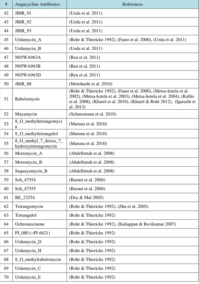
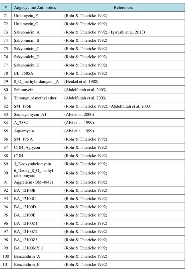
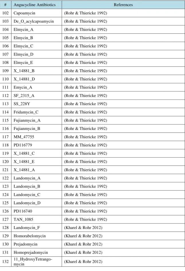
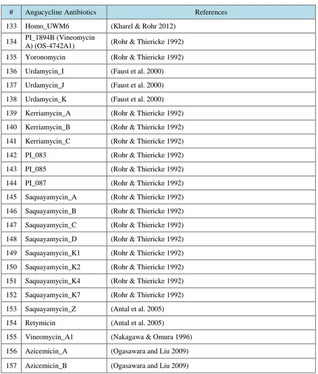
Table No. Description Page
Table 3.1. Angucycline Groups Antibiotics and their references ……….….. 31 Table 4.1. Average and maximum dock scores of ligands docked to CrtM (3ACW) …… 44 Table 4.2. Average of three dock score with maximum dock score of ligands docked to
(3W7F) protein with MG ……….………... 48 Table 4.3. Average of three dock score with maximum dock score of ligands docked to
VII
LIST OF FIGURES
Figure No. Description Page
Figure 2.1. Biosynthetic pathways. Staphyloxanthin biosynthesis in S. aureus …………... 10
Figure 2.2. Tetrangomycin structure ………... 16
Figure 2.3. The planar peptide group ……….. 22
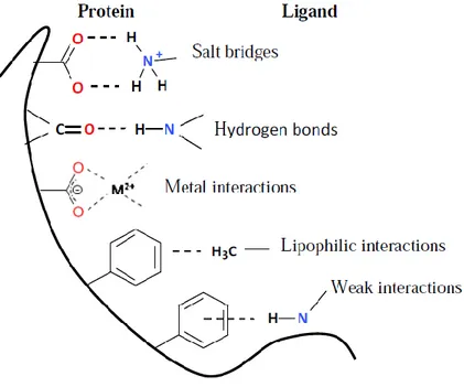
Figure 2.4. Major types of non-bonded interactions in protein-ligand complexes …………. 25
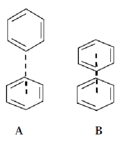
Figure 2.5. Aryl-aryl interactions in protein structure and protein-ligand complexes. A. Edge-to-face geometry, B. Parallel stacking geometry ………. 26
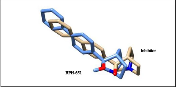
Figure 4.1. Superimpose structure of docked inhibitor with its original BPH-651 coordinates in the complex with CrtM ……….. 39
Figure 4.2. Superimpose structure of docked substrate with its original farnesyl diphosphate coordinates in the complex with CrtM ……… 40
Figure 4.3. Superimpose structure of docked inhibitor BPH-830 with original farnesyl diphosphate coordinates in the complex with CrtM ………... 41
Figure 4.4. Sructures of (a) The tetracyclic benz[a]anthracene frame; (b) Moromycin A (56); (c) Saquayamycin B (58); (d) Saquayamycin A (145); and (e) Saprolmycin E (29) ………... 42
Figure 4.5. Group 1 ligands (28, 29, 155) docked to (3ACW) protein ……….. 45
Figure 4.6. Group 2 ligands (47, 49, 84, 88, 102) docked to (3ACW) protein ……….. 45
Figure 4.7. Group 3 ligands (92, 93, 95, 98) docked to (3ACW) protein ……….. 46
Figure 4.8. Group 4 ligands (37, 94) docked to (3ACW) protein ……….. 46
Figure 4.9. Group 5 ligands (11, 54, 115, 118, 119) docked to (3ACW) protein …………... 47
Figure 4.10. Group 6 ligands (33, 40, 87, 133, 154) docked to (3ACW) protein ………. 47
Figure 4.11. Group 1 ligands (2, 13, 59, 98) docked to (3W7F) protein with MG. ………….. 50
Figure 4.12. Group 2 ligands (19, 28, 29, 30, 38) docked to (3W7F) protein with MG. …….. 51
Figure 4.13. Group 3 ligands (41, 42, 69, 124) docked to (3W7F) protein with MG. ………. 51
Figure 4.14. Group 4 ligands (47, 49, 56, 58) docked to (3W7F) protein with MG. ………… 52
Figure 4.15. Group 5 ligands (65, 93, 123, 124, 134) docked to (3W7F) protein with MG. .... 52
Figure 4.16. Group 6 ligands (84, 92, 145, 146, 147, 148, 151) docked to (3W7F) protein with MG. ………. 53
VIII
Figure 4.17. Group 7 ligands (6, 7, 11, 80, 81, 102, 116, 119, 120) docked to (3W7F) protein
with MG. ………. 53
Figure 4.18. Group 8 ligands (31, 32, 33, 51, 62, 137) docked to (3W7F) protein with MG. .. 54 Figure 4.19. Group 9 ligands (55, 107, 108, 109, 110) docked to (3W7F) protein with MG. .. 54 Figure 4.20. Group 10 ligands (4, 34, 136, 154) docked to (3W7F) protein with MG. ……… 55 Figure 4.21. Group 11 ligands (86, 111, 115, 118) docked to (3W7F) protein with MG. …… 55 Figure 4.22. Group 1 ligands (28, 29, 94, 141) docked to (3W7F) protein without MG. ……. 58 Figure 4.23. Group 2 ligands (38, 56, 134, 146, 147, 148, 151, 155) docked to (3W7F)
protein without MG. ………. 58
Figure 4.24. Group 3 ligands (46, 47, 49) docked to (3W7F) protein without MG. …………. 59 Figure 4.25. Group 4 ligands (143_PI_085, 144_PI_087) docked to (3W7F) protein without
MG. ……….……. 59
Figure 4.26. Group 5 ligands (34, 42) docked to (3W7F) protein without MG. ……….. 60 Figure 4.27. Group 6 ligands (6, 7, 11, 80, 81, 88, 116, 119, 120) docked to (3W7F) protein
without MG. ………. 60
Figure 4.28. Group 7 ligands (31, 32, 33, 51, 62, 82) docked to (3W7F) protein without MG. 61 Figure 4.29. Group 8 ligands (107, 108, 109, 110) docked to (3W7F) protein without MG…. 61 Figure 4.30. Group 9 ligands (111, 115, 118) docked to (3W7F) protein without MG……….. 62 Figure 4.31. Group 10 ligands (137, 156, 157) docked to(3W7F) protein without MG…...…. 62
IX
LIST OF APPENDIX
Appendix No. Description Page
APPENDIX I Docking score and interacted residue of protein (3ACW) with the Angucycline Group Antibiotics (ligands), and the arrangements are according to dock scores (kcal/mol) ……….………... 75 APPENDIX II Docking score and interacted residue of protein (3W7F with MG) with the
Angucycline Group Antibiotics (ligands), and the arrangements are according to dock scores (kcal/mol) ………..………... 90 APPENDIX III Docking score and interacted residue of protein (3W7F without MG) with
the Angucycline Group Antibiotics (ligands), and the arrangements are according to dock scores (kcal/mol) ………. 105 APPENDIX IV The 2D Interactions Between Docked Angucycline Group Antibiotics
X
ABBREVIATION
2D
: Two-Dimention
3D
: Three-Dimention
A
°
: Angstrom
Ala
: Alanine
Arg
: Arginine
Asn
: Asparagine
Asp
: Aspartic Acid
AV.
: Average
CrtM
: Dehydrosqualine Synthase Enzyme
Cys
: Cysteine
Gln
: Glutamine
Glu
: Glutamic Acid
Gly
: Glycine
His
: Histidine
Ile
: Isoleucine
ITC
: Isothermal Titration Calorimetry
Leu
: Leucine
Lys
: Lysine
Max.
: Maximum
Met
: Methionine
MG.
: Magnesium
MGL Tools
: Molecular Graphics Laboratory Tools
MRSA
: Methicillin Resistant Staphylococcus aureus
MSSA
: Methicillin Sensitive Staphylococcus aureus
ORSA
: Oxacillin Resistant Staphylococcus aureus
XI
PDB
: Protein Data Bank
PDBQT
: Protein Data Bank, Partial Charge (Q), & Atom Type (T)
Phe
: Phenylalanine
Pro
: Proline
QSAR
: Quantitative Structural Activity Relationship
RMSD
: Root Mean Square Deviation
S. aureus
: Staphylococcus aureus
Ser
: Serine
TCSs
: Two-component Systems
T.B.
: Tuberculosis
Thr
: Threonine
Trp
: Tryptophan
Tyr
: Tyrosine
Val
: Valine
Hazem A. T. AL-BUSTANY
1
1. INTRODUCTION
The discovery of penicillin in the 1940s revolt the fight against bacterial colony
mainly Staphylococcus aureus infections. Shortly after its introduction isolates of
penicillin-resistant Staphylococcus aureus were identified. Methicillin was first used in
1959 to treat these penicillin-resistant strains of S. aureus (Wilkinson 2010), and
methicillin-resistant S. aureus (MRSA) were reported in the early 1960's first and are
now viewed as a major hospital acquired pathogen worldwide (Batabyal et al. 2012).
This infection is especially troublesome in hospitals, prisons and nursing homes, where
patients with open wounds, invasive devices, and weakened immune systems are at
greater risk of infection compared with the general public (Batabyal et al. 2012).
The swift rise of antimicrobial resistance among pathogens has led to a renewed
interest to search for novel antimicrobial agents. The history of new drug discovery
processes shows that novel skeletons have, in the majority of cases come from natural
sources. They have been the source of, or inspiration for the development of chemical
entities introduced as pharmaceutical. The evolution of microbial natural product
collections and expansion of high-throughput screening methods have brought over
researchers to use the natural product libraries in drug discoveries. Actinomycetes
continue to be a productive and successful focus for natural products research, with
many novel compounds with prominent pharmacological valuable (Adegboye and
Babalola 2013).
Microbial natural products nowadays are the origin of most of the antibiotics in
the market. There is an alarming scarcity of new antibiotics currently under
development in the pharmaceutical industry. Microbial natural products still remain the
most promising source of novel antibiotics, although new methods are required to
improve the efficiency of the discovery process (Singh et al. 2014). The golden
carotenoid pigment staphyloxanthin by S. aureus acts as a virulence factor, mainly by
acting as a bacterial antioxidant which helps to the microbe avoid the reactive oxygen
species which the host immune system uses to kill pathogens (Liu et al. 2008).
1. INTRODUCTION
2
In earlier times, secondary metabolites were defined as substances with a low
molecular weight, which were not products of the primary metabolic pathway of the
producing organism. As a matter of fact, it was thought that these products did not have
a roll in the microbial primary functions or growth. Therefore, it was thought that
production of secondary metabolites did not represent any advantage for the producing
microorganism (Tabarez 2005).
Nowadays it is considered that cell investment in secondary metabolite
production is almost the confirmation of a function that should give the organisms
certain advantage against other members of the microbial community. In fact, secondary
metabolites are accepted to be essential for the producing cell as inhibitors of other
organisms that compete for the same food supply or as regulators of cellular
differentiation processes. In addition, it is reported that they are indeed products of
biosynthetic pathways, which have evolved to give these types of advantages (Tabarez
2005).
Actinomycetes are enthralling resource among microorganisms due to their
capability to produce novel bioactive secondary metabolites with antimicrobial
activities. They have proven to be effective antimicrobial agents, especially against
pathogenic organisms (Adegboye and Babalola 2013). In 1940, Selman A. Waksman
isolated an effective T.B. antibiotic, actinomycin and for this he got success in 1944,
with the discovery of spectromycin (Demain 1998). Scientists have discovered that
actinomycetes have an enormous potential to produce valuable natural products
(Raczkowski 2010). Hence, they produce useful secondary metabolites of high
commercial value and continue to be routinely screened for new bioactive compounds.
These searches have reaches a success and approximately they provide two thirds of
naturally occurring antibiotics, including many of medical importance, mainly
producing over 70 % of the natural product scaffolds found in clinically used
anti-infective agents (Bhat et al. 2013), (Gomez-Escribano et al. 2015). Consequently they
remain essential source of new chemical diversity and main part of drug discovery.
Their ingenuity and immense industrial value is extremely noteworthy (Adegboye and
Babalola 2013).
Hazem A. T. AL-BUSTANY
3
There are a long time consuming and costly way in drug discovery process,
computational methods attempts have been made to increase the efficiency of random
screening to select a typical subset of compounds from a compound collection. This
usually entails grouping (clustering) compounds with similar structure, and then
choosing a few members from each cluster for screening (Silverman and Holladay
2014). Computational approaches have successfully been applied to narrow the time and
cost involved in the process, with the quick increase in computational power, in silico
methods became commonly used in the fields of structural molecular biology and
structure-based drug design. Molecular docking is one of these computational
techniques (Ryska 2011). Molecular docking is a computer‐based, high‐throughput
screening method for identifying compounds of a certain structure or size (Ascencio
2010), Molecular docking may be defined as a problem of lock-and-key, where one is
interested in finding the suitable orientation of a key (ligand) that will open the lock
(protein) (Ryska 2011).
Knowledge about one or more known ligands or about the structure of the target
itself may be used to narrow a large screening collection to a smaller set of compounds
that may be more likely to hit the target. Virtual screening is the most common
computational method for selection of the compounds, which involves the rapid in silico
(by computer) assessment of large libraries of chemical structures to identify those
structures that most likely bind to a drug target, such as enzyme or protein receptor.
The goal is to identify new scaffolds, chiefly ones that may be in the existing collection.
In computer-based analysis two components are needed: (1) a database of structures in a
form that can be computationally analyzed for structural attributes and (2) a hypothesis
or model of the structural attributes that are important for activity, for example, the
hypothesis that structural similarities to a known active ligand should yield similarly
active compounds or a hypothesis of the shape and charge density of a binding pocket
that defines what features a complementary ligand structure should have (Silverman and
Holladay 2014).
Furthermore, one of main tools for virtual screening procedures is docking,
where a library of several compounds is “docked” against one drug target and returns
the best hit. The procedure of virtual screening through docking has become essential
when it is needed to test a database of thousands (or even millions) of compounds
1. INTRODUCTION
4
against one or more targets in a short period of time. This search would be impossible to
be reproduced experimentally at a so small economic and time cost. For this reason
docking has been found to be a beneficial step in Quantitative Structural Activity
Relationship (QSAR) studies, where statistical analysis is applied to thousands of drug
candidates (Novotarskyi, 2013).
Docking method predicts favored orientation of one molecule to the second
when they bind to form a stable complex. In the field of drug design, first molecule is
usually protein (macromolecule) and the second one is ligand (small organic molecule)
which is potential drug candidate. Information of favored orientation of ligand and
protein can be used to predict binding affinity, and this discriminating high-affinity drug
candidate from the low-affinity compounds (Ryska 2011).
Usually scoring function used in docking programs in order to recreate the
chemical potentials which predict the conformation of binding. Need superficial
physics-based (Coulombs energy and van der Waals forces) experimentally weighted to
account for the difference in energy and free energy. Usually protein (receptor) have to
be prepared by adding hydrogens and charges select site and eliminate of water and
cofactors included (Pétursson 2014). Improvements in structure determination methods
along with quick advances in molecular visualization tools have led to the rise of
structure-aided drug design or rational drug design as an integral part of the drug
discovery and development process (Dias 2011). Using of computer based analysis of
molecular interaction for example protein-ligand binding becomes more necessary with
the availability and expanding of molecular biological data. For this purpose, docking
algorithms included a reasonably accurate model of energy and the flexibility of
molecules (Pétursson 2014).
Moreover docking calculations simulate the interactions between the protein’s
binding site and the ligand, and these interactions give a qualitative score, and therefore
the outcomes may be compared to those of biochemical assays (Tunca 2012). As a
consequence, hundreds of thousands of compounds can be screened by using in silico
methods. In addition these computer programs can generate the crystal structure and
NMR solution structures of the target or related proteins, as well as calculating atomic
homology models. Candidate drug‐binding pockets can be identified as well (Ascencio
2010).
Hazem A. T. AL-BUSTANY
5
2.
LITERATURE REVIEW
2.1.
Current Status of MRSA
Methicillin-resistant Staphylococcus aureus (MRSA) is a Gram positive
non-motile, non-facultative coccus whose infections in humans. Best environmental
conditions for growth are temperatures between 15
oC and 45
oC. High concentrations of
sodium chloride do not change the growth, even when concentrations reach up to 15%
(Stuczen 2013). Consequently the universal occurrence of MRSA has become problem
for public health (Howe et al. 2004), (Moghadam et al. 2015). It is also known as
multidrug-resistant S. aureus and oxacillin-resistant S. aureus (ORSA). S. aureus was
first identified in the late 19th century and has since been recognised as part of the
natural flora of humans. It frequents the face, hands and perineum, with the most
common site being the nares (nostril) (Stuczen 2013). S. aureus species developing,
resistance to beta-lactam antibiotics are known as MRSA, they include the penicillins
like methicillin, dicloxacillin, nafcillin, oxacillin, etc. and the cephalosporins (Batabyal
et al. 2012), (Rashid et al. 2015). This organism is part of the natural microbiota of
humans, from which clones of epidemic drug-resistant S. aureus have emerged (Hsu et
al. 2015). MRSA is reported as the leading cause of wound infections in most parts of
the world (Stuczen 2013). Hospital acquired infections and infections in the community
(Onelum et al. 2015). Between 30% and 60% of the healthy population carry S. aureus,
of which between 10% and 20% are chronically colonized (ongoing, persistent
population of S. aureus on or in the body but in the absence of infection) (Stuczen
2013). The appearance and spread of bacterial pathogens that have become modified for
existence in hospitals poses a major threat to global health systems (Hsu et al. 2015).
The high prevalence of MRSA colonization in diabetic foot ulcers is a
consequence of antibiotic overuse and the selection of broad rather than narrow
spectrum agents (Stuczen 2013).
There is another classification which known as methicillin-sensitive S. aureus or
MSSA if the strain unable to resist these antibiotics. The development of such resistance
does not cause more intrinsically virulent for the organism than strains of S. aureus that
2. LITERATURE REVIEW
6
have no antibiotic resistance, but resistance does make MRSA infection more difficult
to treat with typical types of antibiotics and thus more dangerous (Batabyal et al. 2012).
The glycopeptides, particularly vancomycin, have been the mainstays of therapy for
MRSA, and the emergence of resistance to these agents is of great concern (Howe et al.
2004).
People not involved in health care backgrounds are often less aware of this silent
and lethal epidemic. This lack of awareness lies in significance of hazard, perils of
hospital acquired MRSA infection, and potential risk to overall health care system
(Rashid et al. 2015). Healthy people may carry MRSA asymptomatically for long
periods of time but patients with compromised immune system are at a significant
greater risk of symptomatic infections (Onelum et al. 2015). A significant public
behavioral alteration is needed in order to control this global risk as well as a
well-informed public. (Rashid et al. 2015) The range of infections due to MRSA are
manifold and are linked with worse outcome in addition to extended hospital stay,
higher cost of treatment and increased mortality (Onelum et al. 2015).
The concept of “anti-infectious drugs” includes not only compounds that inhibit
the growth of pathogenic microorganisms statically or kill them (so called
chemotherapeutics or antibiotics) and vaccines but also compounds that control
microbial adaptation/survival or pathogenicity, potentiate the activities of known
antibiotics, or enhance the host immune system against microbial infection (Koyama et
al. 2013). Production of β – lactamase enzyme in the affected area is the main cause of
microbial resistance (Rashid et al. 2015). For example, β-lactamase inhibitors such as
clavulanic acid, sulbactam, and tazobactam themselves show very weak or no
antimicrobial (non-antibiotic) activity, but these compounds dramatically potentiate the
antimicrobial activity of β-lactam antibiotics against β-lactamase-producing bacteria
(Koyama et al. 2013). The choice of antibiotics as active treatment is reduced after the
maturity of infection. Such methods are possibly show harmful side effects to the
patient and expensive. Recent evidence supports that domestic animals like cat, dog and
hen can transmit MRSA to their owners (Rashid et al. 2015).
Hazem A. T. AL-BUSTANY
7
In recent years, active anti-infectious compounds against MRSA have been
widely searched for. Several compounds have been found to have new mechanisms of
action against MRSA and are expected to be potential leads for the treatment of
infection (Koyama et al. 2013). S. aureus is an extremely transmittable bacterial species
found in the ecosystem. The microorganism invades the skin and enters deeper tissues.
As in septicemia, it multiplies to cause a localized or systemic response (Rashid et al.
2015). MRSA produce a number of virulence factors which cause suppurative infections
and toxinosis. Surface proteins allow bacterial attachment to the extracellular matrix of
the host, specifically the proteins laminin and fibronectin, found in epithelial and
endothelial tissue. Toxins produced by the bacteria damage host cell membranes and
allow cell invasion. Alpha toxin is produced as a monomer that binds to the membrane
of the susceptible cell. Sub-units then combine to form heptameric rings with a central
pore, through which the cellular contents leak (Stuczen 2013). MRSA infections cause a
huge number of deaths every year worldwide (Koyama et al. 2013). The patient
becomes infected with the growth of its population. People who are weaker, older and
sicker have weaker immune system and may get infected easily. It is also described that
people may bring this infection without having any noticeable indications (Rashid et al.
2015).
2.2.
Treatment approaches for MRSA
Microbiologists are observing an exponential growth in infectious human
diseases through S. aureus, which is precisely known as Methicillin Sensitive S. aureus
(MSSA). In this case, bacterial skin infections are because of the strains of Methicillin
Resistant S. aureus (MRSA) (Rashid et al. 2015). According to drug bank only three
antibiotics namely arbekacin, meticillin and linezolid, are approved for the treatment of
MRSA. Drug targets are inadequate and there is an urgent need to find out the novel
drug targets. Apart from drug target, there is also a need for good ligand preparation that
would act as effective inhibitors to the novel targets without affecting human proteome.
Any contaminated surfaces is the source of several kind of infections, MRSA is one of
them (Balaji SR et al. 2014). As observed in the last decade the microbe has latency to
severely resist antibiotics. The human race is facing significant morbidity caused by
these lethal infections (Rashid et al. 2015).
2. LITERATURE REVIEW
8
Extermination (Eradication) or inhibition of staphylococcal colonization is still
considered as main strategy to prevent infection and transmission of these strains. Basis
behind such a strategy is that the most staphylococcal infections are caused by
endogenous strains; so, existence of S. aureus is a major risk factor for consequent
infections (Moghadam et al. 2015)
Increasing multiple resistances of S. aureus to antibiotics makes the
development of new treatment routes for serious infections a matter of urgent concern
(Kuroda et al. 2007).
MRSA is one of typical resistant strains, and research for its prevention and
treatment was carried out. The idea (concept) that MRSA infection presents different
signs for treatment and diagnoses of colonization is controversial, but the differential
diagnosis between bacterial infection and bacterial colonization is essential to avoid
unnecessary use of anti-MRSA drugs (Shigemura et al. 2013).
The last-resort antibiotic for the treatment of MRSA infections was vancomycin,
but MRSA resistance to vancomycin has been reported too. This suggests that MRSA
will likely obtain more resistance to vancomycin in the near future. Therefore, it is
increasingly essential to discover new antibiotics or to devise new actions that are
effective against MRSA infections (Koyama et al. 2013). As a consequence, clinicians
across the world faced clinical challenge in controlling MRSA (Rashid et al. 2015).
The main component of the bacterial cell wall is peptidoglycan which is an
attractive target for the development of anti-infectious agents. It forms a huge
macromolecule that surrounds the cell as a single, flexible meshwork and is closely
involved in cell division. The structure determines the cell shape and maintains cell
integrity by protecting it against the high internal osmotic pressure. Important
antibiotics have been clinically used including β-lactams and glycopeptides that target
cell wall peptidoglycan synthesis (Koyama et al. 2013).
Hazem A. T. AL-BUSTANY
9
2.3.
Targeted pathway
It is well known that MRSA produces a yellow pigment called staphyloxanthin
(STX). Recently, several research groups described that STX is a virulent factor acting
as an antioxidant, with its various conjugated double bonds enabling detoxification of
host immune system-generated reactive oxygen species (Sakai et al. 2012). The first
committed step in staphyloxanthin biosynthesis by S. aureus is dehydrosqualene
synthase (CrtM) enzyme (Pelz et al. 2005), which catalyzes the condensation of two
farnesyl diphosphates to produce the C30 species, presqualene diphosphate (Liu et al.
2008), (Song et al. 2008), which then undergoes skeletal rearrangement and further loss
of diphosphate to produce dehydrosqualene (Song et al. 2008). Successive
dehydrogenations yield 4,4′-diaponeurosporene (Pelz et al. 2005), which is then further
oxidized, glycosylated, and esterified to give the carotenoid, staphyloxanthin (Figure
2.1) (Liu et al. 2008). Hence forgetting the biosynthesis of STX may provide an
alternative way to develop new drug for preventing a treatment, staphyloxanthine
remain infections since the lack of STX is susceptible to neutrophil killing.
The orange carotenoid staphyloxanthin is produced by most S. aureus strains.
The staphyloxanthin biosynthesis genes are organized in an operon, crtOPQMN, with a
B-dependent promoter upstream of acyl transferase (crtO) and a termination region
downstream of dehydrosqualine desaturase (crtN). The functions of the five encoded
enzymes are predicted on the basis of their sequence similarity to known enzymes and
by product analysis of gene deletion mutants (Pelz et al. 2005).
2. LITERATURE REVIEW
10
Figure 2.1. Biosynthetic pathways. Staphyloxanthin biosynthesis in S. aureus (Pelz et al. 2005), (Song et al. 2008).
The dehydrosqualene desaturase (CrtN) dehydrogenates dehydrosqualene to
form the yellow, main intermediate 4,4-diaponeurosporene. Diaponeurosporene oxidase
(CrtP), very likely a mixed function oxidase, oxidizes the terminal methyl group of
4,4-diaponeurosporene to form 4,4-diaponeurosporenic acid. Glycosyl transferase (CrtQ), a
glycosyltransferase, esterifies glucose at the C1 position with the carboxyl group of
4,4-diaponeurosporenic acid to yield glycosyl 4,4-diaponeurosporenoate; this compound
was the major product in the clone expressing crtPQMN. In the final step, the
acyltransferase (CrtO) esterifies glucose at the C6 position with the carboxyl group of
12-methyltetradecanoic acid to yield staphyloxanthin (Pelz et al. 2005). Hence targeting
are of these step is STX biosynthesis may provide a way to develop the anti-MRSA
drugs.
Hazem A. T. AL-BUSTANY
11
2.4.
Secondary Metabolites
Secondary metabolites are metabolic products that are not critical for vegetative
growth of the producing organisms but they are considered differentiation compounds
conferring adaptive roles, for example, by functioning as resistance compounds or
signaling molecules in ecological interactions. They are produced at the end of the
exponential phase of growth and their syntheses seriously depend on the growth
conditions. Production is usually when growth is limited by the exhaustion of
fundamental nutrients such as carbon or nitrogen. They are structurally different and
most of them are endowed with biological activities, such as antimicrobial agents,
toxins, pesticides, ionophores, bioregulators, and quorum signalling. These bioactive
metabolites are extremely used as antimicrobial agents for the treatment of different
diseases (Adegboye and Babalola 2013). One third of the 22,500 known microbial
metabolites are the secondary metabolites of actinomycetes, mainly Streptomyces
species (Miyaoka et al. 2014). Microbial secondary metabolites have been in the fırst
appearance in the discovery of novel antimicrobial agents for pharmaceutical industry,
and nowadays all indications suggests that novel compounds with potential therapeutic
applications are still waiting to be discovered from secondary metabolites mainly those
produced by actinomycetes. Actinomycetes are abundant producers of secondary
metabolites with biological activities (Adegboye and Babalola 2013).
Secondary metabolites are provided by the producer organism with survival
advantages in various ways, such as improving nutrient availability, acting as a
metabolic defense mechanism, protecting against environmental stressors, enhancing
competitive interactions with other organisms (Breitling et al. 2013). Secondary
metabolites are organic compounds that often play an important role in defense systems
of different organisms and are not directly involved in the normal growth, development
or reproduction of an organism. (Davati and Najafi 2013).
Secondary metabolites usually include various chemical moieties, such as
polyketide backbones, amino acid derivatives and sugars. Biosynthesis of secondary
metabolite is catalyzed by a number of enzymes, usually encoded by genes. These genes
occur nearby to one another in cluster. The gene cluster contains all the needed genes
for the synthesis of a particular secondary metabolite. This includes: the genes that
encode the biosynthetic enzymes, regulatory proteins, genes for resistance to the toxic
2. LITERATURE REVIEW
12
action of secondary metabolites and genes for secretion of the metabolites. Enzymes
such as synthase (PKS) and non-ribosomal peptide synthetase (NRPS) are take part in
the synthesis of secondary metabolites. Other enzymes responsible for the synthesis of
other constitutive compounds, such as sugars, are often encoded by genes nearby to the
gene cluster. Through processes such as elongation, synthesis, glycosylation, alkylation
and oxidation, structurally different and complex metabolites are produced. The
complete process of production and transportation of secondary metabolites are severely
regulated by transcriptional regulators and transporters. The genes encoding for
tailoring enzymes, transcriptional regulators and transporters are often located nearby to
PKS and NRPS genes. The gene cluster size responsible for the synthesis of each
secondary metabolite is usually between 10 -100 kb. (Adegboye and Babalola 2013).
The secondary metabolites frequently have unusual structures and their
formation is regulated by nutrients, feedback control, growth rate, enzyme induction,
and enzyme inactivation. These events generate signals which affect a cascade of
regulatory actions resulting in chemical differentiation (secondary metabolism) and
morphological differentiation (morphogenesis). The signal is often a low molecular
weight inducer which acts by negative control, by binding to and inactivating a
regulatory protein which normally prevents secondary metabolism and morphogenesis
during rapid growth and nutrient sufficiency (Demain 1998). Regulation is affected by
unique low molecular mass compounds, sigma factors, transfer RNA, and gene products
formed during post-exponential growth. The synthases of secondary metabolism are
often coded by clustered genes on chromosomal DNA and infrequently on plasmid
DNA (Davati and Najafi 2013).
2.4.1. Function and Importance of Secondary Metabolites
Several hypotheses exist about the origin and function of secondary metabolites.
The most accepted considers secondary metabolites as waste products that under the
pressure of natural selection have evolved as messenger molecules which must endure
long enough to shuttle between the various components of the microbial community.
This fact, would explain the secondary metabolites tendency to be small organic
molecules, as a natural consequence of their functions.
Hazem A. T. AL-BUSTANY
13
The presence of complex biosynthetic pathways for production of complex
antibiotics suggest that they must have an important role in microbial survival, either as
inhibitors of other competing organisms or as regulatory effectors during some stage of
the cell differentiation process, since sensitive organisms need to evolve only a single
enzyme of any several reaction types to inactivate most antibiotics (Tabarez 2005).
Nowadays, several arguments support the hypothesis that secondary metabolites
improve the survival of the producer in competition with other living species. These
arguments are as follows:
a) Secondary metabolites act as an alternative defense mechanism, because only the
organisms lacking an immune system are prolific producers of these compounds.
b) They have sophisticated structures, complex and mechanisms of action, and
energetically expensive pathways.
c) They act in the competition between microorganisms, plant and animals.
d) They are produced by biosynthetic genes clusters, which would only be selected if
the product conferred a selective advantage. Some exactitudes of these genes clusters
are the absence of non-functional genes and the presence of regulatory and resistance
genes.
e) The production of them with antibiotic activities is temporarily related with
sporulation when the cells are particularly sensitivity to competitors and requiring
special protection when a nutrient runs out.
Furthermore, the wide diversity of secondary metabolites suggests a broad range
of functions. Nevertheless, these functions could depend on the conditions, optimal or
not, surrounding the producer microorganism. Finally, due to their crucial importance
the study and exploitation of secondary metabolites continue to progress despite the lack
of agreement regarding why microbes produce such chemical diversity of antimicrobial
compounds (Tabarez 2005).
2. LITERATURE REVIEW
14
2.4.2. Genetics of Secondary Metabolites
The genes regulating and ensuring synthesis of secondary metabolites and their
expression can be grouped in 5 classes: first, structural genes, that code for enzymes
involved in the biosynthesis; second, regulatory genes, that determine the repression or
induction of the structural genes; third, genes that determine the resistance of the
generating organism; fourth, genes controlling the compound permeability and fifth,
genes that control primary pathways. The genetic regulation of all above mentioned
genes is highly complicated because many environmental and microbial factors affect
the production of these compounds (Tabarez 2005)
Previous studies showed that the gene cluster responsible for the production of
secondary metabolites is not found in all bacteria and even in those surviving it is not
regularly distributed among them. For example Streptomyces coelicolor has more than
20 gene clusters for the synthesis of secondary metabolites while Streptomyces
avermitilis has 30 gene clusters. Genome mining for new candidate secondary metabolic
pathways built on clustering and co-expression has proved to be a highly successful
method in microbes. This useful to expect the types of antibiotic one might expect to
find after extraction and purification. With the increasing number of genome nucleotide
sequence information in the Gen Bank and coming of next generation sequencing it will
be possible to search for secondary metabolite gene cluster candidate in a wide range of
actinomycetes (Adegboye and Babalola 2013).
Functionally related genes are on the chromosome suggesting that at least part of
their evolution has occurred as a unit. Evolutionally, two types of gene clusters that
produce secondary metabolites can be described: first, a gene cluster might contain
many genes, some of which give rise to chemical transformations of substrates and
many other that do not. This type of cluster would be indicative of a natural product
which has not been selected for and which is not functional. Second, a gene cluster
might contain only genes that code for enzymes of a particular biosynthetic pathway,
resistance genes and regulatory factors, and no “junk” genes. This type of gene cluster
indicate a natural product that has been selected for and which has had a beneficial
function for the producer organism (Tabarez 2005).
Hazem A. T. AL-BUSTANY
15
2.5.
Angucycline Compounds
The angucyclines are a large group of natural products (Song et al. 2015). They
are a large family of actinomycete-derived polyketide antibiotics with a four-ring
skeleton that constitutes the aglycone part. Angucyclines structure consist of a
benz[a]anthracene core (Ren et al. 2011), (Abdelmuhsen et al. 2014), which is
commonly C- or O-glycosylated with sugar (single deoxyoligosaccharide) chains of
various lengths with up to six carbohydrates, as in landomycin A, the carbohydrate
composition of landomycin is a repeated sequence of β(4-1)-β
-D-olivose-(3-1)-α-L-rhodinose attached to a phenolic oxygen of the angucycline core. The
biological activities were mainly found to be depending on the length of the saccharide
chain (Helaly et al. 2013). They have been shown to exhibit different activities of
medical interest (Rohr and Thiericke, 1992) including antitumor (Kirschning et al.
2000), (Kharat et al. 2009) and antibacterial activities (Kawasaki et al. 2010a),
(Kormanec et al. 2014) as well as platelet aggregation inhibitory effects (Kirschning et
al. 2000). The first reported compounds of this class were tetrangomycin and tetrangulol
(Song et al. 2015).
The angucycline family of antibiotics is a large and ever-growing group of
secondary metabolites of microbial origin comprising more than 100 members (Ren et
al. 2011). The first member of this group of secondary metabolites was tetrangomycin
(Figure 2.2) (Kalyon et al. 2013). These compounds are found widely in nature and are
mainly produced by soil-dwelling Streptomyces bacteria. The biosynthesis of
angucyclines diverges from other type II aromatic polyketides by the action of the
specific cyclase that closes the fourth ring of the polyketide into an angular orientation
producing UWM6 (Kallio et al. 2008), (Patrikainen et al. 2012). Aromatic polyketides
include a large class of natural secondary metabolites produced by certain fungi,
bacteria and plants. Many of these compounds have medically important properties, and
several are in clinical use (Koskiniemi et al. 2007).
2. LITERATURE REVIEW
16
Figure 2.2. Tetrangomycin structure (Kalyon et al. 2013)
Aromatic polyketides are complex natural compounds well established for their
biological activities and applications in medicine. They are produced by a number of
different organisms, but especially Streptomyces bacteria have been a rich source of
natural products. Angucyclines comprise a distinct group of aromatic polyketides (Rohr
and Thiericke 1992), and first member of group was discovered in 1966 (Kawasaki et
al. 2010a), but still only a few hundred structures have been described. New genetic
screening studies have indicated that angucycline gene clusters are much more abundant
than expected depending on traditional cultivation methods (Metsa-Ketela et al. 2002),
which proposes that many potentially important angucyclines are yet to be discovered
(Kallio et al. 2008).
Many of the new compounds, mainly described most recently, are from
marine-derived actinomycetes, and represent new structures arising from new biosynthetic
pathways. This suggests that even if marine actinomycetes are derived from terrestrial
sources, they have been exposed to a new environment long enough to have evolved
exceptional biosynthetic pathways of their own. The discovery of new actinomycete
species producing novel chemical compounds suggests that they are metabolically
active in the marine environment. Furthermore, it has become more obvious that there is
great diversity of new actinomycete species within the marine environment with
evidence that stems from phylogenetic analysis, metabolic requirements, morphological
characteristics, and new secondary metabolite production (Raczkowski 2010).
Hazem A. T. AL-BUSTANY
17
2.6.
Actinomycetes, especially genus Streptomyces (Angucyline producers)
Actinomycetes belong to the phylum Actinobacteria that consist of Gram
positive, aerobic bacteria, were first thought to be fungi for their mycelium producing
capabilities, for which the suffix “mycetes” is used (Raczkowski 2010). They are
filamentous, characterized by a complex life cycle (Sharma 2014) and produce
extensive branching vegetative (substrate) mycelium and aerial mycelium bearing
chains of arthrospores. The substrate mycelium and spores can be pigmented, but also
diffusible pigments are produced. On agar plates, they form lichenoid, leathery or
butyrous colonies (Rintala 2013).
The high densities of actinomycetes in soils revealed their importance, which are
one of the most significant decomposers for recalcitrant polymers such as cellulose. In
addition to cellulose, Streptomyces spp. have shown to degrade chitin, keratin, pectin,
and other hemicelluloses. These compounds can also be used as elements for their
growth on artificial media. Although some pesticides inhibit their proliferation,
Nocardiopsis spp., are known to degrade chlordane insecticides. With the ability to
produce numerous antifungal compounds, actinomycetes are suspected to be associated
with plant roots and hence they are believed to protect roots by using these compounds
to inhibit the growth of fungal pathogens (Raczkowski 2010).
Actinomycetes produce perhaps the most diverse and most unique,
unprecedented, sometimes very complicated compounds exhibiting excellent
antimicrobial potency and usually low toxicity. The metabolic diversity of the
actinomycetes is due to their extremely big genome, which has hundreds of
transcription factors that control gene expression, allowing them to respond to specific
needs (Singh et al. 2014). The GC-content of the DNA is 69-78 %. L-diaminopimelic
acid is the characteristic compound present in the cell wall peptidoglycan of
streptomycetes. The streptomycetes are able to use a wide range of organic compounds
as a carbon source, with complex biological materials, such as cellulose and lignin, and
can also use an inorganic nitrogen source (Rintala 2013).
Actinomycetes have provided important bioactive compounds of great
commercial value and continue to be routinely screened for new bioactive substances. It
2. LITERATURE REVIEW
18
is generally accepted that the streptomycetes have a specific capacity to produce a large
variety of different bioactive compounds with a wide range of activity (Singh et al.
2014). There are over 500 species of Streptomyces bacteria described by Euzeby (2008)
(Raja and Prbakarana 2011). They undergo a complex process of morphological and
physiological differentiation that leads to the production of exospores and specialized
metabolites possessing a wide range of biological activities. While the function of many
of these molecules in the natural environment is not always evident, they are believed to
provide a competitive advantage to the producing organism (Gomez-Escribano et al.
2015).
Among the 140 defined Actinomycetes genera, only a few are responsible for
the more than 10,000 bioactive compounds in clinical use (Adegboye and Babalola
2013), more than half of the 10,000 documented bioactive compounds, have offered
over 50 years of interest to industry and academia (Anderson and Wellington 2001). It
is representing 45% of known bioactive microbial metabolites were isolated from
various actinomycetes species (Sharma 2014), and 7600 compounds are produced by
Streptomyces species (74% of all actinomycetales), while the rare actinomycetes
represent 26%, altogether 2500 compounds (Raja and Prbakarana 2011), (Singh et al.
2014).
The composition of cell wall in actinomycetes differs greatly among different
groups and is of considerable taxonomic value. Four major cell wall types are
distinguished in these filamentous bacteria depends on three features of peptidoglycan
composition and structure, which are: diaminopimelic acid isomer on tetrapeptide side
chain position 3, sugar content of peptidoglycan, and the presence of glycine in
interpeptide bridges (Adegboye and Babalola 2012).
Microorganisms included in the genus Streptomyces that inhabit soil niches, thus
facing ever changing environmental conditions and nutrient scarcity. Along evolution,
this challenging environment has pushed the genus Streptomyces towards complex
adaptive responses. Among them, two-component systems (TCSs) are the most
important transduction signal mechanism in bacteria, allowing the translation of these
rapid environmental or nutritional changes into a regulatory readout. Typically, TCSs
Hazem A. T. AL-BUSTANY
19
comprise a membrane-bound histidine kinase, which senses specific environmental
stimuli, and a cognate regulator, which mediates the cellular response, mainly through
the transcriptional regulation of target genes (Rodríguez et al. 2013).
It is now evident that as new molecular approaches are used to unravel the
microbial community of unique marine environments, new species of actinomycetes
have continued to reappear. However, these new species of actinomycetes indigenous to
the marine habitat, or they derived from terrestrial run-off that are in fact a source of
dormant actinomycete spores that can survive for many years (Raczkowski 2010).
Bacteria belonging to the genus Streptomyces harbor a high number of TCSs in
comparison with other bacterial genera, probably due to the changing environment that
these organisms must inhabit (Rodríguez et al. 2013).
Approaches for overproduction of microbial products can be based on microbial
response (elicitors, quorum sensing), genetic, ribosome, and metabolic engineering.
Also, molecular genetic improvement methods include amplification of secondary
metabolites biosynthetic genes, inactivation of competing pathways, disruption or
amplification of regulatory genes, manipulation of secretory mechanisms, expression of
a convenient heterologous protein and combinatorial biosynthesis (Davati and Najafi
2013). In 1995 more undoubted data, through the use of 16S rRNA genus-specific probe
with hybridization experiments, suggested that salt marsh Streptomyces sp. strains were
indigenous to the marine environment by being important proportion of the microbiota
of the salt marsh environment through an entire annual cycle. Scientists revealed that
Streptomyces sp. occurred dominantly in the salt marsh community and that dormant
spores were not the source of their abundance (Raczkowski 2010).
2.7.
Protein (Receptor) Structure and Function
2.7.1. Introduction to Proteins
The highly specific protein complex formation is an important process in
biology and an integral constituent to all major biochemical pathways. The structures of
these protein complexes can provide detailed understanding into the mechanisms of
function, starting from disease pathogenesis, to drug action, to promoting our
2. LITERATURE REVIEW
20