ABSTRACT
This work is licensed under a Creative Commons Attribution-NonCommercial 4.0 International License.
Pınar Çimen1 , Dursun Alizoroğlu1 , Mehmet Ünlü1 , Cenk Kıraklı1 , Özlem Ediboğlu1 , Ahmet Emin Erbaycu2
Safety and Effectiveness of Thrombolytic Therapy
Compared with Standard Anticoagulation in
Subjects with Submassive Pulmonary Embolism
Objective: Thrombolytic and anticoagulation therapy modalities are the possible treatment for submassive pulmonary
em-bolism (PE). However, the indications are still the subject of debate. The aim of the present study was to compare the effi-cacies of thrombolytic and standard anticoagulation treatment modalities on mortality and also to determine the safety of thrombolytic treatment in subjects with submassive PE.
Materials and Methods: Subjects with submassive PE were recruited from the intensive care unit (ICU). Demographic
data, comorbidity, bedside echocardiography (ECHO) findings, treatment procedure, treatment-related side effects, and total length of stay in the hospital and ICU were collected. Control ECHO was performed 48 h after the initiation of treatment. Short-term and 1-year mortality rates were recorded. The correlation between the increased risk for major bleeding and thrombolytic treatment was assessed.
Results: A total of 54 subjects were enrolled during the study period. The median age of the subjects was 66 (54–73) years.
Of the 54 subjects, 18 (33.3%) underwent thrombolytic treatment, and 36 (66.7%) received standard anticoagulation ther-apy. Short-term and 1-year mortality rates were statistically lower in subjects who received thrombolytic therapy (p=0.02 and p=0.04, respectively). The reduction in mean pulmonary arterial pressure was significantly higher in the thrombolytic treatment group (p<0.001). Risk for major bleeding was similar between the two.
Conclusion: Thrombolytic therapy may reduce the mortality rates in subjects with submassive PE without an increase in
the risk of major bleeding.
Keywords: Submassive pulmonary embolism, thrombolytic therapy, anticoagulation treatment
INTRODUCTION
Pulmonary embolism (PE) is a blockage of the main artery of the lung or one of its branches by a substance that has traveled from elsewhere in the body throughout the bloodstream. It is subdivided into massive, submassive, and nonmassive categories. The classification of acute PE severity is based on the risk of early death, which is influenced by demographic factors, comorbidity, and the functional status of the right ventricle (RV) under acute pressure overload (1). Shock or persistent arterial hypotension, indicating overt RV failure at presentation, has long been identified as a key determinant of poor prognosis and represents the only widely accepted indication for (systemic) thrombolytic therapy to date. In contrast, anticoagulation remains the primary treatment option for patients who were normotensive who present with imaging findings that indicate RV dysfunction and biochemical evidence of myocardial injury (2–4).
Right ventricular failure and myocardial injury are the main pathophysiological changes of acute PE that are di-rectly associated with the prognosis of the subjects. Subjects with acute PE with right ventricular dysfunction have higher mortality rates than those with normal right ventricular function (5).
Anticoagulation is the main treatment modality for submassive PE. On the other hand, although the rapid reso-lution of PE is accompanied by an improvement in right ventricular function and prognosis, the indications for thrombolytic treatment are still under debate in subjects with submassive PE. The severity of symptoms and sur-vival are strongly associated with right ventricular function in a case of PE. Despite its importance, little is known about the mechanisms of right ventricular failure in subjects with submassive PE, and only few trials have been conducted in hemodynamically stable subjects to address the clinical outcomes (6). The establishment of the indica-tors of right ventricular function and structure, in particular those measured non-invasively, may be used to assess the prognosis and response to therapy. Currently, echocardiography (ECHO) is the most widely used non-invasive technique for this purpose, and the measurement of mean pulmonary arterial pressure (mPAP) by ECHO may be an appropriate indicator of right ventricular function. In addition to this, it is necessary for physicians to evaluate the benefits of thrombolytic therapy against the increased risk of bleeding (7, 8).
Cite this article as:
Çimen P, Alizoroğlu D, Ünlü M, Kıraklı C, Ediboğlu Ö, Erbaycu AE. Safety and Effectiveness of Thrombolytic Therapy Compared with Standard Anticoagulation in Subjects with Submassive Pulmonary Embolism. Erciyes Med J 2019; 41(2): 175–9.
1Department of Intensive
Care, University of Health Sciences İzmir Training and Research Hospital for Thoracic Medicine and Surgery,
İzmir, Turkey
2Department of Thoracic
Disease, University of Health Sciences, İzmir Training and Research Hospital for Thoracic Medicine and Surgery,
İzmir, Turkey
Submitted
27.10.2018
Accepted
12.02.2019
Available Online Date
09.05.2019
Correspondence
Ahmet Emin Erbaycu, Department of Thoracic Disease, University of Health Sciences, İzmir Training and Research Hospital for Thoracic Medicine and Surgery,
İzmir, Turkey Phone: +90 232 458 72 62 e.mail: [email protected] ©Copyright 2019 by Erciyes University Faculty of Medicine - Available online at www.erciyesmedj.com
The aim of the present study was to compare the efficacies of thrombolytic and standard anticoagulation therapy modalities on mortality and also to determine the safety of thrombolytic therapy in subjects with submassive PE.
MATERIALS and METHODS
PatientsIn this single-center study, data of subjects with submassive PE re-ferred to the intensive care unit (ICU) at a tertiary, academic train-ing hospital between January 2012 and December 2014 were ret-rospectively reviewed. The study was approved by the local ethics committee (February 8, 2010/263). Written informed consent was obtained from all subjects for thrombolytic therapy.
Exclusion criteria were subjects with a blood platelet count of
<100×109/L, known neoplasia, renal insufficiency in dialysis,
un-controlled heart failure, and an active or a history of intracranial hemorrhage or visceral bleeding.
The criteria for thrombolytic and standard anticoagulation therapy modalities were defined as massive PE presenting with sustained hypotension (systolic blood pressure (SBP) <90 mm Hg for at least 15 min or requiring inotropic support, not due to a cause other than PE), pulselessness, or persistent profound bradycardia. Submassive PE refers to patients with acute PE without systemic hypotension (SBP ≥90 mm Hg) but with evidence of either RV dys-function or myocardial necrosis. RV dysdys-function parameters refer to the presence of at least one of the following (9):
• RV dilation (apical four-chamber RV diameter divided by left ventricle (LV) diameter >0.9) or RV systolic dysfunction on ECHO,
• RV dilation (four-chamber RV diameter divided by LV diameter >0.9) on computed tomography (CT),
• elevation of brain-type natriuretic peptide (>90 pg/mL), • elevation of N-terminal pro-brain-type natriuretic peptide
(>500 pg/mL),
• electrocardiographic changes (new complete or incomplete right bundle branch block, anteroseptal ST elevation or depres-sion, or anteroseptal T wave inversion).
Subjects with confirmed recent submassive PE (symptoms onset <15 days), significant perfusion defects, mPAP ≥50 mm Hg re-vealed by ECHO, and with no contraindication for thrombolytic agent were given thrombolytic therapy.
Measurements
Demographic data, such as age and gender, risk factors of PE, comorbidities, bedside ECHO findings, treatment procedure, treatment-related side effects, and total length of stay in the hos-pital and ICU were recorded. The diagnosis was confirmed by the multidetector CT angiography of the thorax. Control ECHO was performed 48 h after the initiation of treatment. Short-term and 1-year mortality rates were calculated separately. The term ‘’short-term mortality” was used to de‘’short-termine the mortality rate in the first 2 weeks after the administration of treatment.
Thrombolytic/Anticoagulation Therapy
For the thrombolytic therapy group, a total of 100 mg recombinant tissue plasminogen activator were added to 100 mL physiological saline solution that was infused by using an infusion pump for 2 h. After the completion of thrombolytic therapy, following recovery to less than twofold the basic value of activated partial thromboplas-tin time (aPTT), intravenous unfractionated heparin infusion (80 units/kg as a loading dose and then 18 units/kg/h continuous infu-sion) was started. The dose of unfractionated heparin infusion was adjusted according to the aPTT of the subjects that was checked every 4 h. Warfarin was added to unfractionated heparin therapy in the first 24 h of treatment. Once the international normalized ratio (INR) reached the therapeutic level (INR=2.0–3.0) for at least 2 days, heparin was discontinued, and warfarin was administered as an oral anticoagulant therapy for at least 3 months.
For the standard anticoagulation group, unfractionated heparin infusion (80 units/kg as a loading dose and then 18 units/kg/h continuous infusion) was initiated on admission, and warfarin was added to this infusion in the first 24 h of treatment. The dose of unfractionated heparin infusion was regulated according to the aPTT of the subjects that was checked every 4 h. Once the INR reached the therapeutic range (INR=2.0–3.0) for at least 2 days, unfractionated heparin infusion was discontinued, and warfarin was administered as a single oral anticoagulant agent for at least 3 months.
Statistical Analysis
All analyses were performed using IBM SPSS Statistics version 17.0 (IBM, Armonk, NY, USA). Shapiro–Wilk test was used to test the normality of data. Continuous variables were expressed
as medians (25th–75th percentiles). Mann–Whitney U test was used
for comparison of continuous variables. Categorical variables were expressed as numbers (%). Fisher’s exact test was used for compar-ison of categorical variables. A p-value of <0.05 was considered to be statistically significant.
RESULTS
A total of 54 subjects were analyzed during the study period. The study included 29 (53.7%) male subjects. The median age of the subjects was 66 (54–73) years.
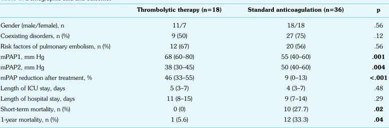
Of the 54 subjects, 18 (33.3%) underwent thrombolytic treatment, and 36 (66.7%) received standard anticoagulation therapy. Risk factors of PE, comorbid diseases, and length of stay in the ICU and hospital were comparable between the two groups. The median mPAP value on admission was significantly higher in subjects who received thrombolytic therapy (p=0.001). Control ECHO that was performed 48 h after the initiation of treatment revealed a signifi-cantly higher reduction of mPAP value in the thrombolytic therapy group than in the standard anticoagulation group. Short-term and 1-year mortality rates were significantly lower in the thrombolytic therapy group than in subjects who received standard anticoagula-tion therapy (Table 1).
No treatment-related side effect was observed in the anticoagu-lation group. However, minor bleeding (epistaxis) occurred only in 2 (11.1%) subjects secondary to thrombolytic therapy, and bleeding could be controlled by symptomatic treatment. There
was no statistically significant increase in major bleeding with thrombolytic therapy when compared with standard anticoagula-tion therapy (p>0.05).
DISCUSSION
The study revealed that thrombolytic treatment may reduce short-term mortality and is also associated with lower 1-year mortality in subjects with submassive PE than standard anticoagulation therapy. In addition, it was shown that thrombolysis leads to more reduction in the median of mPAP values, reverses right ventricular function, and restores pulmonary tissue perfusion. The present study failed to demonstrate the reduction of both total length of stay in the hospital and length of stay in the ICU in the thrombolysis group. Another finding was that the differences in the bleeding rates of the two treatment modalities were not statistically significant.
Acute PE often occurs rapidly and unpredictably, and it is a po-tentially fatal disorder with highly varying mortality rates. Fast and accurate diagnostic procedure and proper treatment may reduce the rate of mortality (10–12). Thrombolytic therapy is able to di-rectly dissolve thrombi and appears to accelerate the resolution of PE that helps to improve right ventricular dysfunction and myocar-dial damage. However, the usage of thrombolytic agents in the treatment of submassive PE remains controversial. The European Society of Cardiology guidelines for the treatment of acute PE rec-ommend anticoagulation therapy for nonmassive PE (13), whereas the 2008 American College of Chest Physicians evidence-based clinical practice guidelines consider thrombolytic therapy as an op-tion for subjects with a low risk of bleeding (14).
Increased afterload leads to right ventricular dysfunction in a case of submassive PE. Increased PAP is one of the hemodynamic factors directly related to right ventricular function and has been identified as a significant predictor of mortality in a case of right heart failure. In the present study, thrombolytic agents were especially adminis-tered to subjects with higher PAP values for the rapid resolution of clots. As a result, PAPs demonstrated by control ECHO after 48 h were significantly lower in the thrombolytic therapy group than in the anticoagulation group. Consistent with our data, Fei et al. an-nounced significantly lower PAP values in subjects who underwent
thrombolysis than in those who underwent standard anticoagula-tion treatment (15). It is possible that thrombolytic treatment is more effective in the reduction of mPAP and improvement of right ventricular function than standard anticoagulation therapy. The length of stay in the hospital and the length of stay in the ICU are possible indicators to demonstrate the effectiveness of throm-bolytic treatment in subjects with submassive PE. In the present study, there was no difference in the reduction of both these pa-rameters when two treatment modalities were compared. Berghaus et al. demonstrated a reduction of the total median length of stay in the hospital in subjects who underwent thrombolytic treatment. However, they could not demonstrate the reduction in the length of stay in the ICU, consistent with the results of the present study. The possible explanation for this difference is that there were some differences in the parameters that may affect these durations, such as age and comorbidities, between the subjects of the present study and the subjects they analyzed. Another possible reason of this situation is the tendency of the physicians for a longer and close medical follow-up of the thrombolytic group, which originates from the fear of bleeding (16).
The short-term mortality rate of submassive PE ranges from 3% to 15% (17, 18). Different in-hospital mortality rates were demonstrated in different studies of subjects with submassive PE to whom the thrombolytic agent was administered. Meneveau et al. observed the overall in-hospital mortality rate as 8.8% in their study, which was the first study to evaluate the short- and long-term effects of thrombolytic therapy in a large cohort of subjects with massive and submassive PE (19). The International Cooperative Pulmonary Embolism Registry investigators announced the mor-tality rate as 11.4% at 2 weeks of thrombolytic treatment (20). The thrombolytic arm of the Management Strategy and Prognosis of Pulmonary Embolism registry reported the short-term mortality rate as 4.7% at 4 weeks of thrombolytic treatment (21). As a new approach, some studies revealed that low-dose, brief duration infu-sions of alteplase may improve the efficacy of anticoagulation alone for submassive PE, without conferring a high risk of bleeding, par-ticularly in patients who have not had recent major surgery (22). In the treatment of PE, recanalization procedures do not appear to offer a clear advantage compared with standard anticoagulation.
Table 1. Demographic data and outcomes
Thrombolytic therapy (n=18) Standard anticoagulation (n=36) p
Gender (male/female), n 11/7 18/18 .56
Coexisting disorders, n (%) 9 (50) 27 (75) .12
Risk factors of pulmonary embolism, n (%) 12 (67) 20 (56) .56
mPAP1, mm Hg 68 (60–80) 55 (40–60) .001
mPAP2, mm Hg 38 (30–45) 50 (40–60) .004
mPAP reduction after treatment, % 46 (33–55) 9 (0–13) <.001
Length of ICU stay, days 5 (3–7) 4 (3–7) .48
Length of hospital stay, days 11 (8–15) 9 (7–14) .29
Short-term mortality, n (%) 0 (0) 10 (27.7) .02
1-year mortality, n (%) 1 (5.6) 12 (33.3) .04
Low-dose thrombolysis was associated with the lowest probability of dying and bleeding (23). The short-term mortality rate observed in the present study was 0% at 2 weeks of thrombolytic treatment. This rate is expressively lower than the short-term mortality rates demonstrated in the majority of previous studies.
It was shown that thrombolytic therapy preserves the diffusing ca-pacity of the lung and improves the pulmonary capillary blood flow volume at the end of the first year (1, 3). In the present study, thrombolytic treatment was found to be statistically correlated with a higher survival rate at 1 year of treatment than anticoagulation therapy. The survival rate at 1 year of thrombolytic treatment was 94.4% and similar to previous studies. Consistent with previous data, in the present study, the cause of death between week 2 and year 1 of thrombolytic treatment was not associated with throm-bolysis procedure or PE itself. Only one subject died due to acute myocardial infarction. A study containing 249 subjects demon-strated that the survival rate at 1 year of thrombolytic treatment was 92%, and the main reason of death was cancer. Only 20% of the deaths were directly related to PE and thrombolytic treatment (18% recurrent PE and 2% bleeding events) (19).
As a result, it may be possible to hypothesize that thrombolytic treatment has a capability to reduce the short-term and 1-year mor-tality rates in subjects who survived after an acute episode of sub-massive PE. In addition, the majority of the reasons for the deaths are not related to the complication or insufficiency of thrombolysis (recurrent PE), supporting the efficacy of this treatment modality. Although thrombolytic treatment dissolves clots to accelerate the resolution of PE, some authors do not support the routine use of thrombolytic agents in subjects who were normotensive with signs of right ventricular dysfunction (24–26). The bleeding com-plications of thrombolytic treatment have been demonstrated to be notably higher than those of anticoagulation treatment. The use of thrombolytic agents should be weighed against the risk of severe bleeding (27). The overall major bleeding may reach up to 20% (28). Especially the fear of bleeding complications is still the main reason to avoid the administration of thrombolytic agents. In the present study, only two subjects suffered from minor bleeding (epistaxis) related to thrombolytic treatment, whereas no bleeding was observed in the anticoagulation group. A study consisting of 50 subjects reported that the bleeding rate of thrombolytic ment was significantly higher than that of anticoagulation treat-ment; however, the authors predominantly observed dermatorrha-gia, particularly at the sites of blood vessel paracentesis (15). Cao et al. (29) and Nakamura et al. (30) also could not demonstrate the increased risk of major bleeding related to thrombolytic agents. Conversely, Geibel et al. reported a more than threefold increase in major bleeding related to thrombolytic treatment especially in female subjects with submassive PE. These different rates indicate the importance of assessing the risk of bleeding before the admin-istration of thrombolytic agents. In addition, it may be possible to hypothesize that thrombolysis is not a very dangerous process as it is feared of (31).
Despite the high incidence of acute PE worldwide, many questions on the optimal management of severe PE remain to be answered. Owing to the large spectrum of clinical presentation and outcomes, treat-ment has to be adapted according to the initial risk stratification (32).
As a limitation, first, this was not a randomized control trial that is the best study design to compare the effects of two treatment modalities. Second, these data reflect the attitudes and outcomes of subjects of a single center that limits the generalizability of the results. Third, the sample size was small due to the limited period of the study.
CONCLUSION
Thrombolytic therapy appears to be an effective and safe modality for the treatment in subjects with submassive PE. It should be ad-ministered especially to those selected subjects who have increased right ventricular afterload, low risk of bleeding, and an expecta-tion for a long survival time. In addiexpecta-tion, bleeding risk should be assessed carefully and individually prior to the administration of thrombolytic treatment. Further randomized study with a large sample size is needed for final conclusion.
Ethics Committee Approval: The study was approved by the local ethics committee (February 8, 2010/263).
Informed Consent: Written informed consent was obtained from all sub-jects for thrombolytic therapy.
Peer-review: Externally peer-reviewed.
Author Contributions: Designed the study: PÇ, DA. Collected the data: MÜ, CK, DA. Analyzed the data: ÖE, AEE, CK. Wrote the paper: CK, ÖE, AEE. All authors have read and approved the final manuscript.
Conflict of Interest: The authors have no conflict of interest to declare. Financial Disclosure: The authors declared that this study has received no financial support.
REFERENCES
1. Konstantinides SV, Barco S, Lankeit M, Meyer G. Management of Pul-monary Embolism: An Update. J Am Coll Cardiol 2016;67(8):976–90. 2. Sekhri V, Mehta N, Rawat N, Lehrman SG, Aronow WS. Manage-ment of massive and nonmassive pulmonary embolism. Arch Med Sci 2012;8(6):957–69. [CrossRef]
3. Konstantinides SV, Torbicki A, Agnelli G, Danchin N, Fitzmaurice D, Galiè N, et al. 2014 ESC guidelines on the diagnosis and management of acutepulmonary embolism. Eur Heart J 2014;35(43):3033–69. 4. Kearon C, Akl EA, Ornelas J, Blaivas A, Jimenez D, Bounameaux H,
et al. Antithrombotic Therapy for VTE Disease: CHEST Guideline and ExpertPanel Report. Chest 2016;149(2):315–52. [CrossRef]
5. Grifoni S, Olivotto I, Cecchini P, Pieralli F, Camaiti A, Santoro G, et al. Short-term clinical outcome of patients with acute pulmonary em-bolism, normal blood pressure, and echocardiographic right ventricular dysfunction. Circulation 2000;101(24):2817–22. [CrossRef]
6. Todd JL, Tapson VF. Thrombolytic therapy for acute pulmonary em-bolism: a critical appraisal. Chest 2009;135(5):1321–9. [CrossRef]
7. Wan S, Quinlan DJ, Agnelli G, Eikelboom JW. Thrombolysis com-pared with heparin for the initial treatment of pulmonary embolism: a meta-analysis of the randomized controlled trials. Circulation 2004;110(6):744–9. [CrossRef]
8. Dalen JE. The uncertain role of thrombolytic therapy in the treatment of pulmonary embolism. Arch Intern Med 2002;162(22):2521–3. 9. Jaff MR, McMurtry MS, Archer SL, Cushman M, Goldenberg N,
Gold-haber SZ, et al. Management of massive and submassive pulmonary embolism, iliofemoraldeep vein thrombosis, and chronic
thromboem-bolic pulmonary hypertension: a scientific statement from the American Heart Association. Circulation 2011;123(16):1788–830. [CrossRef]
10. Heit JA, Melton LJ 3rd, Lohse CM, Petterson TM, Silverstein MD, Mohr DN,et al. Incidence of venous thromboembolism in hospitalized patients vscommunity residents. Mayo Clin Proc 2001;76(11):1102– 10. [CrossRef]
11. Sanchez O, Planquette B, Meyer G. Update on acute pulmonary em-bolism. Eur Respir Rev 2009;18(113):137–47. [CrossRef]
12. Ryan MG, Westrich GH, Potter HG, Sharrock N, Maun LM, Macaulay W, et al. Effect of mechanical compression on the prevalence of proxi-mal deep venous thrombosis as assessed by magnetic resonance venog-raphy. J Bone Joint Surg Am 2002;84-A(11):1998–2004. [CrossRef]
13. Torbicki A, Perrier A, Konstantinides S, Agnelli G, Galiè N, Pruszczyk P, et al. Guidelines on the diagnosis and management of acute pul-monaryembolism: the Task Force for the Diagnosis and Management of AcutePulmonary Embolism of the European Society of Cardiology (ESC). Eur Heart J 2008;29(18):2276–315.
14. Kearon C, Akl EA, Comerota AJ, Prandoni P, Bounameaux H, Gold-haber SZ, et al. Antithrombotic therapy for VTE disease: Antithrom-botic Therapy and Prevention of Thrombosis, 9th ed: American Col-lege of Chest PhysiciansEvidence-Based Clinical Practice Guidelines. Chest 2012;141(2 Suppl):e419S–e496S. [CrossRef]
15. Fei J, Tang Y, Wu J, Kang L, Zhao J, Dai H, et al. Thrombolytic and anticoagulant therapy for acute submassive pulmonary embolism. Exp Ther Med 2014;7(1):103–8. [CrossRef]
16. Berghaus TM, Thilo C, Bluethgen A, von Scheidt W, Schwaiblmair M. Effectiveness of thrombolysis in patients with intermediate-risk pulmonary embolism: influence on length of hospital stay. Adv Ther 2010;27(9):648–54. [CrossRef]
17. Tapson VF, Carroll BA, Davidson BL, Elliott CG, Fedullo PF, Hales CA, et al. The diagnostic approach to acute venous thromboembolism. Clinical practice guideline. American Thoracic Society. Am J Respir Crit Care Med 1999;160(3):1043–66. [CrossRef]
18. American College of Emergency Physicians Clinical Policies Com-mittee; Clinical Policies Committee Subcommittee on Suspected Pul-monary Embolism. Clinical policy: critical issues in the evaluation and management of adultpatients presenting with suspected pulmonary embolism. Ann Emerg Med 2003;41(2):257–70. [CrossRef]
19. Meneveau N, Ming LP, Séronde MF, Mersin N, Schiele F, Caulfield F, et al. In-hospital and long-term outcome after sub-massive and mas-sivepulmonary embolism submitted to thrombolytic therapy. Eur Heart J 2003;24(15):1447–54. [CrossRef]
20. Goldhaber SZ, Visani L, De Rosa M. Acute pulmonary embolism:
clin-ical outcomes in the InternationalCooperative Pulmonary Embolism Registry (ICOPER). Lancet 1999;353(9162):1386–9. [CrossRef]
21. Konstantinides S, Geibel A, Olschewski M, Heinrich F, Grosser K, Rauber K, et al. Association between thrombolytic treatment and the prognosis of hemodynamically stable patients with major pulmonary embolism: resultsof a multicenter registry. Circulation 1997;96(3):882– 8. [CrossRef]
22. Lozier JN, Elinoff JM, Suffredini AF, Rosing DR, Sidenko S, Sherry RM, et al. Low-dose, short course alteplase treatment of submassive pulmonaryembolism: a case series from the National Institutes of Health ClinicalCenter. Blood Coagul Fibrinolysis 2018;29(8):701–7. 23. Jimenez D, Martin-Saborido C, Muriel A, Zamora J, Morillo R, Barrios
D, et al. Efficacy and safety outcomes of recanalisation procedures in patients with acute symptomatic pulmonary embolism: systematic re-view and network meta-analysis. Thorax 2018;73(5):464–71. [CrossRef]
24. Perlroth DJ, Sanders GD, Gould MK. Effectiveness and cost-effective-ness of thrombolysis in submassive pulmonary embolism. Arch Intern Med 2007;167(1):74–80. [CrossRef]
25. Zamanian RT, Gould MK. Effectiveness and cost effectiveness of thrombolysis in patients with acute pulmonary embolism. Curr Opin Pulm Med 2008;14(5):422–6. [CrossRef]
26. Ramakrishnan N. Thrombolysis is not warranted in submassive pul-monary embolism: a systematic review and meta-analysis. Crit Care Resusc 2007;9(4):357–63.
27. Liew J, Stevens J, Slatore C. Refractory Hypoxemia in a Patient with Submassive Pulmonary Embolismand an Intracardiac Shunt: A Case Report and Review of the Literature. Perm J 2018;22:17–061. 28. Fiumara K, Kucher N, Fanikos J, Goldhaber SZ. Predictors of major
hemorrhage following fibrinolysis for acute pulmonary embolism. Am J Cardiol 2006;97(1):127–9. [CrossRef]
29. Cao Y, Zhao H, Gao W, Wang Y, Cao J. Systematic review and meta-analysis for thrombolysis treatment in patients with acute submassive pul-monary embolism. Patient Prefer Adherence 2014;8:275–82. [CrossRef]
30. Nakamura S, Takano H, Kubota Y, Asai K, Shimizu W. Impact of the efficacy of thrombolytic therapy on the mortality of patientswith acute submassive pulmonary embolism: a meta-analysis. J Thromb Haemost 2014;12(7):1086–95. [CrossRef]
31. Geibel A, Olschewski M, Zehender M, Wilsch M, Odening K, Heinrich F, et al. Possible gender-related differences in the risk-to-benefit ratio of thrombolysis for acute submassive pulmonary embolism. Am J Cardiol 2007;99(1):103–7. [CrossRef]
32. Engelberger RP, Kucher N. Reperfusion Treatment for Acute Pul-monary Embolism. Hamostaseologie 2018;38(2):98–105. [CrossRef]