Design and development of a collimator mechanism that will be utilized in radiation therapy
Tam metin
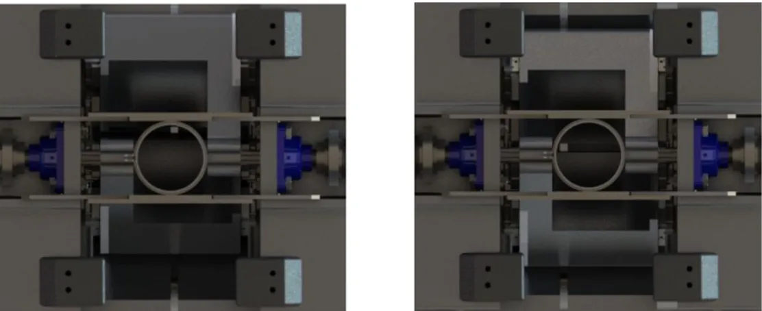
Şekil
![Figure 1.3: Leaf arrangement examples of MLCs [5,6].](https://thumb-eu.123doks.com/thumbv2/9libnet/3708077.24890/31.892.221.801.486.712/figure-leaf-arrangement-examples-of-mlcs.webp)
![Figure 1.20: Illustration of LINAC [21].](https://thumb-eu.123doks.com/thumbv2/9libnet/3708077.24890/46.892.131.767.181.592/figure-illustration-of-linac.webp)


Benzer Belgeler
Shirley Jackson’s famous story “The Lottery” takes place in an American town and like many of her works it includes elements of horror and mystery.. Name symbolism,
Bevacizumab Advanced colon kcancers, NSCLC, ovarium, renal cancers, glioblastoma multiforme mTOR pathway HIF1a>VEGFA inhibition. Ramucirumab Advanced gastric and eosophagal
Based on the analysis of the modern market for the development of innovative industrial products, as well as on the basis of a symbiosis of domestic
In case, there is no other charging station nearby and the primary battery is drained completely the system will automatically switch to the secondary battery and it is being
Fecal Occult Blood test (FOBT) in CRC screening: With the formation of cancer tissue or adenomas reaching a certain size, bleeding occurs into the lumen. In this case,
After performing normalization of the skeletal joint positions to achieve user independence and extraction of mean and standard deviation of the inertial data, the data obtained
In this paper, we propose a facial emotion recognition approach based on several action units (AUs) tracked by a Kinect v2 sensor to recognize six basic emotions (i.e., anger,
The outcomes are nothing out of ordinary while the same problems with thick papers being jammed persist and the best printing quality is achieved with 250 g/m 2
