echocardiography, tissue Doppler imaging,
carotid intima-media thickness, and
asymmetric dimethyl arginine levels in obese
adolescents with metabolic syndrome
Eyup Aslan1, Ahmet Sert2, Muammer Buyukinan3, Mustafa Ozgur Pirgon4, Huseyin Kurku5, Hakan Yilmaz6and Dursun Odabas7
1
Department of Pediatric Cardiology, Denizli State Hospital, Denizli, Turkey,2Department of Pediatric Cardiology, Faculty of Medicine, Selcuk University, Konya, Turkey,3Department of Pediatric Endocrinology and Diabetes, University of Health Sciences, Konya Training and Research Hospital, Konya, Turkey,4Department of Pediatric Endocrinology and Diabetes, Faculty of Medicine, Suleyman Demirel University, Isparta, Turkey,
5
Department of Biochemistry, University of Health Sciences, Konya Training and Research Hospital, Konya, Turkey,6Department of Radiology, Agri State Hospital, Agri, Turkey and7Department of Pediatric Cardiology, Faculty of Medicine, Necmettin Erbakan University Konya, Konya, Turkey
Abstract
Purpose: The aim of our study was to assess left ventricle and right ventricle systolic and diastolic functions in obese adolescents with metabolic syndrome using conventional echocardiography and pulsed-wave tissue Doppler imaging and to investigate carotis intima-media thickness, and asymmetric dimethyl arginine levels. Methods: A total of 198 obese adolescents were enrolled in the study. The obese patients were divided into metabolic syndrome group and non-metabolic syndrome group. All subjects underwent laboratory blood tests, including asymmetric dimethyl arginine, complete two-dimensional, pulsed, and tissue Doppler echocardiography, and measurement of the carotid intima-media thickness. Results: Obese adolescents were characterised by enlarged left end-diastolic, end-systolic and left atrial diameters, thicker left and right ventricular walls compared with non-obese adolescents. The metabolic syndrome group had normal left ventricle systolic function, impaired diastolic function, and altered global systolic and diastolic myocardial performance. In the metabolic syndrome obese group patients, left ventricle mass was found positively correlated with body mass index, waist and hip circumferences, diastolic blood pressure, age, and waist-to-hip circumference ratio. The carotid intima-media thickness was found positively correlated with waist and hip circumferences and total cholesterol levels. Asymmetric dimethyl arginine levels were found positively correlated with systolic blood pressure, waist-to-hip circumference ratio, and diastolic blood pressure. Conclusions: The results of this study demonstrate that metabolic syndrome in adolescence is associated with significant changes in myocardial geometry and function. In addition, it has been associated with a high level of asymmetric dimethyl arginine concentration and thicker carotid intima-media thickness reflecting endothelial dysfunction.
Metabolic syndrome is a cluster of risk factors including abdominal obesity, hypertension, insulin resistance, dyslipidaemia, which directly increase the risk of cardiovascular disease. Myocardial and vascular structural alterations may influence both left1and right ventricular2 function as well as an increased risk of atherosclerosis.3 Increased carotid intima-media thickness of the common carotid artery measured by carotid artery ultrasound has been a useful non-invasive marker of atherosclerosis in patients with metabolic syndrome.3
Nitric oxide is the main vasorelaxing and antithrombotic factor produced by endothelial cells through the action of endothelial nitric oxide synthase. Asymmetric dimethyl arginine is the major endogenous inhibitor of nitric oxide synthase leading to endothelial dysfunction and atherosclerotic vascular disease.4 Increased plasma asymmetric dimethyl arginine concentrations mainly occur following inhibition of the enzyme responsible for asymmetric dimethyl arginine catabolism, dimethylarginine dimethylaminohydrolase, through oxidative stress triggered by several cardiovascular risk factors.5 It has been shown in adults that patients with metabolic syndrome had higher plasma asymmetric dimethyl arginine levels and tissue Doppler imaging values than the controls, reflecting the presence of endothelial dysfunction.6
cambridge.org/cty
Original Article
Cite this article:Aslan E, Sert A, Buyukinan M, Pirgon MO, Kurku H, Yilmaz H, Odabas D (2019) Left and right ventricular function by echocardiography, tissue Doppler imaging, carotid intima-media thickness, and asymmetric dimethyl arginine levels in obese adolescents with metabolic syndrome. Cardiology in the Young29: 310–318. doi: 10.1017/S1047951118002329
Received: 30 July 2018 Revised: 19 November 2018 Accepted: 28 November 2018 First published online: 28 January 2019 Key words:
Adolescent; asymmetric dimethyl arginine; cardiovascular disease; carotid intima-media thickness; metabolic syndrome
Author for correspondence:
E. Aslan, Department of Pediatric Cardiology, Denizli State Hospital, Denizli, Turkey. Tel: +902582639311; Fax: +902582619206; E-mail: [email protected]
The aim of our study was to assess left ventricle and right ventricle systolic and diastolic functions in obese adolescents with metabolic syndrome using conventional echocardiography and pulsed-wave tissue Doppler imaging and to investigate asym-metric dimethyl arginine levels. We also studied left ventricle and right ventricle structural remodelling, and cardiovascular risk profiles in obese adolescents with metabolic syndrome.
Materials and method
A total of 198 obese adolescents, consisting of 118 females and 80 males with mean age of 13.9± 1.5 years; range 12–17; mean body mass index 31.1± 3.8 kg/m2, were recruited from obese adoles-cents admitted to the paediatric endocrinology unit between June 2013 and June 2014. The obese group was divided into two subgroups; one group is patients with metabolic syndrome con-sisting of 60 females and 40 males, with mean age of 14.0± 1.4 years; mean body mass index 32.4± 3.5 kg/m2; and the other is non-metabolic syndrome consisting of 58 females and 40 males, with mean age of 13.9± 1.3 years; mean body mass index 29.9± 3.6 kg/m2. Control adolescents – 52 females, 50 males; mean age 14.0± 1.3 years, range 12–17; mean body mass index 19.5± 2.3 kg/m2 – were selected from non-obese healthy adolescents.
Metabolic syndrome was defined according to the modified WHO criteria adapted for children. Subjects were diagnosed as having metabolic syndrome if they met three of four WHO cri-teria, which are obesity where body mass index is>95th percentile for age and sex; dyslipidaemia caused by high triglycerides (>136 mg/dl) in children ⩾ 10 years of age, low high-density lipoprotein cholesterol (<35 mg/dl), or high total cholesterol (>95th percentile); hypertension where systolic blood pressure is >95th percentile for age and sex and height; and abnormal glucose homeostasis caused by fasting hyperinsulinemia, impaired fasting glucose, or impaired glucose tolerance).7
Patients with clinical or laboratory signs of any systemic disease, including type 1 or type 2 diabetes mellitus; taking medications; or that had conditions known to affect insulin action or insulin secretion, for example glucocorticoid therapy, hypothyroidism, and Cushing disease; CHD; valvular heart disease; chronic renal failure; and smokers were excluded from the study. The study protocols were approved by our hospital’s ethics committee (approval number: 0.28.00.00/130-304). Signed informed consent forms were obtained from the parents of the adolescents.
Height and weight were measured with an empty bladder in post-absorptive conditions. Body mass index was calculated as weight in kilograms divided by the square of height in meters. Patients with body mass index ⩾ 95th percentile according to reference curves for Turkish adolescents were accepted as obese.8 Waist circumference was measured midway between the lowest rib and the top of the iliac crest at the end of gentle expiration. Hip circumference was measured over the great trochanters. Pubertal developmental stage was assessed by a single paediatric endocrinologist using the Tanner criteria. Staging for sexual maturation was >2 in all patients; Tanner stages II–IV. After resting for⩾ 5 minutes, systolic and diastolic blood pressures were measured in the sitting position, using a mercury-gravity manometer and a cuff appropriate for body size.
Blood samples were obtained in the morning by venipuncture after an overnight fast, that is ⩾ 12 hours fasting. Serum
concentrations of total cholesterol, high-density lipoprotein cho-lesterol, and triglycerides were measured using routine enzymatic methods with an Abbott Diagnostics c16000 chemistry analyser (Abbott Diagnostics, Lake Forest, IL, United States of America). Serum insulin levels were measured using the IMMULITE immunoassay (Siemens Healthcare Diagnostics, Camberley, United Kingdom). Insulin resistance was estimated by home-ostasis model assessment of insulin resistance using the formula: fasting insulin concentration (mU/ml) × fasting glucose con-centration (mmol/L)/22.5.9
Measurement of asymmetric dimethyl arginine levels was accomplished by high-performance liquid chromatography, using the method described by Chen et al.10
Carotis ultrasound studies were performed by a single radi-ologist, who was blinded to the clinical and laboratory status of the patients, using high-resolution B-mode ultrasonography (Logiq 7) and a high-resolution linear-array vascular transducer (14 MHz). An optimal two-dimensional image of the common carotid artery was obtained in which the near and far wall intima-media complex was well visualised. After a 10-minute rest, the M-mode cursor was placed 1 cm proximal to the beginning of the carotid artery bulb during end-diastole. Carotid intima-media thickness was calculated by taking the mean value of three measurements. There was no evidence of carotid plaque forma-tion in all obese and control groups.
All echocardiographic and Doppler examinations were per-formed by a single expert paediatric cardiologist, who was blinded to the clinical and laboratory results of the study group. ProSound Alpha 7 (Aloka, Hitachi-Aloka Medical, Tokyo, Japan) with a 3-MHz phased-array transducer was used for each study subject. Conventional echocardiographic evaluation from the parasternal long-axis view included left ventricle diastolic and end-systolic diameter, septal and left ventricle posterior wall thick-nesses in diastole and systole, right ventricle free wall thickthick-nesses in diastole and systole, the left ventricle ejection fraction, and left ventricle fractional shortening. Teichholz’s M-mode formula was used to calculate the left ventricle ejection fraction and left ven-tricle fractional shortening. All data were determined according to the recommendations of the American Society of Echocardio-graphy.11 Relative posterior wall thickness was calculated as (interventricular septal thickness in diastole + left ventricle pos-terior wall thickness in diastole)/left ventricle end-diastolic dia-meter. Left ventricular mass was calculated using the Devereux12 formula and indexed to height.2,7
Transmitral and transtricuspid flow patterns were obtained by pulsed-wave Doppler echocardiography from the apical four-chamber view with the sample volume placed at the mitral and tricuspid valves leaflets tips. Peak early (Em, Et) and late (Am,
At) diastolic velocities, the (E/A)m and (E/A)t ratios,
isovolu-metric relaxation times, isovoluisovolu-metric contraction times, and ejection times were measured. The Doppler-derived index of combined systolic and diastolic myocardial performance (Tei index; [isovolumetric relaxation time + isovolumetric con-traction time]/ejection time) was used to quantify global left and right ventricle functions.13
A 5-mm pulsed-wave Doppler tracing was placed at the septal and lateral segments of the mitral valve, and lateral segment of the tricuspid valve and peak myocardial systolic (sm, st), early
(e′m, e′t), and late diastolic (a′m, a′t) velocities were measured
from the apical four-chamber view. (E/e′)mand (E/e′)tratios of
the left and the right ventricle were determined by using pre-viously estimated Doppler values.
The left and right ventricle Tei index was calculated according to the formula: (Isovolumic relaxation time + Isovolumic con-traction time)/Ejection time.
Right ventricle global systolic function was assessed as the tricuspid annular plane systolic excursion, which was measured as the difference between the distance among the tricuspid annulus and right ventricle apex end-diastole and end-systole of the same cardiac cycle.14
Continuous variables are expressed as mean± SD (range). Normality assumptions were assessed before conducting para-metric tests. When all groups were compared for parameters, analyses of variance were used; post-hoc analysis was performed using Tukey’s honestly significantly different test. Bivariate associations of continuous variables were assessed using Pearson’s correlation coefficients.
Stepwise multivariate linear regression was then used to determine which determinants independently explained a significant (p< 0.05) fraction of the variance of the dependent variables. A p-value <0.05 was considered to indicate statistical significance. Statistical analyses were performed using SPSS version 15.0 for Windows (SPSS, Inc., Chicago, IL, United States of America).
Results
The characteristics of the patients and healthy controls are listed in Table 1. Metabolic syndrome was present in 100 of 198 of all
obese adolescents (50.5%). There was no statistically important difference in heart rate between the obese subjects metabolic or non-metabolic syndrome and controls. The metabolic syndrome obese group had significantly higher body mass index, waist and hip circumferences, and systolic and diastolic blood pressure than the non-metabolic syndrome obese and control groups.
The levels of serum triglyceride, fasting glucose, and insulin were significantly higher and levels of high-density lipoprotein cholesterol were significantly lower in the metabolic syndrome obese group compared with controls. The control group had significantly lower homeostasis model assessment of insulin resistance values than the metabolic and non-metabolic syndrome obese groups. We also found that asymmetric dimethyl arginine levels were significantly lower in the control group than the metabolic syndrome and non-metabolic syndrome obese groups. Two-dimensional, M-mode, and pulsed-wave Doppler con-ventional echocardiographic findings and carotid intima-media thickness results of the patients and controls are listed in Table 2. Left ventricle M-mode measurements were significantly larger in the metabolic and non-metabolic syndrome obese groups com-pared with controls. The metabolic syndrome obese group had significantly higher interventricular septal thickness in systole and left ventricle posterior wall thickness in diastole than the non-metabolic syndrome obese and control groups. The left atrial and aortic diameters were significantly higher in the metabolic syn-drome group than the control group. By conventional echo-cardiography, the left ventricle ejection fraction and left ventricle shortening fraction were similar in the obese and control groups. Table 1. Clinical features of healthy controls and obese adolescents.
Variable Healthy controls Obese Non-MS Subjects MS
Subjects (female/male) 102 98 100
Age (years) 14.0 ± 1.3 (12–17) 13.9 ± 1.3 (12–17) 14.0 ± 1.4 (12–17)
BMI (kg/m2) 19.5 ± 2.3 (16.1–24.5) 29.9 ± 3.6 (25.1–39.7)ˠ 32.4 ± 3.5 (26.5–39.9)δ*
Waist circumference (cm) 73 ± 7 (56–93) 95 ± 10 (68–128)ˠ 99 ± 11 (69–132)δ*
Hip circumference (cm) 88 ± 7 (71–106) 106 ± 10 (70–144)ˠ 112 ± 9 (94–157)δ*
Waist circumference/hip circumference ratio 0.82 ± 0.06 (0.67–1) 0.89 ± 0.10 (0.71–1.71)ˠ 0.88 ± 0.07 (0.52–1.05)δ
Systolic BP (mmHg) 102 ± 8 (80–120) 114 ± 9 (100–150)ˠ 125 ± 12 (100–150)δ*
Diastolic BP (mmHg) 63 ± 7 (40–80) 73 ± 7 (60–100)ˠ 78 ± 9 (65–100)δ*
Heart rate (beats/min) 89 ± 12 (61–117) 89 ± 11 (62–125) 90 ± 14 (58–133)
Total cholesterol (mg/dl) 152 ± 23 (104–213) 166 ± 27 (111–246)ˠ 163 ± 31 (98–269)δ
Triglycerides (mg/dl) 86 ± 30 (24–154) 100 ± 36 (25–192)ˠ 167 ± 78 (54–398)δ*
Low-density lipoprotein cholesterol (mg/dl) 86 ± 19 (38–138) 96 ± 23 (49–168)ˠ 91 ± 30 (22–171)
High-density lipoprotein cholesterol (mg/dl) 50 ± 8 (33–74) 48 ± 8 (30–70) 38 ± 7 (25–62)δ*
Fasting glucose (mg/dl) 81 ± 5 (69–92) 88 ± 11 (62–125)ˠ 94 ± 10 (81–146)δ*
Fasting insulin (U/ml) 7.3 ± 2.4 (2–15) 14.5 ± 9.0 (3–59)ˠ 25.2 ± 15.8 (7–102)δ*
HOMA-IR 1.4 ± 0.4 (0.4–3) 3.1 ± 2.0 (0.7–12.8)ˠ 6.1 ± 4.9 (1.6–36)δ*
ADMA 0.35 ± 0.22 (0.1–1.00) 0.54 ± 0.23 (0.16–1.29)ˠ 0.60 ± 0.25 (0.16–1.57)δ
ADMA= asymmetric dimethylarginine; BMI = body mass index; BP = blood pressure; HOMA-IR = homeostasis model assessment of insulin resistance; MS = metabolic syndrome Data are expressed as mean ± SD (range)
*p< 0.05, obese subjects with non-MS versus obese subjects with MS ˠp < 0.05, controls versus obese subjects with non-MS
The right ventricle free wall thicknesses in systole and diastole were significantly higher in the metabolic and non-metabolic syndrome obese groups than the controls. Tricuspid annular plane systolic excursion was completely preserved in all groups. The metabolic and non-metabolic syndrome obese groups had significantly higher carotid intima-media thickness and left ventricle mass than the controls.
The parameters of the left ventricle myocardial systolic func-tion obtained by the tissue Doppler– s′septaland s′lateral– seemed
normal in interventricular septum and left ventricle lateral wall in all groups. The parameters of the left ventricle diastolic function such as E/Amitralwere lower in the metabolic and non-metabolic
syndrome obese groups than the controls, but E/e′septal and
E/e′lateralvalues were similar in each group. The left ventricle Tei
index was also significantly higher in subjects in the metabolic and non-metabolic syndrome obese groups than the controls. The parameters of the right ventricle myocardial systolic function seemed normal in interventricular septum and right ventricle lateral wall in all groups. However, the parameters of the right ventricle diastolic function such as E/Atricuspid and E/e′tricuspid
were lower and higher, respectively, in the metabolic and non-metabolic syndrome groups as compared with the controls. The right ventricle Tei index was also significantly higher in the
metabolic and non-metabolic syndrome obese groups than the controls (Table 3).
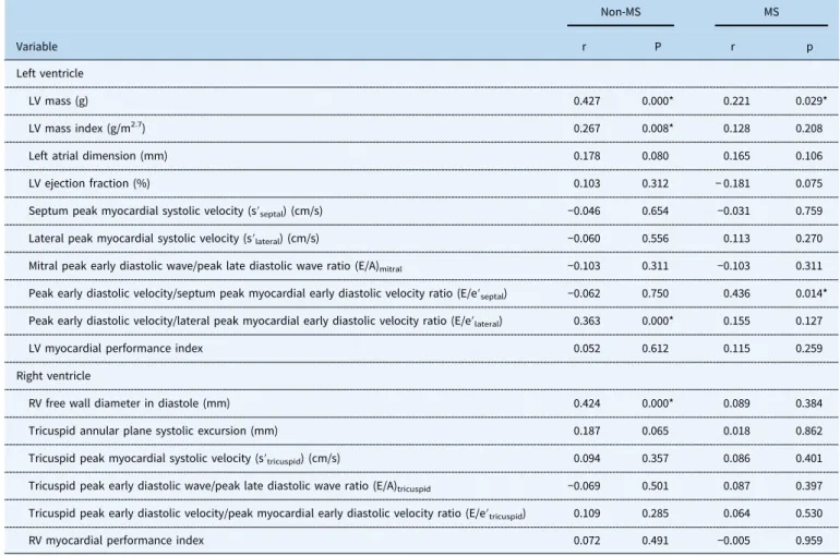
In the metabolic syndrome obese group, left ventricle mass was positively correlated with body mass index, waist and hip circumferences, diastolic blood pressure, age, and waist to hip circumference ratio. In addition, carotid intima-media thickness was positively correlated with waist and hip circumferences, total cholesterol levels, left ventricle mass, and E/e′septal(Table 4), and
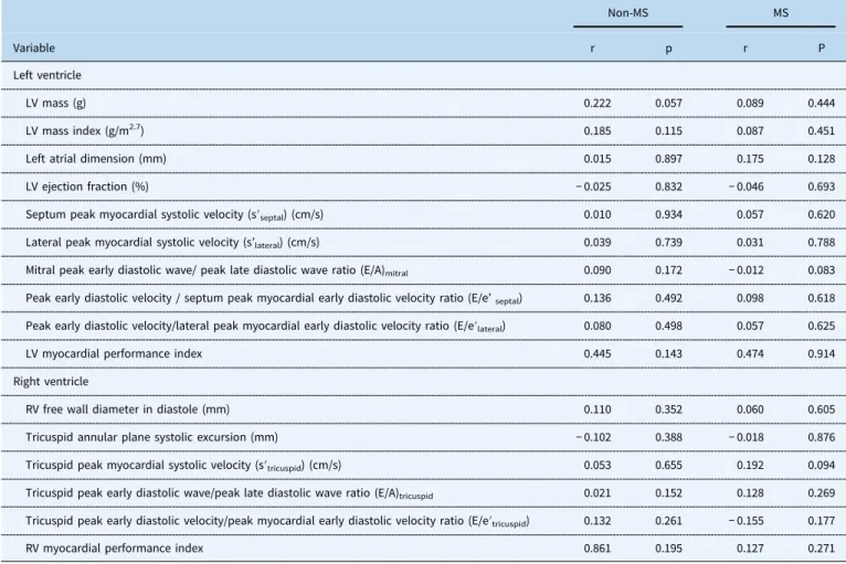
asymmetric dimethyl arginine was positively correlated with systolic and diastolic blood pressure and, waist to hip cir-cumference ratio in the metabolic syndrome obese group (Table 5).
To evaluate which variables independently influenced left ventricle mass, a multiple regression analysis model was per-formed in the metabolic syndrome obese group. A multiple stepwise regression was conducted to disclose the independent contributions of age, the ratio of waist-to-hip circumference, systolic and diastolic blood pressures, homeostasis model assessment of insulin resistance, total cholesterol, high-density lipoprotein cholesterol, asymmetric dimethyl arginine and regional tissue Doppler imaging myocardial performance indexes of left ventricle mass. The independent variables for left ventricle mass were age (β: 0.344, p: 0.006), and diastolic Table 2. Conventional echocardiographic findings and carotid intima-media thickness measurements in controls and obese adolescents.
Controls (n= 102) Non-MS (n= 98) MS (n= 100)
Left ventricle
LV end-diastolic diameter (mm) 42.8 ± 4.1 (34–55) 44.0 ± 3.5 (35–53)ˠ 44.6 ± 4.2 (35–56)δ
LV end-systolic diameter (mm) 26.8 ± 3.7 (19–35) 27.2 ± 3.7 (18–35) 28.0 ± 3.6 (18–40)δ
Interventricular septal diameter in diastole (mm) 7.2 ± 0.6 (5–9) 8.4 ± 1.1 (6–13)ˠ 9.3 ± 1.5 (7–14)*δ
Interventricular septal diameter in systole (mm) 8.3 ± 1.1 (6–12) 9.3 ± .1.3 (7–14)ˠ 10.0 ± .1.4 (6–14)*δ
LV posterior wall diameter in diastole (mm) 6.7 ± 1.2 (4–12) 7.9 ± 1.2 (6–14)ˠ 8.0 ± 1.1 (6–12)δ
LV posterior wall diameter in systole (mm) 10.9 ± 2.2 (8–18) 12.1 ± 2.4ˠ(8–29)ˠ 12.5 ± 1.8 (9–16)δ
LV ejection fraction (%) 67 ± 3.8 (60–79) 67 ± 4.0 (60–80) 67 ± 4.5 (60–83)
LV fractional shortening (%) 37 ± 3.3 (31–48) 37 ± 3.0 (32–48) 37 ± 5.1 (30–51)
Relative wall thickness (mm) 3.26 ± 0.37 (2.40–4.10) 3.72 ± 0.51 (2.64–5.29)ˠ 3.90 ± 0.58 (2.80–5.36)*δ
LV mass (g) 99 ± 28.6 (44–213) 124 ± 26.0 (68–209)ˠ 133 ± 34.2 (75–234)*δ
LV mass index (g/m2.7) 28 ± 5.7 (15–48) 35 ± 7.7 (17–67) 36 ± 9.0 (23–68)
Left atrial dimension (mm) 25 ± 3 (18–37) 28 ± 2.7 (21–34)ˠ 30 ± 3.6 (22–41)δ
Aortic dimension (mm) 19 ± 2.2 (15–26) 21 ± 2.7 (16–33)ˠ 22 ± 2.9 (11–29)δ
Right ventricle
RV free wall diameter in diastole (mm) 4.19 ± 0.64 (3–6) 5.67 ± 0.87 (4–8)ˠ 5.80 ± 0.82 (4–7)*δ
RV free wall diameter in systole (mm) 5.39 ± 0.81 (3–7) 7.05 ± 0.95 (5–9)ˠ 7.60 ± 0.88 (6–10)δ
Tricuspid annular plane systolic excursion (mm) 22.8 ± 2.8 (17–29) 24.5 ± 2.7 (19–32)ˠ 25.4 ± 2.89(19–33)*δ
CIMT (mm) 0.370 ± 0.025 (0.32–0.45) 0.421 ± 0.042 (0.32–0.58)ˠ 0.444 ± 0.052 (0.34–0.65)*δ
CIMT= carotid intima-media thickness; LV = left ventricle; MS = metabolic syndrome; RV = right ventricle Data are expressed as mean ± SD (range)
*p< 0.05, obese subjects with non-MS versus obese subjects with MS ˠp < 0.05, controls versus obese subjects with non-MS
blood pressure (β: 0.122, p: 0.036) in the metabolic syndrome group.
Discussion
A considerable number of studies exist, which show the harmful effects of metabolic syndrome and its components on the struc-ture and function of the heart. On the contrary, it was shown in previous studies that asymmetric dimethyl arginine values, which negatively influence vascular haemodynamics in metabolic syn-drome patients, were found high. In addition, it is known that the measurement of carotid intima-media thickness is a useful method in assessing the development of atherosclerosis. However, there are no studies on asymmetric dimethyl arginine and carotid intima-media thickness that relate to the structure and function of the heart in adolescents with metabolic syndrome. Our study is the first study in literature to assess the effects of metabolic syndrome on the right and left ventricular structure and function and carotid intima-media thickness as well as asymmetric dimethyl arginine levels.
The present study demonstrates that patient with metabolic syndrome have significantly changed left and right ventricles geometry and diastolic functions. Furthermore, we found increased carotid intima-media thickness and asymmetric dime-thyl arginine levels in metabolic syndrome obese adolescents compared to non-metabolic syndrome group and controls.
In obesity, which is the main parameter of the metabolic syndrome, left and right ventricular dilatation and hypertrophy have been shown in previous studies.15,16 The Bogalusa Heart Study revealed that obesity was associated with left ventricle dilatation and hypertrophy, and they also found that body mass index was the only independent predictor of left ventricle struc-ture.17 In our study, we found that left ventricle diameters, left
ventricular mass, relative posterior wall thickness, and right ventricle free wall thickness were significantly higher in metabolic and non-metabolic syndrome obese groups compared with con-trols. Moreover, we also identified positive correlation between body mass index, waist and hip circumference measurements, and left ventricular mass and right ventricle free wall thicknesses.
Hypertension, often present in metabolic syndrome, has an adverse effect on intima-media and increase afterload of the heart. Tadic et al18 reported that left ventricle relative posterior wall
thickness, left ventricular mass index, and right ventricle free wall thickness were significantly higher in non-dippers hypertensive patients irrespective of the presence of metabolic syndrome. Despite that, the meta-analysis by Li et al19 reported that the metabolic syndrome patients combined with hypertension showed higher left ventricular mass, and relative posterior wall thickness when compared to those with hypertension alone. In addition, they claimed that metabolic syndrome played an important role in the development of left ventricular hypertrophy, and metabolic syndrome amplified hypertension-related cardiac changes. In our study, metabolic and non-metabolic syndrome Table 3. Comparison of left and right ventricular systolic and diastolic functions by pulse wave Doppler and tissue Doppler imaging measurements in controls and obese adolescents.
Controls (n= 102) Non-MS (n= 98) MS (n= 100)
Left ventricle
Mitral peak early diastolic wave (E) (m/s) 0.94 ± 0.13 (0.59–1.35) 0.94 ± 0.15 (0.66–1.40) 0.91 ± 0.14 (0.57–1.30) Mitral peak late diastolic wave (A) (m/s) 0.53 ± 0.12 (0.31–0.81) 0.53 ± 0.11 (0.32–0.96) 0.53 ± 0.11 (0.29–0.83) Mitral peak early diastolic wave/peak late diastolic wave ratio (E/A)mitral 1.84 ± 0.35 (1.18–2.90) 1.79 ± 0.30 (1.18–2.87) 1.74 ± 0.27 (1.26–2.46)δ
Septum peak myocardial systolic velocity (s′septal) (cm/s) 10.6 ± 1.5 (7.8–18.4) 10.9 ± 1.7 (7.5–16.6) 11.1 ± 1.8 (8.0–18.2)δ
Septum peak myocardial early diastolic velocity (e′septal) (cm/s) 15.8 ± 2.1 (5.6–22.1) 15.4 ± 1.9 (11.8–20.4) 15.4 ± 1.8 (10.9–21.2)
Lateral peak myocardial systolic velocity (s′lateral) (cm/s) 11.4 ± 2.0 (7.6–21.5) 11.1 ± 1.8 (8.0–17.3) 11.6 ± 1.7 (8.6–17.0)
Lateral peak myocardial early diastolic velocity (e′lateral) (cm/s) 19.2 ± 3.2 (11.1–27.2) 18.8 ± 3.1 (10.9–27.2) 18.6 ± 2.3 (13.6–24.1)
LV global performance index 0.650 ± 0.113 (0.3–1.2) 0.776 ± 0.160 (0.4–1.1)ˠ 0.767 ± 0.167 (0.4–1.2)δ*
Right ventricle
Tricuspid peak early diastolic wave (E) (m/s) 64.8 ± 8.3 (44–82) 65.5 ± 11.0 (44–90) 66.4 ± 9.9 (46–101)
Tricuspid peak late diastolic wave (A) (m/s) 38.8 ± 8.4 (22–60) 40.6 ± 9.2 (26–84) 40.9 ± 8.2 (25–73)
Tricuspid peak early diastolic wave/ peak late diastolic wave ratio (E/A)tricuspid 1.72 ± 0.3 (1.2–2.6) 1.65 ± 0.3 (0.88–2.3) 1.66 ± 0.3 (1.09–2.4)
Tricuspid peak myocardial systolic velocity (s′tricuspid) (cm/s) 14.8 ± 1.9 (10.6–21.0) 14.3 ± 2.6 (9.1–22.4) 14.2 ± 3.1 (9.1–22.4)
Tricuspid peak myocardial early diastolic velocity (e′tricuspid) (cm/s) 17.5 ± 3.8 (12.5–32.0) 16.3 ± 2.8 (8.4–24.4) 16.7 ± 3.1 (8.8–28.0)*
RV global performance index 0.60 ± 0.10 (0.28–0.91) 0.72 ± 0.12 (0.36–1.03)ˠ 0.75 ± 0.12 (0.33–1.00)*
LV= left ventricle; MS = metabolic syndrome; RV = right ventricle Data are expressed as mean ± SD (range)
*p< 0.05, obese subjects non-MS versus obese subjects with MS ˠp < 0.05, controls versus obese subjects with non-MS
obese groups had significantly higher systolic and diastolic blood pressure than the controls. Furthermore, we found that there is a positive correlation between systolic and diastolic blood pressure and left ventricular mass, and right ventricle free wall thickness in metabolic and non-metabolic syndrome obese groups, which agreed with the previous studies.
There are conflicting results regarding the effects of insulin resistance on the heart structure. In African-American adoles-cents, Gidding et al20 found that, in cases, where patients with severe obesity and chronic hypertension were excluded, a weak relationship of left ventricle relative wall thickness with insulin resistance was estimated by homeostasis model assessment of insulin resistance. Karamitsos et al21 found normal systolic function but impaired diastolic function in both ventricles in the patient with type 1 diabetes mellitus, and were unable to find any correlations between diabetes and right ventricle structure. In turn, some researchers22–24stated that it had a negative effect on the ventricle structure. In our study, the homeostasis model assessment of insulin resistance values in obese groups were higher as compared to the control group; however, no correla-tions between homeostasis model assessment of insulin resistance and left ventricular mass were found in the study groups.
In the majority of studies, systolic function of the left ventricle was reported to be in the normal range in metabolic syndrome. Mangner et al15found comparable left ventricle ejection fraction, decreased tissue Doppler-derived systolic peak velocity, and
regional basoseptal strain in obese children compared with non-obese children. Faganello et al25found that left ventricle systolic function measured as stress-corrected mid-wall shortening and s′ was frequently impaired in type 2 diabetes mellitus patients without coronary artery disease; however, the coexistence of metabolic syndrome is not associated with more severe left ven-tricle systolic dysfunction. In our study, it was seen that left ventricle myocardial normal sseptal, lateral values and systolic
function measured by ejection fraction and tissue Doppler ima-ging were preserved in the obese and control groups. In addition, tricuspid lateral wall myocardial stricuspidand tricuspid annular
plane systolic excursion were found to be within normal ranges in obese and control groups. While Karakurt et al26measured lower tricuspid annular plane systolic excursion values in metabolic syndrome, Tadic et al2 found that the right ventricle tricuspid annular plane systolic excursion, stricuspid and right ventricle
ejection fraction values were normal in metabolic syndrome and reported preservation of the systolic functions.
In contrary to the preservation of systolic functions, it was stated that there was left ventricle diastolic dysfunction with metabolic syndrome. Ayalon et al1 reported that preclinical left
ventricle diastolic dysfunction could occur independently of left ventricle hypertrophy in metabolic syndrome with lower E/A ratio and mean e′. While Tadic et al27
found non-dipping pattern and metabolic syndrome separately cause left ventricle diastolic dysfunction in hypertensive patients, Akçay et al28 found lower Table 4. Pearson’s correlation between carotid intima-media thickness and cardiac geometry and functions in obese adolescents.
Non-MS MS
Variable r P r p
Left ventricle
LV mass (g) 0.427 0.000* 0.221 0.029*
LV mass index (g/m2.7) 0.267 0.008* 0.128 0.208
Left atrial dimension (mm) 0.178 0.080 0.165 0.106
LV ejection fraction (%) 0.103 0.312 − 0.181 0.075
Septum peak myocardial systolic velocity (s′septal) (cm/s) −0.046 0.654 −0.031 0.759
Lateral peak myocardial systolic velocity (s′lateral) (cm/s) −0.060 0.556 0.113 0.270
Mitral peak early diastolic wave/peak late diastolic wave ratio (E/A)mitral −0.103 0.311 −0.103 0.311
Peak early diastolic velocity/septum peak myocardial early diastolic velocity ratio (E/e′septal) −0.062 0.750 0.436 0.014*
Peak early diastolic velocity/lateral peak myocardial early diastolic velocity ratio (E/e′lateral) 0.363 0.000* 0.155 0.127
LV myocardial performance index 0.052 0.612 0.115 0.259
Right ventricle
RV free wall diameter in diastole (mm) 0.424 0.000* 0.089 0.384
Tricuspid annular plane systolic excursion (mm) 0.187 0.065 0.018 0.862
Tricuspid peak myocardial systolic velocity (s′tricuspid) (cm/s) 0.094 0.357 0.086 0.401
Tricuspid peak early diastolic wave/peak late diastolic wave ratio (E/A)tricuspid −0.069 0.501 0.087 0.397
Tricuspid peak early diastolic velocity/peak myocardial early diastolic velocity ratio (E/e′tricuspid) 0.109 0.285 0.064 0.530
RV myocardial performance index 0.072 0.491 −0.005 0.959
*Statistically significant (p< 0.05) LV= left ventricle; RV = right ventricle
E/A ratio, higher E/e′ and myocardial performance index values in normotensive prediabetics. Furthermore, Karakurt et al26 found that adult metabolic syndrome patients with preserved left ventricle ejection fraction had low E, E/A ratio in right ventricle, and high right ventricle global myocardial performance index as compared to the control group and identified diastolic dysfunc-tion. In our study, we found lower E/Amitral and higher
myo-cardial performance index in obese adolescents, but nearly similar values in E/e′lateraland E/e′septalin obese groups as compared to
the controls.
Similarly with left ventricle diastolic dysfunction, Tadic et al18 reported that metabolic syndrome, hypertension with non-dipping pattern, and their interaction had a huge impact on right ventricle wall thickness, parameters of right ventricle dia-stolic function (E/At, E/e′t, e′/a′t), as well as right ventricle global function estimated by the myocardial performance index. Gokdeniz et al29assessed right ventricle in metabolic syndrome patients with strain and reported deterioration in right ventricle systolic and diastolic functions in comparison with the control group. In our study, we also found right ventricle diastolic dys-function in obese adolescents with lower E/Atricuspid, and higher
E/e′tricuspid and right ventricle global myocardial performance
index than the controls.
High carotid intima-media thickness measurement in children with metabolic syndrome was shown in studies.3,30 Elshorbagy et al31 identified a significant increase in carotid intima-media
thickness in metabolic syndrome patients as compared to the non-metabolic syndrome obese and control groups. Hirata et al32 demonstrated in a study, which included 1727 subjects, that an elevated number of metabolic syndrome components, with or without central obesity, was associated with higher intima-media thickness and, hypertension had the strongest association with higher intima-media thickness. Alp et al33 found that carotid artery intima-media thickness was strongly associated with Tei index values. In our study, we found significant increased carotid intima-media thickness in metabolic syndrome group as com-pared to the non-metabolic syndrome obese and control groups. Moreover, carotid intima-media thickness is correlated with waist and hip circumference measurements, total cholesterol, left ven-tricular mass and E/e′septalratio in metabolic syndrome patients.
Increased plasma asymmetric dimethyl arginine concentra-tions cause impaired nitric oxide synthesis leading to endothelial dysfunction and atherosclerotic vascular disease.4 Bai et al34 showed a significant association of asymmetric dimethyl arginine with carotid intima-media thickness. Palomo et al35 found that asymmetric dimethyl arginine levels were significantly increased in the metabolic syndrome group and the levels of asymmetric dimethyl arginine were modestly but significantly correlated only with waist circumference among components of metabolic syn-drome. Similar to Palomo et al,35 we found that asymmetric dimethyl arginine levels were significantly higher, both in meta-bolic and non-metameta-bolic syndrome obese groups compared to the Table 5. Pearson’s correlation between asymmetric dimethyl arginine and cardiac geometry and functions in obese adolescents.
Non-MS MS
Variable r p r P
Left ventricle
LV mass (g) 0.222 0.057 0.089 0.444
LV mass index (g/m2.7) 0.185 0.115 0.087 0.451
Left atrial dimension (mm) 0.015 0.897 0.175 0.128
LV ejection fraction (%) − 0.025 0.832 − 0.046 0.693
Septum peak myocardial systolic velocity (s′septal) (cm/s) 0.010 0.934 0.057 0.620
Lateral peak myocardial systolic velocity (s’lateral) (cm/s) 0.039 0.739 0.031 0.788
Mitral peak early diastolic wave/ peak late diastolic wave ratio (E/A)mitral 0.090 0.172 − 0.012 0.083
Peak early diastolic velocity / septum peak myocardial early diastolic velocity ratio (E/e’septal) 0.136 0.492 0.098 0.618
Peak early diastolic velocity/lateral peak myocardial early diastolic velocity ratio (E/e′lateral) 0.080 0.498 0.057 0.625
LV myocardial performance index 0.445 0.143 0.474 0.914
Right ventricle
RV free wall diameter in diastole (mm) 0.110 0.352 0.060 0.605
Tricuspid annular plane systolic excursion (mm) − 0.102 0.388 − 0.018 0.876
Tricuspid peak myocardial systolic velocity (s′tricuspid) (cm/s) 0.053 0.655 0.192 0.094
Tricuspid peak early diastolic wave/peak late diastolic wave ratio (E/A)tricuspid 0.021 0.152 0.128 0.269
Tricuspid peak early diastolic velocity/peak myocardial early diastolic velocity ratio (E/e′tricuspid) 0.132 0.261 − 0.155 0.177
RV myocardial performance index 0.861 0.195 0.127 0.271
*Statistically significant (p<0.005) LV= left ventricle; RV = right ventricle
controls. Furthermore, we found a correlation between asym-metric dimethyl arginine and waist/hip ratio, systolic and diastolic blood pressure in metabolic syndrome group.
Limitations
There are some limitations to our study. As this was a cross-sectional study design, it does not allow us making conclusions about the causal relationships among variables and potential reversibility of the changes with weight loss over time. Therefore, the clinical significance of these changed values in obese persons remains unknown and will require extensive longitudinal follow-up to ultimately determine their predictive value. In addition, our healthy controls were patients who were free from any metabolic syndrome factors but referred to our department for echo-cardiography. For this reason, they were perfectly healthy sub-jects. This resulted by design in baseline differences of clinical characteristics between participants with metabolic and non-metabolic syndrome. It is therefore possible that residual con-founding could in part account for our findings.
Conclusion
The results of the present study demonstrate that left and right ventricular structure, diastolic, and global functions are sig-nificantly influenced by the metabolic syndrome, but that the left and right ventricular systolic functions are completely preserved in patients with metabolic syndrome. Conducting the study with a longitudinal design in the future should explain what prognostic significance the structural and functional changes of the left and right ventricle have in patients with the metabolic syndrome. Moreover, asymmetric dimethyl arginine concentrations have been found to be significantly higher in obese adolescents. In light of this data, asymmetric dimethyl arginine might be proposed as a cardiovascular risk marker to be used extensively in populations that are at risk.
Acknowledgements. None.
Financial Support. This research received no specific grant from any funding agency, or from commercial or not-for-profit sectors.
Conflicts of Interest. None.
References
1. Ayalon N, Gopal DM, Mooney DM, et al. Preclinical left ventricular diastolic dysfunction in metabolic syndrome. Am J Cardiol 2014; 114: 838–842.
2. Tadic M, Ivanovic B, Grozdic I. Metabolic syndrome impacts the right ventricle: true or false? Echocardiography 2011; 28: 530–538.
3. Kawada T, Andou T, Fukumitsu M. Metabolic syndrome showed significant relationship with carotid atherosclerosis. Heart Vessels 2016; 31: 664–670.
4. De Gennaro Colonna V, Bianchi M, Pascale V, et al. Asymmetric dimethylarginine (ADMA): an endogenous inhibitor of nitric oxide synthase and a novel cardiovascular risk molecule. Med Sci Monit 2009; 15: 91–101.
5. Bouras G, Deftereos S, Tousoulis D, et al. Asymmetric Dimethylarginine (ADMA): a promising biomarker for cardiovascular disease? Curr Top Med Chem 2013; 13: 180–200.
6. Sen N, Poyraz F, Tavil Y, et al. Carotid intima-media thickness in patients with cardiac syndrome X and its association with high
circulating levels of asymmetric dimethylarginine. Atherosclerosis 2009; 204: 82–85.
7. Alberti KG, Eckel RH, Grundy SM, et al. International Diabetes Federation Task Force on Epidemiology and Prevention; National Heart, Lung, and Blood Institute; American Heart Association; World Heart Federation; International Atherosclerosis Society; International Associa-tion for the Study of Obesity. Harmonizing the metabolic syndrome: a joint interim statement of the International Diabetes Federation Task Force on Epidemiology and Prevention; National Heart, Lung, and Blood Institute; American Heart Association; World Heart Federation; Interna-tional Atherosclerosis Society; and InternaInterna-tional Association for the Study of Obesity. Circulation 2009; 120: 1640–1645.
8. Ozturk A, Mazicioglu MM, Hatipoglu N, et al. Reference body mass index curves for Turkish children 6 to 18 years of age. J Pediatr Endocrinol Metab 2008; 21: 827–836.
9. Matthews DR, Hosker JP, Rudenski AS, Naylor BA, Treacher DF, Turner RC. Homeostasis model assessment: insulin resistance and betacell function from fasting plasma glucose and insulin concentrations in men. Diabetologia 1985; 28: 412–429.
10. Chen XM, Hu CP, Li YJ, Jiang JL. Cardiovascular risk in autoimmune disorders: role of asymmetric dimethylarginine. Eur J Pharmacol 2012; 696: 5–11.
11. Sahn DJ, De Maria A, Kisslo J, Weyman A. The committee on M-mode standardization of the American Society of Echocardiography: results of a survey of echocardiographic measurements. Circulation 1978; 58: 1072–1083.
12. de Simone G, Daniels SR, Devereux RB, et al. Left ventricular mass and body size in normotensive children and adults: assessment of allometric relations and impact of overweight. J Am Coll Cardiol 1992; 20: 1251–1260.
13. Tei C, Ling LH, Hodge DO, et al. New index of combined systolic and diastolic myocardial performance: a simple and reproducible measure of cardiac function—a study in normals and dilated cardiomyopathy. J Cardiol 1995; 26: 357–366.
14. Jurcut R, Giusca S, La Gerche A, Vasile S, Ginghina C, Voigt JU. The echocardiographic assessment of the right ventricle: what to do in 2010? Eur J Echocardiogr 2010; 11: 81–96.
15. Mangner N, Scheuermann K, Winzer E, et al. Childhood obesity: impact on cardiac geometry and function. JACC Cardiovasc Imaging 2014; 7: 1198–1205.
16. Porcar-Almela M, Codoñer-Franch P, Tuzón M, Navarro-Solera M, Carrasco-Luna J, Ferrando J. Left ventricular diastolic function and cardiometabolic factors in obese normotensive children. Nutr Metab Cardiovasc Dis 2015; 25: 108–115.
17. Toprak A, Wang H, Chen W, Paul T, Srinivasan S, Berenson G. Relation of childhood risk factors to left ventricular hypertrophy (eccentric or concentric) in relatively young adulthood (from the Bogalusa Heart Study). Am J Cardiol 2008; 101: 1621–1625.
18. Tadic M, Ivanovic B, Celic V, Cuspidi C. Are the metabolic syndrome, blood pressure pattern, and their interaction responsible for the right ventricular remodeling? Blood Press Monit 2013; 18: 195–202. 19. Li NY, Yu J, Zhang XW, et al. Features of left ventricular hypertrophy in
patients with metabolic syndrome with or without comparable blood pressure: a meta-analysis. Endocrine 2013; 43: 548–563.
20. Gidding SS, Palermo RA, DeLoach SS, Keith SW, Falkner B. Associations of cardiac structure with obesity, blood pressure, inflammation, and insulin resistance in African-American adolescents. Pediatr Cardiol 2014; 35: 307–314.
21. Karamitsos TD, Karvounis HI, Dalamanga EG, et al. Early diastolic impairment of diabetic heart: the significance of right ventricle. Int J Cardiol 2007; 114: 218–223.
22. Straznicky NE, Grima MT, Sari CI, et al. The relation of glucose metabolism to left ventricular mass and function and sympathetic nervous system activity in obese subjects with metabolic syndrome. J Clin Endocrinol Metab 2013; 98: 227–237.
23. Widya RL, van der Meer RW, Smit JW, et al. Right ventricular involvement in diabetic cardiomyopathy. Diabetes Care 2013; 36: 457–462.
24. Korkmaz O, Gursu HA, Karagun BS. Comparison of echocardiographic findings with laboratory parameters in obese children. Cardiol Young 2015; 14: 1–6. 25. Faganello G, Cioffi G, Faggiano P, et al. Does metabolic syndrome worsen
systolic dysfunction in diabetes? The shortwave study. Acta Diabetol 2015; 52: 143–151.
26. Karakurt O, Oztekin S, Yazıhan N, Akdemir R. Impaired right ventricular functions in metabolic syndrome patients with preserved left ventricular ejection fraction. Turk Kardiyol Dern Ars 2011; 39: 549–556.
27. Tadic M, Ivanovic B, Celic V, Neskovic A. Do nondipping pattern and metabolic syndrome impact left ventricular geometry and global function in hypertensive patients? Clin Exp Hypertens 2013; 35: 637–644. 28. Akçay M, Aslan AN, Kasapkara HA, et al. Assessment of the left
ventricular function in normotensive prediabetics: a tissue Doppler echocardiography study. Arch Endocrinol Metab 2016; 60: 341–347. 29. Gökdeniz T, Erkol A, Kalaycıoğlu E, et al. Relation of epicardial fat
thickness to subclinical right ventricular dysfunction assessed by strain and strain rate imaging in subjects with metabolic syndrome: a two-dimensional speckle tracking echocardiography study. Echocardiography 2015; 32: 248–256.
30. Juonala M, Singh GR, Davison B, et al. Childhood metabolic syndrome, inflammation and carotid intima-media thickness. The Aboriginal Birth Cohort Study. Int J Cardiol 2016; 203: 32–36.
31. Elshorbagy HH, Fouda ER, Kamal NM, Bassiouny MM, Fathi WM. Evaluation of epicardial fat and carotid intima-media thickness in obese children. Iran J Pediatr 2016; 26: 2968.
32. Hirata C, Miyai N, Idoue A, et al. Effect of metabolic syndrome components and their clustering on carotid atherosclerosis in a sample of the general Japanese population. Hypertens Res 2016; 39: 362–366. 33. Alp H, Eklioğlu BS, Atabek ME, et al. Evaluation of epicardial adipose
tissue, carotid intima-media thickness and ventricular functions in obese children and adolescents. J Pediatr Endocrinol Metab 2014; 27: 827–835.
34. Bai Y, Sun L, Du L, et al. Association of circulating levels of asymmetric dimethylarginine (ADMA) with carotid intima-media thickness: evidence from 6168 participants. Ageing Res Rev 2013; 12: 699–707.
35. Palomo I, Contreras A, Alarcón LM, et al. Elevated concentration of asymmetric dimethylarginine (ADMA) in individuals with metabolic syndrome. Nitric Oxide 2011; 24: 224–228.