FUNCTIONAL IDENTIFICATION OF RASGEF1 FAMILY OF
EXCHANGE FACTORS AS ACTIVATORS OF RAP2, AND AS
INTERACTING PARTNERS OF CCDC124
A THESIS SUBMITTED TO
THE DEPARTMENT OF MOLECULAR BIOLOGY AND GENETICS AND THE INSTITUTE OF ENGINEERING AND SCIENCE OF
BILKENT UNIVERSITY
IN PARTIAL FULFILLMENT OF THE REQUIREMENTS FOR THE DEGREE OF DOCTOR OF PHILOSOPHY
BY ELİF YAMAN DECEMBER 2009
ii
I certify that I have read this thesis and that in my opinion it is fully adequate, in scope and in quality, as a thesis for the degree of Doctor of Philosophy.
Prof. Dr. Ediz Demirpençe
I certify that I have read this thesis and that in my opinion it is fully adequate, in scope and in quality, as a thesis for the degree of Doctor of Philosophy.
Assoc. Prof. Dr. Rengül Çetin Atalay I certify that I have read this thesis and that in my opinion it is fully adequate, in scope and in quality, as a thesis for the degree of Doctor of Philosophy.
Assist. Prof. Dr. Ali Güre I certify that I have read this thesis and that in my opinion it is fully adequate, in scope and in quality, as a thesis for the degree of Doctor of Philosophy.
Assist. Prof. Dr. Uygar H. Tazebay
Approved for the Institute of Engineering and Science
Director of Institute of Engineering and Science
iii
ABSTRACT
FUNCTIONAL IDENTIFICATION OF RASGEF1 FAMILY OF EXCHANGE FACTORS AS ACTIVATORS OF RAP2, AND AS INTERACTING
PARTNERS OF CCDC124
Elif Yaman
Ph.D. in Molecular Biology and Genetics Supervisor: Dr. Uygar H. Tazebay
December 2009, 105 Pages
Coiled coil domain-124 gene is highly conserved among eukaryotes and the human counterpart encodes a protein with no domain similarities with any previously characterized eukaryotic proteins. In this study, we aimed to identify biological functions and interaction partners of human Ccdc-124. A yeast-two-hybrid analysis carried in this study has revealed that Ccdc-124 interacts with RasGEF1B which was predicted to be a member of Ras guanine exchange factors. The highly conserved RasGEF1 family of proteins contain C-terminal CDC25-homology domain (CDC25-HD) and an N-terminal RasGEF-N domain (Ras Exchange Motif, REM), and is of unknown function and specificity. In this thesis, the interaction of Ccdc-124 and RasGEF1 family of proteins was also established with co-immunoprecipitation and GST pull down assays. On the other hand, by using purified RasGEF1A and RasGEF1B proteins, as well as a large number of Ras family of G-proteins, we established that RasGEF1A and RasGEF1B function as very specific exchange factors for Rap2, a member of the Rap subfamily of Ras-like G-proteins. They do not act on Rap1 or other members of the Ras subfamily. On the other hand, Ccdc-124 protein did not change the stimulatory effect of RasGEF1 family of proteins on any of the tested G proteins in vitro. Furthermore, using reciprocal site-directed mutagenesis, we analyzed residues that allow RasGEF1 proteins to discriminate between Rap1 and Rap2, and we were able to identify Phe39 in the switch I region of Rap2 as a specificity residue. Mutation of the corresponding Ser39 in Rap1 changed the specificity and allowed the nucleotide exchange of Rap1(S39F) to be stimulated by RasGEF1B. This study describes for the first time GEFs that are uniquely specific for Rap2 among Rap family of G-proteins.
iv
ÖZET
RAS GUANİN DEĞİŞİM FAKTÖRÜ-1 (RASGEF1) AİLESİ PROTEİNLERİN RAP2 AKTİVATÖRLERİ OLARAK TANIMLANMASI VE CCDC-124 İLE
ETKİLEŞİMİNLERİNİN BELİRLENMESİ Elif Yaman
Moleküler Biyoloji ve Genetik Doktorası Tez Yöneticisi: Dr. Uygar H. Tazebay
Aralık 2009, 105 Sayfa
Ccdc-124 geni bütün ökaryot canlılarda korunmuştur. Ccdc-124 proteininde bilinen protein motiflerine gözlemlenmemiştir. Bu tez kapsamında yapılan çalışmalarda, maya ikili hibrit analizleri sonucunda, guanin nükleotid değişim faktörü olduğu düşünülen RasGEF1B‟nin Ccdc-124 ile etkileştiği bulunmuştur. Fonksiyonu ve özelliği bilinmeyen, ancak yüksek oranda korunmuş olan RasGEF1 ailesi proteinlerinin, C-ucunda CDC25-Homoloji Bölgesi, N-ucunda ise RasGEF-N, Ras Değişim Motifi, bölgesi bulunmaktadır. Ccdc-124 ve RasGEF1 ailesi proteinlerinin birbirlerine bağlanması immünopresipitasyon ve GST çöktürme yöntemleri ile de doğrulanmıştır. Saflaştırılmış RasGEF1A, RasGEF1B ve birçok Ras ailesi proteinleri kullanılarak yapılan deneylerde, RasGEF1A ve RasGEF1B proteinlerinin Ras benzeri G poteinlerinden sadece Rap2‟ye özgün değiştirici faktör olarak ekileştiği bulunmuştur. Bu proteinler Rap1 ve diğer Ras ailesi proteinlerine etki etmemişlerdir. Diğer yandan, Ccdc-124 proteini, in vitro deneylerde, RasGEF1 ailesinin hiçbir G proteini stimülasyonunu da etkilememiştir. Karşılıklı bölge-yönelimli mutagenez ile RasGEF1 proteinlerinin Rap1 ve Rap2 proteinlerini ayırt edici bölgesi analiz edilmiş ve Rap2‟nin switch I bölgesinde yeralan fenilalanin39‟un özgüllüğü sağlayan amino asit olduğu bulunmuştur. Rap1 üzerinde bu bölgeye karşılık gelen serin39‟un mutasyonu ise RasGEF1B ile stimülasyonunu sağlamaktadır. Bu çalışma sonucunda insan hücrelerinde sadece Rap2‟ye özgün bir nükleotid değiştirici faktörün varlığı ilk defa belirlenmiştir.
v
DEDICATION PAGE
vi
ACKNOWLEDGEMENTS
I would like to thank to my supervisor Dr. Uygar Tazebay for giving me the oppotunity to work with him and for his guidance and support.
I would like to thank to Hani Alotaibi for teaching me every technique he knows and big support on every aspect and especially for his great friendship.
I would like to thank to Pelin Telkoparan, Serap Erkek, Işıl Nalbant Çevik for their technical support and friendship.
I would like to thank all members of Molecular Biology and Genetics Department for their helps, especially to Onur Emre Onat for his great friendship.
I would like to thank to Alfred Wittinghofer‟s lab at Max Planck Institute of Molecular Physiology, Dortmund, Germany for their collobarations and supports.
I would like to thank to the thesis committee; Ediz Demirpeçe, Elif Erson, Regül Çetin-Atalay and Ali Güre for reading my thesis and for their valuable feedbacks. I would like to thank to my family and Onur Arıkök for their support and patience. This project was supported by TUBITAK-SBAG 104-T-231 and DPT-Kaniltek.
Mobility grant to Germany was supported by EU 7th Framework Programme Project UNAM-REGPOT (grant no. 203953).
vii
TABLE OF CONTENTS
Abstract ... iii Özet ... iv Dedication page ... v Acknowledgements ... viTable of contents ... vii
1 INTRODUCTION ... 1
1.1 RAS family of small G proteins ... 1
1.2 GTPase Activating Proteins (GAPs) ... 1
1.3 Guanine Nucleotide Exchange Factors (GEFs) ... 2
1.3.1 General Mechanism of GEF Function ... 2
1.3.2 Regulation of GEF Activity ... 3
1.4 RAP (Ras-Proximal) family of small G proteins ... 5
1.4.1 RAP1 ... 5
1.4.2 RAP2 ... 7
1.5 RAP Guanine Exchange Factors ... 8
1.6 RasGEF1 Family ... 8
viii
2 MATERIAL & METHODS ... 12
2.1 Yeast Two Hybrid ... 12
2.2 Northern Blotting ... 13
2.3 Cell Culture ... 14
2.3.1 Transfection... 14
2.4 Western blot analysis ... 14
2.5 Immunostaining ... 15
2.6 GST-Pull Down Assay ... 15
2.7 Polarization Assay ... 16
2.7.1 IAEDANS Labelling ... 16
2.7.2 Fluorescence Polarization ... 16
2.8 Protein Imunoprecipitation Assay ... 17
2.9 Plasmid Constructs ... 17
2.10 Protein Purification ... 20
2.10.1 Small Scale Expressions of pQe80L-RasGEF1 and pQe81L-Ccdc-124 20 2.10.2 Large Scale Expression of Ccdc-124 ... 21
2.10.3 Protein expression and purification of wild type G proteins... 22
2.10.4 Site Directed Mutagenesis... 23
2.10.5 Mant-GppNHp Exchange Reaction ... 24
2.11 Nucleotide exchange reaction (GEF Assay) ... 24
ix
3 RESULTS ... 26
3.1 Identification of Ccdc 124 as a Ubiquitously Expressed Gene Conserved in Lower and Higher Eukaryotes ... 26
3.1.1 History of Identification ... 26
3.1.2 Expressional Analysis of Ccdc 124 ... 27
3.2 Characterization of the protein encoded by Ccdc124 ... 29
3.2.1 Generation of polyclonal Antibody ... 29
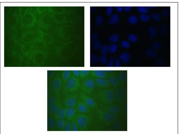
3.2.2 Subcellular localization of Ccdc-124 ... 30
3.3 Identification of RasGEF1 Family Members as Proteins Interacting with Ccdc124 in vitro and in vivo ... 36
3.3.1 Identification of proteins that interact with human CCDC124 by yeast two-hybrid (Y2H) screening. ... 36
3.3.2 RasGEF1 family was validated as interacting partners of Ccdc124. ... 37
3.3.2.1 Small Scale Protein Expressions... 37
3.3.2.2 Large Scale Culture of CCDC-124 ... 42
3.3.2.3 Large Scale Culture of pGEX-RasGEF1B ... 43
3.3.2.4 Large Scale Culture of RasGEF1A ... 45
3.3.3 GST Pull Down Assay ... 46
3.3.4 In vitro Interaction of RasGEF1B and Ccdc-124: Polarization ... 48
3.3.5 In Vivo Detection of Interaction between Ccdc-124 and RasGEF1B with Co-Immunoprecipitation ... 52
3.3.6 Studies towards Crystalization of Ccdc-124 ... 55
x
3.5 Mutational Analysis of RasGEF1/Rap2 Interaction and Determination of
Phe 39 as a specification residue. ... 79
4 Discussion ... 92
5 Future Perspectives ... 98
xi
LIST OF TABLES
Tablo 2.1: PCR Reaction of RasGEF1A and RasGEF1B ………..13
Table 2.2: RasGEF1A and RasGEF1B primers ……….13
Table 2.3: Digestion and ligation reactions of RasGEF1A, RasGEF1B …………...14
Table 2.4: Ccdc-124 Mutagenesis Oligos……….…..15
Table 2.5: Mutagenesis Oligos of Rap2A and Rap1B………15
Table 2.6: Northern blotting probes………...20
Table 2.7: First antibodies used in Western Blot experiments………...22
Table 3.1: Representative nucleotide exchange rates of Rap1 and Rap2…………...69
xii
LIST OF FIGURES
Figure 1.1: The switch mechanism of Nucleotide Exchange ………..4
Figure 1.2: The RasGEF proteins possess two domains ………10
Figure 3.1: High conservation of ccdc-124 gene protein sequence in eukaryotes …26 Figure 3.2: Northern blotting probes for Ccdc-124, Gapdh and β-actin. mRNA size ……….27
Figure 3.3: Human tissue expression analysis of Ccdc-124 by northern blotting ….27 Figure 3.4: Western Blot Analysis of Ccdc-124 ………28
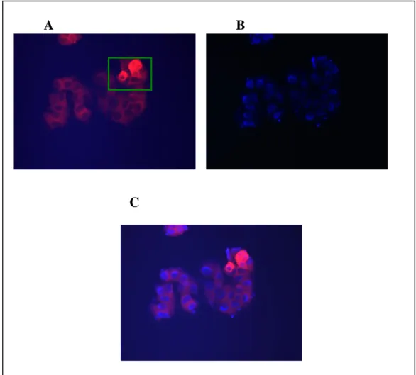
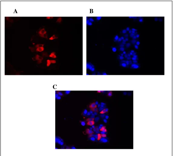
Figure 3.5: Immunocytochemistry of Ccdc-124 ………30
Figure 3.6: Immunocytochemistry of Ccdc-124 ………31
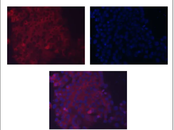
Figure 3.7: Immunocytochemistry of Ccdc-124 without DAPI stain ………32
Figure 3.8: Immunocytochemistry of Ccdc-124 ………32
Figure 3.9: Immunocytochemistry of Ccdc-124 ………33
Figure 3.10: Immunocytochemistry of Ccdc-124 ………..……34
Figure 3.11: Immunocytochemistry of Ccdc-124 without DAPI stain ………..34
Figure 3.12: Yeast-two-hybrid analysis revealed RasGEF1B as a potential interaction partner of Ccdc-124 ………...35
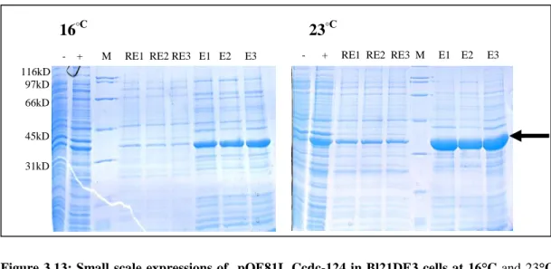
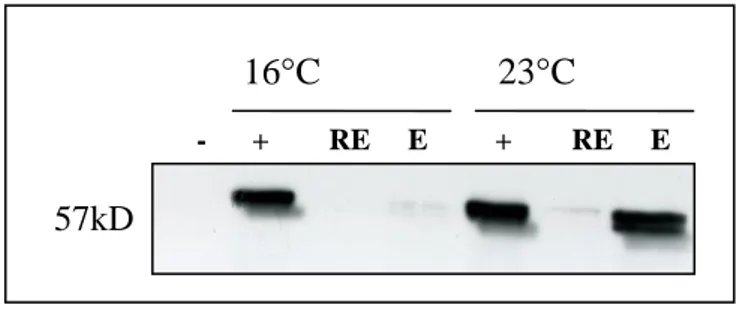
Figure 3.13: Small scale expressions of pQE81L Ccdc-124 in Bl21DE3 cells at 16°C and 23°C ………36
xiii
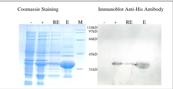
Figure 3.14: Coomassie and Western Blot analysis of small scale pQE81L Ccdc-124
expression ………..37
Figure 3.15: Overnight small scale expression of pQE80L RasGEF1B in Bl21DE3 cells……….38
Figure 3.16: 2 hour and 3 hour small scale expression of pQE80L RasGEF1B in Bl21DE3 cells ………38
Figure 3.17: Western blot analysis of pQE80L RasGEF1B with anti-Histidine antibody ……….39
Figure 3.18: Overnight small scale expression of pGEXTEVn-RasGEF1A in Bl21DE3 cells ………40
Figure 3.19: Purification of pQE81L-Ccdc-124 ………41
Figure 3.20: Purification of RasGEF1B protein ………...42
Figure 3.21: TEV Cleavage of GST-RasGEF1B protein ………...43
Figure 3.22: Purification of RasGEF1B protein ………...43
Figure 3.23: Purified GST tagged RasGEF1B protein pool ………..44
Figure 3.24: RasGEF1A protein purification ……….45
Figure 3.25: GST-Pull Down Assay of GST-RasGEF1A and GST-RasGEF1B …...46
Figure 3.26: Purification of Ser12, Ser92 and Ser156 Ccdc-124 proteins ………….48
Figure 3.27: Elution of Ccdc-124 S12C was subjected to gel filtration ………48
Figure 3.28: Elution of Ccdc-124 S92C was subjected to gel filtration ………49
Figure 3.29: Elution of Ccdc-124 S155C was subjected to gel filtration …………..49
xiv
Figure 3.31: Polarization assay of IAEDANS labeled Ccdc-124 protein with
RasGEF1B protein ……….…51
Figure 3.32: Western Blot analysis of RasGEF1B ………52
Figure 3.33: FLAG-IP of Hela cells ………..53
Figure 3.34: Myc IP of HeLa cells with FLAG-HRP antibody ………54
Figure 3.35: Diagram of sitting drop method ………55
Figure 3.36: Some examples from crystallization results ………..57
Figure 3.37: Guanine Nucleotide Exchange Assay of RasGEF1A on H-Ras and R-Ras ………..60
Figure 3.38: Guanine Nucleotide Exchange Assay of RasGEF1A on Rap1A and M-Ras ………..60
Figure 3.39: Guanine Nucleotide Exchange Assay of RasGEF1A on Rheb and RhebL ……….61
Figure 3.40: Guanine Nucleotide Exchange Assay of RasGEF1A on Rerg and RalB ……….61
Figure 3.41: Guanine Nucleotide Exchange Assay of RasGEF1A on Rap1B and DiRas2 ………62
Figure 3.42: RasGEF1A concentrations on Rap2A ………...62
Figure 3.43: Guanine Nucleotide Exchange Assay of RasGEF1A with Ccdc-124 on Rap2A ………63
Figure 3.44: Guanine Nucleotide Exchange Assay of RasGEF1B with Ccdc-124 on R-Ras ……….63
Figure 3.45: Guanine Nucleotide Exchange Assay of RasGEF1B with Ccdc-124 on H-Ras ……….64
xv
Figure 3.46: Guanine Nucleotide Exchange Assay of RasGEF1B with Ccdc-124 on M-Ras ……….64
Figure 3.47: Guanine Nucleotide Exchange Assay of RasGEF1B with Ccdc-124 on Rheb ………...65 Figure 3.48: Guanine Nucleotide Exchange Assay of RasGEF1B with Ccdc-124 on RhebL ……….65 Figure 3.49: Guanine Nucleotide Exchange Assay of RasGEF1B with Ccdc-124 on RalB ………...66
Figure 3.50: Guanine Nucleotide Exchange Assay of RasGEF1B with Ccdc-124 on Rerg ………66
Figure 3.51: Guanine Nucleotide Exchange Assay of RasGEF1B with Ccdc-124 on Rap1B ………67
Figure 3.52: Guanine Nucleotide Exchange Assay of RasGEF1B with Ccdc-124 on DiRas1 ………67 Figure 3.53: Guanine Nucleotide Exchange Assay of RasGEF1B with Ccdc-124 on DiRas2 ………68 Figure 3.54: Guanine Nucleotide Exchange Assay of RasGEF1B with Ccdc-124 on Rap2A ………68
Figure 3.55: Guanine nucleotide exchange rates of Ras family of G-proteins in the presence of RasGEF1A and RasGEF1B ………70
Figure 3.56: Guanine nucleotide exchange assays of Rap family of G-proteins in the presence of RasGEF1A and RasGEF1B ………70
Figure 3.57: Positions of residues selected for mutational analysis ………..72
xvi
Figure 3.59: Purifications of Rap2A T27I, Rap2A S66A and Rap2A F39S proteins
……….74
Figure 3.60: HLPC reaction of Rap2A S66A ………75
Figure 3.61: HLPC reaction of Rap2A T27I ……….75
Figure 3.62: HLPC reaction of Rap2A F39S ……….76
Figure 3.63: Gel filtrations of Rap2A T27I, S66A and F39S proteins after mGppNHp exchange reaction ………...77
Figure 3.64: Mant-GppNHp retention time on HPLC ………...77
Figure 3.65: Mant-GppNHp retention time of Rap2A S66A on HPLC ………78
Figure 3.66: Mant-GppNHp retention time of Rap2A T27I on HPLC ……….78
Figure 3.67: Mant-GppNHp retention time of Rap2A F39S on HPLC ………79
Figure 3.68: Guanine nucleotide exchange rates of wild-type and mutant Rap2A proteins ………..80
Figure 3.69: Purification of Rap1B S39F protein ……….81
Figure 3.70: Mant-GppNHp retention time of Rap1B S39F on HPLC ……….81
Figure 3.71: Guanine nucleotide exchange rates of wild-type and mutant Rap1B proteins ………...82
Figure 3.72: Guanine nucleotide exchange (GEF) assay results of increasing concentrations of RasGEF1A on Rap1B(S39F) ………83
1
1 INTRODUCTION
1.1 RAS family of small G proteins
Small GTP binding proteins together with their effectors and regulators have an important role in cell signaling pathways which affects almost all parts of the cell system. Most of these small GTP binding proteins belong to a superfamily called RAS (Colicelli 2004). Ras genes are found mutated in 20% of all human tumors.The highest incidences are found in adenocarcinomas of the pancreas (90%), colon (50%), and lung cancers (30%), thyroid tumors (50%), and myeloid leukemia (30%). The human Ras superfamily consists of at least 154 members divided into five principal families: the Ras, Rho, Rab, Arf, and Ran families (Wennerberg et al. 2005). This family of proteins has roles in the regulation of cell growth, proliferation, differentiation, apoptosis, adhesion and gene expression (Ehrhardt et al. 2002). For example, Ran G proteins are responsible for nuclear import and export, the regulation of nuclear envelope formation, and the control of spindle formation. Members of the Rab and Arf families play important roles in vesicle-associated processes, ranging from vesicle formation and transport to exocytosis. The Rho family is mainly involved in the regulation of cell shape, the cytoskeleton, and cell migration, whereas Ras family members regulate a variety of signaling pathways, resulting in transcription and cellular differentiation and proliferation (Bos et al. 2007).
1.2 GTPase Activating Proteins (GAPs)
Small G-Proteins are molecular switches that cycle between an active GTP-bound state and an inactive GDP-bound state. Only in the GTP-bound form they interact with effector proteins and activate target pathways. For activation, GDP has to be released and a new GTP molecule has to be bound. To become inactivated, the bound GTP molecule has to be hydrolysed. Although G proteins are also called GTPases, the actual GTP hydrolysis reaction is in fact very slow for most of the G proteins, and efficient hydrolysis requires the interaction with a GAP, which
2
accelerates the cleavage step by several orders of magnitude. Several structural and biophysical studies have unraveled the reaction mechanism. The GTP-bound state is stabilized by backbone interactions with their switch I and switch II regions, and the γ-phosphate of GTP. Efficient GTP hydrolysis requires the presence of a trans element (typically an Arg residue) that interacts with the nucleotide to promote hydrolysis (Fig.1.1). The main contribution of different GAPs to catalysis is the stabilization of the intrinsically mobile catalytic machinery of the G protein and, in most cases, the insertion of a catalytic residue in trans (Weirich et al. 2008).
1.3 Guanine Nucleotide Exchange Factors (GEFs)
When G proteins are GTP bound, they create a binding site for their downstream effector proteins. Activation of RAS proteins is regulated by the catalytic action of GEFs. This diverse family includes GEFs acting on p21 Ras proteins (mSos, RasGRF, RasGRP) on Rap proteins (RasGRP, Epac, C3G, MR-GEF and RA-GEF), and on Ral proteins (RalGDS, Rlf, Rgl) (Quilliam et al. 2002); notably, most GEFs can act on more than one GTPase. The GDP/GTP cycle is highly regulated by GEFs that induce the release of the bound GDP to be replaced by the more abundant GTP. The large number of G proteins requires a multitude of GEFs to ensure signaling specificity. GEFs active on the Ras family of proteins share a catalytic domain of about 250 amino acids that is homologous with the catalytic domain of CDC25 in
Saccharomyces cerevisiae and a Ras exchange motif (REM) domain. GEFs are
usually multidomain proteins, many of which are protein or lipid interaction domains, indicating that they serve as localization signals and/or as scaffolds for the formation of protein complexes.
1.3.1 General Mechanism of GEF Function
The affinity of most small G proteins for GDP/GTP is in the lower nanomolar to picomolar range. The direct consequence of this high affinity is a slow dissociation rate of nucleotides with a half-life on the order of one or more hours. Because
3
exchange of GDP for GTP and, thus, activation of G proteins in biological processes occur within minutes or even less, exchange of GDP for GTP requires the activity of GEFs. Indeed, GEFs accelerate the exchange reaction by several orders of magnitude (Vetter and Wittinghofer 2001). The GEFs are often the targets of biological signals, which induce, inhibit, or modulate their catalytic activity. GEFs catalyze the dissociation of the nucleotide from the G protein by modifying the nucleotide-binding site such that the nucleotide affinity is decreased and, thus, the nucleotide is released and subsequently replaced. In general the affinity of the G protein for GTP and GDP is similar, and the GEF does not favor rebinding of GDP or GTP. Thus the resulting increase in GTP-bound over GDP-bound is due to the approximately ten times higher cellular concentration of GTP compared to GDP.
In contrast, the affinities of the exchange factor for the nucleotide-bound G protein and of the nucleotide for the exchange-factor-bound G protein (the ternary complexes) are much lower (Vetter and Wittinghofer 2001). Thus, the interaction of a GEF weakens the affinity for the nucleotide, and vice versa, the nucleotide weakens the affinity for the GEF. In the course of the exchange reaction the GEF displaces the bound nucleotide, and subsequently a new nucleotide displaces the GEF. The G-protein-bound nucleotide is sandwiched between two loops called switch 1 and switch 2. The switch regions together with the phosphate-binding loop (P loop) interact with the phosphates and a coordinating magnesium ion. Both phosphates and the magnesium ion are essential for the high-affinity binding of the nucleotide to the G protein (Vetter and Wittinghofer 2001). GEF binding induces conformational changes in the switch regions and the P loop, while leaving the remainder of the structure largely unperturbed.
1.3.2 Regulation of GEF Activity
Almost all GEFs are multidomain proteins regulated in a highly complex fashion. This regulation includes protein-protein or protein-lipid interactions, binding of second messengers, and posttranslational modifications. These interactions and modifications induce either one or more of three major changes: a translocation to a
4
specific compartment of the cell where the small G protein is located, the release from autoinhibition by a flanking domain or region, which covers the binding site for the small G protein, or the induction of allosteric changes in the catalytic domain (Bos et al. 2007).
Figure 1.1: The switch mechanism of Nucleotide Exchange. Mechanism relies on the inherently
low nucleotide-hydrolysis and nucleotide-exchange rates of these proteins. The GTP-bound state (panel a) is stabilized by backbone interactions (red circles) in two sequence motifs, known as switch I and switch II, and the -phosphate of GTP. Efficient GTP hydrolysis requires the presence of a trans element (blue circle; typically an Arg residue) that interacts with the nucleotide to promote hydrolysis (red arrow, panel b). The GDP-bound state is also stable, but the interactions between the switch motifs and the nucleotide are lost (panel c), in some cases leading to significant changers in the structure of the switch regions. Nucleotide exchange requires the contribution of an exchange factor (panel d), which stabilizes a relaxed conformation of the GTP-binding (G) domain (red arrows), causing GDP to be released and a new GTP molecule to enter the active site (Weirich et al. 2008).
5
1.4 RAP (Ras-Proximal) family of small G proteins
Rap proteins are Ras-like small G-proteins, which functions as molecular switches by cycling between a GDP-bound inactive and a GTP-bound active state. They control a wide variety of cellular process, most notably cell adhesion and cell junction formation, which play a crucial role in cell migration and tumor formation. On the basis of the sequence homology, the Rap family is divided into two subgroups, Rap1 and Rap2, with a total of five members: Rap1A, Rap1B, Rap2A, Rap2B, and Rap2C (Paganini et al. 2006). No functional differences have been reported between the members of each group. There is about 95% sequence identity between Rap1A and Rap1B, and about 90% identity between Rap2A, Rap2B, and Rap2C. Effector-binding regions, including switch I, show the highest conservation, and C-terminal regions the lowest. Similarly, Rap1 proteins are closely related (about 70% identical) to Rap2 subfamily members. Although identical or overlapping functions have been described for Rap1 and Rap2 proteins (Christian et al. 2003; McLeod et al. 2002; McLeod et al. 2004), a number of studies showing functional distinctions and different effector–protein interactions have been reported for Rap1 and Rap2 proteins (Fu et al. 2007; Huang et al. 2004; Imamura et al. 2003; Zhu et al. 2005).
1.4.1 RAP1
Rap-GEF/Rap signaling regulates the formation of adherens junctions. RAP1 was first identified as a gene that could reverse the loss of adhesion observed in NIH3T3 cells transformed by K-Ras (Kitayama et al. 1989). Subsequently, activation of Rap GTPases has been shown to increase cell adhesion and spreading and also to play a role in migration (Arthur et al. 2004; Enserink et al. 2004; Price and Bos 2004; Price
et al. 2004). Although the mechanism of Rap activity is not completely understood,
there is convincing evidence that activated Rap promotes adhesion through integrin activation (Bos et al. 2003; McLeod et al. 2004; Reedquist et al. 2000). RAP proteins are activated by mitogenic stimuli and function as regulators of integrin mediated cell adhesion and cell spreading. In cultured cells, RAP proteins do not show transforming activity. Rather, overexpression of RAP1A inhibits RAS
6
mediated transformation. However, RAP1A has been reported to bind and activate BRAF, suggesting that it has the capacity to promote mitogenesis and perhaps transformation in some cases. Two observations suggest contributions of RAP proteins in tumorigenesis, but with possible tissue type specificity. Activation of a RAP directed GEF or inactivation of a RAP directed GAP promotes hematopoietic tumor formation. Conversely the loss of an activator of RAP1 proteins has been found in a mouse osteosarcoma and in several nonhematopoietic human cancer cell lines (Colicelli 2004).
Recent studies have suggested that Rap1 may actually regulate adherens junctions. Cell-cell junctions are formed evenly around the lateral circumference of cells by homophilic interactions between the extracellular domains of E-cadherin, linked by their intracellular tail to catenins and to the actin cytoskeleton (Jamora and Fuchs 2002). Two recent studies in mammals have shed new lights on the connection between Rap1 and adherens junctions. In the first study, a Rap1 GTPase activator, DOCK4, was identified as a tumor suppressor (Yajnik et al. 2003). DOCK4 specifically activates Rap1 and regulates the formation of adherens junctions. In the second study, the authors demonstrated that ligation of the extracellular domain of E-cadherin enhances Rap1 activity, and that active Rap1 regulates the subsequent accumulation of at newly formed cellcell contact sites (Hogan et al. 2004). Data from the second study suggests that formation of adherens junctions is a two-step process. When cells first contact one another, small clusters of E-cadherin ligate through the homophilic interaction, which may, in turn, induce the activation of Rap1; activation of Rap1 may then activate inside-to-outside signaling through stimulating actin polymerization, which mediates the further recruitment of E-cadherin from the cytoplasmic or plasma membrane pool and facilitates the formation of mature E-cadherin-based adherens junctions. In Drosophila, it is reported that, Rap1 regulates the even distribution of adherens junctions of epithelial cells in wing imaginal disc (Knox and Brown 2002); Rap1 null cells have uneven adherens junctions and are dispersed into neighboring cells.
7 1.4.2 RAP2
Knowledge about Rap2 is limited. Rap2 is a member of the Ras family of small GTPases whose effector domain is almost identical to that of Ras, and can therefore bind most Ras effectors. Rap2 inhibits many Ras pathways including Ras-induced Raf activation at the plasma membrane (Ohba et al. 2000). Rap2 also binds to the Ral GEFs, Ral GDS, RGL and RLF (Nancy et al. 1999). These proteins are also Ras effectors and induce nucleotide exchange leading to the formation of active RalA. Rap2 is reported to localizemainly in the endoplasmic reticulum (ER), whereas Rap1 localizesat the Golgi apparatus (Beranger et al. 1991; Beranger et al. 1991). Unlike Rap1, Rap2 cannotreverse Ras-induced transformation of NIH 3T3 cells (Jimenez et
al. 1991), andno biological phenotype has been linked to Rap2 in the literature.The regulation of Rap2 also remains unknown. Rap1GAP stimulates Rap2 GTPase activity in vitro, albeit significantly more weaklythan Rap1 (Janoueix-Lerosey et al. 1992). PDZ-GEF1, also activates Rap2 and that GTP-bound Rap2 makes up more than50% of the Rap2 in A14 and COS1 cells (Ohba et al. 2000).
In Xenopus embryos, Rap2 was shown to regulate activin/nodal signaling by modulating receptor trafficking (Choi et al. 2008). In the absence of ligand, Rap2 directs activin/nodal receptors into a Rab11-dependent recycling compartment, thereby avoiding degradation and maintaining cell-surface levels of receptors. Upon ligand addition, Rap2 no longer directs the receptors for recycling, but rather competes with Smad7 and delays receptor degradation, thus enhancing signaling (Colicelli 2004). Recently, specific Rap2-binding proteins such as RPIP8, TNIK and MAP4K4 were reported as candidate effectors; however, their cellular functions as Rap2 effectors are not fully established(Kardassis et al. 2009).
Further research in another eukaryote model, C. elegans, have revealed that there are three like genes. rap-1 and rap-2 are very similar to vertebrate 1b and
Rap-2a. rap-3 shows less similarity to vertebrate Rap genes but is most similar to Rap-1b.
Rap function in C. elegans involves the function and morphogenesis of hypodermal cells but not their generation or specification (Pellis-van Berkel et al. 2005). rap-1 mutants display defects in hypodermal morphogenesis and function, including
8
formation of adherens junctions, secretion of cuticle and basement membrane, and hypodermal cell integrity and viability. While rap-2 mutations on their own are wild-type, rap-2 mutations enhance the effects of rap-1, suggesting that rap-1 and rap-2 act redundantly and can partially compensate for each other's loss (Pellis-van Berkel
et al. 2005).
1.5 RAP Guanine Exchange Factors
Particularly, GEFs enhance the formation of the GTP-bound active conformation in response to upstream signals mediated by various cell surface receptors. To date, a number of GEFs for Rap proteins have been identified like, C3G, Epac (or cyclic AMP [cAMP]-GEF), CalDAG-GEFI, PDZ-GEF1, and GFR (Repac). These multi-domain proteins are highly regulated and responsible for the temporal and spatial activation of Rap. The Rap-GEFs Epac1 and Epac2 are directly regulated by cAMP and particular Epac2 modulates insulin secretion from pancreatic cells (Rehmann 2006). C3G binds to the adaptor protein Crk, being involved in tyrosine kinase-dependent activation of Rap1 (Gotoh et al. 1995). Epac/CAMPGEF is activated through direct association with cAMP, thereby stimulating Rap-dependent signaling (de Rooij et al. 1998; Kawasaki et al. 1998). Another Rap GEF, CalDAGGEF1, which contains calcium and diacylglycerol binding motifs, has a role in Rap activation in response to these second messengers (Kawasaki et al. 1998). Another Rap GEF is RAGEF-1 (also termed PDZ-GEF1, nRapGEP, or CNrasGEF), which exhibits GEF activity toward Rap1 and Rap2 (Liao et al. 1999; Liao et al. 2001). RA-GEF-1 contains putative cNMP-binding, REM, PDZ, and RA domains as well as the GEF catalytic domain. RA-GEF-2, whose structural features are intimately related to RA-GEF-1, exhibits GEF activity toward Rap1 and Rap2. GFR (Repac), which lacks the cAMP dependent regulatory sequences, is a constitutive activator of both Rap1 and Rap2 (Gao et al. 2001).
1.6 RasGEF1 Family
Proteins that act as Guanine exchange factors of Ras can be classified into at least two families, on the basis of sequence similarities, the CDC24 family and the CDC25
9
family. The size of the proteins of the CDC25 family range from 309 residues (LTE1) to 1596 residues (Sos). The sequence similarity shared by all these proteins is limited to a region of about 250 amino acids generally located in their C-terminal section (currently the only exceptions are sos and ralGDS where this domain makes up the central part of the protein). This domain has been shown, in CDC25 an SCD25, to be essential for the activity of these proteins.
The crystal structure of the GEF region of human Sos1 complexes with Ras has been solved. The structure consists of two distinct alpha helical structural domains: the N-terminal domain which seems to have a purely structural role and the C-N-terminal domain which is sufficient for catalytic activity and contains all residues that interact with Ras. A main feature of the catalytic domain is the protrusion of a helical hairpin important for the nucleotide-exchange mechanism. The N-terminal domain is likely to be important for the stability and correct placement of the hairpin structure. The signature pattern for this entry spans the helical hairpin.
The rasgef genes encode a subgroup of highly conserved Ras guanine nucleotide exchange factors. While EST projects revealed the presence of rasgef genes in organisms that range from nematodes to humans, their functions remain to be elucidated. In zebrafish two rasgef genes, rasgef and rasgef1b, have been identified and high throughput analysis revealed tissue specific embryonic expression for
rasgef1b. The combined data generated from EST projects and genome-scanning
gene-prediction programs suggest that vertebrates have three distinct rasgef genes and that only two of these genes are present in zebrafish. Phylogenetic analysis of the predicted RasGEF proteins show that the fish and mammalian proteins have slightly diverged during evolution. Furthermore, the cDNA of this zebrafish gene has a slightly higher homology to mammalian rasgef1b (73%) than to rasgef1c (66%) and
rasgef1a (64%). In common, the RasGEF proteins possess two domains, a highly
conserved carboxy-terminal RasGEF domain and a slightly less conserved amino-terminal RasGEFN domain (Figxx) with a yet unknown function. At the level of the protein sequence, vertebrate RasGEF1B proteins display 80–95% identity in the RasGEFN domain and 91–98% identity in the RasGEF domain. RasGEF domains
10
normally activate small GTPases of the Ras/Rho family by catalyzing the exchange of the inactive GDP-bound form to the activated GTP-bound form (Quilliam et al., 2002). It is shown that three rasgef1b-transcripts are generated from two transcriptional start sites and by alternative splicing. Detailed expression analyses show that rasgef1b is expressed in a subset of adaxial cells, in the anterior part of somites, in the rostral part of the mid-hindbrain boundary and in the rhombomere boundaries. In the larva, rasgef1b is further expressed in the pallium and the inner nuclear layer of the retina. It is also found that rasgef1b is expressed maternally and that the ubiquitous distribution of maternal transcripts disappears shortly after mid-blastula transition. At early epiboly stages, rasgef1b expression is restricted to the margin with low levels of expression on the ventral and high levels of expression on the dorsal side. It is also showed that early zygotic expression is regulated by Nodal and FGF signals and that these signals have different activities in regulating the level and distribution of early zygotic rasgef1b mRNA expression (Epting et al. 2007).
Whether or not RasGEF1B proteins have a similar function remains to be investigated, particularly because their GEF domains display characteristic differences when compared to the GEF domains of the related Ras activating proteins CDC25 and SOS (Jones et al. 1991). Till now, the only evidence for a biological function of RasGEF1B proteins comes from studies in invertebrates.
Drosophila and Caenorhabditis elegans both contain a single rasgef1b-related gene,
named rasgef, which shows 35% and 36–37% identity to the genes encoding the vertebrate RasGEF1B proteins, respectively (Kiger et al. 2003). In large scale RNAi experiments, knock down of Drosophila RasGEF in Schneider-cells was found to result in a slight change in cell morphology. However, no defects were found when
Drosophila or C. elegans rasgef were knocked down in the entire organism (Rual et al. 2004).
11
Figure 1.2: The RasGEF proteins possess two domains, a highly conserved carboxy-terminal
RasGEF domain and a slightly less conserved amino-terminal RasGEFN domain. RasGEFN domain spans 33th to 161st residues, RasGEF domain through 201th-454th residues.
1.7 Objectives and Rationale
Previously, our laboratory has established the expression of a novel gene of unknown function, Ccdc-124, in a number of cell lines tested. Intrigued by a very high conservation of this gene among eukaryotes, we decided to establish the biological function of Ccdc-124. For this, we have first used the well known yeast-two-hybrid technique, and screened a liver cDNA library in order to identify interaction partners of Ccdc-124. These analysis have revaled RasGEF1B as a potential interaction partner. We have then validated these results by a number of in vitro and in vivo protein-protein interaction assays. Even though proteins belonging to RasGEF1 family were predicted to act as guanine exchange factors for Ras G-proteins, their precise biological functions were ambigious, and G-proteins stimulated by RasGEF1 family members were unknown. Therefore, we decided to test the guanine exchange factor activities of proteins belonging to this family, and we settled to identify Ras family of G-proteins stimulated by these GEFs in in vitro guanine nucleotide exchange assays. We have identified Rap2, a member of Ras family of G-proteins as substrate of RasGEF1A and RasGEF1B, and we then aimed to study residues having discriminatory roles in the interactions between RasGEF1s and Ras Family of G-proteins. In parallel to these studies, we have also studied the effect of Ccdc-124 in RasGEF1 stimulated guanine nucleotide exchange activities in vitro.
12
2 MATERIAL & METHODS
2.1 Yeast Two Hybrid
Bait cloning and Y2H screening were performed by Hybrigenics, S.A., Paris, France (http://www.hybrigenics.com).
Human CCDC124 (aa1-aa223) cDNA was PCR-amplified and cloned in a LexA, C-terminal fusion vector optimized by Hybrigenics. The bait construct was checked by sequencing the entire insert, and was subsequently transformed in the L40 GAL4 yeast strain (Fromont-Racine et al. 1997).A Human Liver random-primed cDNA library, transformed into the Y187 yeast strain and containing ten million independent fragments, was used for mating. High mating efficiency was obtained by using specific mating method (Legrain et al., 1998, 2000, 2002). The screen was first performed on a small scale to adapt the selective pressure tothe intrinsic property of the bait. No autoactivation of the bait was observed. Then, the full-scale screenwas performed in conditions ensuring a minimum of 50 million interactions tested, in order to cover five times the primary complexity of the yeast-transformed cDNA library (Rain et al. 2001). 98 millions of interactions were actually tested with Human CCDC124. After selection on medium lacking leucine, tryptophane, and histidine, 16 positive clones were picked, and the corresponding preyfragments were amplified by PCR and sequenced at their 5' and3' junctions.
Sequences were then filtered and contiged (Formstecher et al. 2005) and compared to the latest release of the GenBank database usingBLASTN (Altschul et al. 1997). A Predicted Biological Score (PBS) was attributed to assess the reliability of each interaction, as described previously (Formstecher et al. 2005). Briefly, the PBS relies on two different levels of analysis: firstly a local score takes into account the redundancy and independency of prey fragments, as well as the distributions of reading frames and stopcodons in overlapping fragments. Secondly, a global score
13
takes into account the interactions found in all the screens performed at Hybrigenics using the same library. In addition, potential false-positives are flagged by a specific “E” PBS score. This is done by discriminating prey proteins containing “highly connected” domains, previously found several times in screens performed on libraries derived from the same organism. The PBS scores have been shown to positively correlate with the biologicalsignificance of interactions (Rain et al. 2001; Wojcik et al. 2002).
2.2 Northern Blotting
Northern blotting was performed on FirstChoice Northern Human Blot1 membrane (each lane containing 2 µg poly (A) RNA) (Ambion). DNA templates for probe preparation were formed via restriction enzyme digestion from PCR product cloned plasmids for Ccdc-124 and RasGEF1B or directly from PCR products for Gapdh and β-actin. PCR products for Gapdh and β-actin formed via primers which were kindly provided by Ayşe Elif Erson and Mehmet Öztürk‟s group, respectively. Then, DNA templates were labeled by north2south biotin random prime labeling kit (Pierce). Probes were synthesized as described in manufacturer‟s protocol. Probes and their sizes are presented in Table 2.6.
Gene Starting material Restriction Enzyme Probe Size
Ccdc-124 p3XFlagCcdc-124 Apa I and Xba I 381 bp
RasGEF1B p3XFlagRasGEF1B Bgl II and BamHI 284 bp
Gapdh Gapdh PCR product - 408 bp
β-actin β-actin PCR product - 539 bp
Table 2.6: Northern blotting probes. Starting DNA material, used restriction enzymes for probe
synthesis and synthesized probe sizes are indicated.
In order to hybridize the nucleic acids and perform the detection by synthesized probes, north2south chemiluminescent hybridization and detection kit (Pierce) was
14
utilized. Kit‟s protocol was exactly followed and resulting blots were exposed to film for approximately 1 min.
2.3 Cell Culture
In this study, Huh-7 (Human hepatocellular carcinoma), HeLA (Human Cervical carcinoma) and MCF-7 (Caucasian female breast adenocarcinoma) cell lines were used for immunoprecipitation, immunostaining and western analysis.
Cell lines were grown in high glucose Dulbecco‟s modified Eagle‟s medium (Gibco) with the addition of 10% fetal bovine serum (FBS), 1% penicillin/streptomycin and 1% L-glutamine (Biochrom) at 37˚C in a 5% CO2 incubator.
2.3.1 Transfection
Transfection was performed via using FuGene-6 reagent (Roche). Optimized transfection reagent (µl) /plasmid DNA (µg) was 3:1. Firstly, transfection reagent was diluted in serum and antibiotics free medium. After 10 minutes of incubation, plasmid DNA was added and FuGene-plasmid DNA mixture was incubated for 30 minutes. During this time interval, medium of the cells was changed. Finally, FuGene-DNA complex was transfected to cells in a drop-wise manner.
2.4 Western blot analysis
Proteins from cell lines were isolated for western blotting analysis. Cell pellets were lysed in 50 µl lysis buffer consisting of 50mM Tris Base, 250mM NaCl, 1X proteinase inhibitor cocktail and 0.1% NP40. Protein concentrations were determined via using Bradford assay. Twenty micrograms of whole cell extracts were denatured in gel loading buffer [50 mM Tris-HCl pH 6.8, 1% SDS, 0.02% bromophenol blue, 10% Glycerol and 5% 2-mercaptoethanol] at 95°C for 5 min, resolved by SDS-PAGE using a 10% gel, and electrotransferred onto PVDF membranes (Millipore). The membranes were blocked in Blotto (Tris-buffered saline containing 0.5% Tween 20 and 5% nonfat milk powder) for 1 hour at room temperature. The membranes were incubated with first antibody (Table 2.7) for 1 hour, washed 3 times with Blotto and incubated with secondary antibody (see below) for 1 hour, immunocomplexes
15
were then detected via Super Signal West Dura (Pierce), and exposed to X-Ray films (AGFA) for 1 minute. The films were then developed using a hyper-processor developer (Amersham).
Primary antibody Description Concentration in blotto
Anti-Ccdc-124 Rabbit, polyclonal 2 µg/ml
Anti-Flag (Sigma) Mouse, monoclonal 1 µg/ml
Anti-Calnexin Rabbit, polyclonal 0.1 µg/ml
Anti-His-Probe(Santa
Cruz) Rabbit, polyclonal 1 µg/ml
Table 2.7: First antibodies used in Western Blot experiments.
2.5 Immunostaining
Cells that cultured on cover slips in 6-weel plates were washed 3 times with cold 1XPBS (137 mM NaCl, 2,7mM KCl, 1,4 mM KH2PO4, 4,3 mM Na2HPO4) then fixed with 4% Para-formaldehyde at room temperature for 15 min. After fixation cells were permeabilized with 1:1000 Triton X100 in 1XPBS for 6 min at room temperature. Cells then blocked with 5% BSA in1XPBS for 1 hour at room temperature, then incubated with first antibody which is diluted in blocking solution for 1 hour. After first antibody incubation cells were washed 3 times with 1XPBS and incubated with TRITC or FITC conjugated secondary antibody diluted in blocking solution for 1 hour at room temperature. Then cells were washed for 3 times with 1XPBS and counter stained with DAPI for 30 sec, washed with dH2O and mounted with glycerol.
2.6 GST-Pull Down Assay
100µl 50% slurry (~50µl packed) GSH beads were washed 3 times with wash buffer (50mM Tris HCl pH 7.5, 100mM NaCl, 3mM β-Mercaptoethanol). First two vials
16
incubated (immobilized) with 200µg purified GST-RasGEF1 protein, second vial incubated (immobilized) with 200µg GST protein, fourth vial just incubated with wash buffer and rotated at 4◦C for 1 hour. After immobilization, beads were washed 3 times. First and third vials incubated with eash buffer only, second and fourth vials incubated with 500µg Ccdc-124 protein and incubated at 4◦C for 1 hour by rotating. Then beads were washed 9-10 times with wash buffer, and samples were eluted with 40µl 4x SDS loading buffer, boiled for 5 min and loaded on an SDS gel. For the control of of protein sizes 10mg/ml from each protein were loaded to the same gel. The gel then stained with coomasie for 15 min and destained with water.
2.7 Polarization Assay
2.7.1 IAEDANS Labelling
Cystein mutants of purified Ccdc-124 proteins were washed with 50mM Tris HCl (pH 7.5), 100mM NaCl, 5mM Ascorbic Acid in eppendorf concentrators and 0.5mg of them incubated with 10 times molar concentration of IAEDANS overnight at 4◦C.
2.7.2 Fluorescence Polarization
Monitoring protein-protein interactions by using fluorescence polarization. After excitation with polarized light, the photons emitted by fluorescent probes are also polarized; however, rotational diffusion of a fluorophore over its excitedstate lifetime causes depolarization of the emitted photons. Fluorescence polarization measures the average angular displacement of a fluorophore over its excited-state lifetime, which is affected by the molecule‟s rate of rotational diffusion. Because the rate of rotational diffusion of a molecule is inversely related to its size, polarization provides a sensitive measure of changes in molecular volume that occur when the fluorescent protein is bound by a nonfluorescent protein (Harrington et al. 2003). Fluorescence polarization was used to monitor the association of IAEDANS-labeled Ccdd-124 with nonfluorescent RasGEF1B. Measurements were performed at 25°C in 20 mM Tris HCl (pH 7.5), 50 mM NaCl, 5 mM MgCl2, 3 mM β-mercaptoethanol in quartz cuvettes. Fluorescence data were recorded with a Fluoromax-2 spectrophotometer, with excitation and emission wavelengths of m-nucleotides at 336 nm and 450 nm,
17
respectively. Firstly the polarizations of 2.5µM IAEDANS labeled Ccdc-124 proteins alone were measured and then 20nM RasGEF1B protein was added each time with time laps.
2.8 Protein Imunoprecipitation Assay
70-90% confluent (100mm dish) cells were rinsed twice with PBS (10mM phosphate, 2.7mM potassium chloride, 137mM sodium chloride, pH 7.4) and 1 ml of lysis buffer (50mM Tris HCl, pH 7.4, with 150mM NaCl,1mM EDTA, and 1% TRITON X-100) was added and incubated for 15−30 minutes on a shaker at 4°C. Then cells were scraped, collected and centrifuged at 12,000Xg for 10 min. Supernatant transferred onto 40µl 50% slurry ANTI-FLAG M2 affinity gel (Sigma) and incubated overnight on shaker at 4°C. Subsequently the resin was washed three times with 1ml TBS. 20 µl of 2Xsample buffer (125mM Tris HCl, pH 6.8, 4% SDS, 20% (v/v) glycerol, 0.004% bromphenol blue) was added to each sample. After denaturation by boiling for 3 min, samples were loaded on SDS-PAGE and immunoblotted with specific antibody against the Ccdc-124 protein and anti-Flag antibody against the fusion RasGEF1B protein.
2.9 Plasmid Constructs
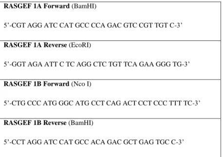
cDNAs corresponding to the RasGEF1A and RasGEF1B genes were cloned into the pQE80L system by Hani Alotaibi and Pelin Telkoparan. Then these cDNAs were transferred into pGEX-4T-1 expression vector: cDNAs corresponding to RasGEF1A and RasGEF1B were amplified by PCR (Table 2.1) with the primers (Table 2.2) suitable for pGEX cloning, and then fragments were purified from agarose gel. RasGEF1A and RasGEF1B were digested at 37◦C overnight with their suitable enzymes. pGEX-TEVn was also digested first with EcoRI at 37◦C overnight and then EcoRI was inactivated at 65◦C for 20 min. and divided into two vials. One was digested with BamHI and the other half was digested with NcoI at 37◦C for 2 hours. Then digested samples were purified from agarose gel and ligated with RasGEF1A and RasGEF1B fragments (Table 2.3). After ligation they were transformed into Top10 competent cells and spread on 50µg/ml Amp included LB agar plates. After
18
incubating the plates overnight at 37◦C colonies were picked and a colony PCR was made in order to select the positive colonies. Then the plasmids from positive colonies were isolated and sequenced.
DiRas2, M-Ras, R-Ras, RalB, TC21, RERG and Rheb were cloned into pGex4T1– TEV and expressed in E. coli BL21 DE3 cells. C-terminally truncated forms of H-Ras, Rap1A, Rap1B and Rap2A were cloned into ptac vectors and expressed in CK600K cells by Raphael Gasper.
Tablo 2.1: PCR Reaction of RasGEF1A and RasGEF1B by using pQE80L-RasGEF1A and pQE80L-RasGEF1B as template. Conditions: 94◦C 4min, (94◦C 30sec, 53◦C 60sec, 72◦C 60sec)x30, 72◦C 4 min.
19 RASGEF 1A Forward (BamHI)
5‟-CGT AGG ATC CAT GCC CCA GAC GTC CGT TGT C-3‟
RASGEF 1A Reverse (EcoRI)
5‟-GGT AGA ATT C TC AGG CTC TGT TCA GAA GGG TG-3‟
RASGEF 1B Forward (Nco I)
5‟-CTG CCC ATG GGC ATG CCT CAG ACT CCT CCC TTT TC-3‟
RASGEF 1B Reverse (BamHI)
5‟-CCT AGG ATC CAT GCC ACA GAC GCT GAG TGC C-3‟
Table 2.2: RasGEF1A and RasGEF1B primers for cloning into pGEX system.
20 2.10 Protein Purification
2.10.1 Small Scale Expressions of pQe80L-RasGEF1 and pQe81L-Ccdc-124
BL21 codon+ RIL competent cells transformed with pQE80L-RasGEF1 and pQE81L-Ccdc-124 for test expression.
200 µl competent cell + 1 µl plasmid DNA on ice for 30 min, heat shock at 42◦C for 1 min then add 500 µl LB and shake for 1 hour at 37◦C. After 1 hour of shaking, 100 µl was spread over an agarose plate which has 50µg/ml ampisilin (Amp) + 25µg/ml chloramphenicol (Cam) in it and incubated overnight at 37◦C. The next day half of the colonies grew on the plates scaped into 400 ml LB flasks which have 50µg/ml Amp and 25µg/ml Cam. Flasks were shaked at 23◦C till they reached the A600 nm of 0.4. When they reached the A600 nm of 0.4, 1 ml of “uninduced” control was taken from each of them and the rest is induced with 100 µM Isopropyl β-D-1-thiogalactopyranoside (IPTG) and divided into two flasks, one was grown at 16 ◦C the other was grown at 23 ◦C. Next day 1 ml of induced control was taken from each flask then they divided into three 50 ml falcons so that there were 3 samples for 3 different lysis buffers, lysis buffer1, 2 and 3 (see below).
Falcons were centrifuged and pellets were lysed with different lysis buffers. After dissolving pellets with lysis buffers, 100 µM phenylmethanesulphonylfluoride or phenylmethylsulphonyl fluoride (PMSF) was added and each was sonicated with microtipped sonicator at the maximum level with 50% amplitude, 50 seconds for 2 times. 10 µl control samples were taken from them, then 1 ml from each sample was centrifuged at 4 ◦C and 14000 rpm for 30 min and 10 µl sample was taken as a Raw Extract (RE) control, the rest is incubated with 50 µl 50% Ni-beads for 30 min at 4 ◦
C. 10 µl Flow Though (FT) control was also taken before the beads were washed. After 5 times washing step with their corresponding washing buffers, beads were eluted with 4x sample buffer. All the samples heated up to 95 ◦C for 5 min then loaded in 15% SDS gels and ran at 70mA/gel for 30 min and gels were stained with coomassie for 15 min then destained with water.
21 2.10.2 Large Scale Expression of Ccdc-124
5L culture of pQE81L Ccdc-124 in BL21 codon + RIL E.coli cells was grown at 23◦C till the A600 nm reached to 0.5. 1ml of “uninduced” control sample was taken and the rest was induced with 100mM IPTG overnight and pelletted by centrifuging at 4400 rpm at 16 ◦C for 15 min. Pellets were resuspended with lysis buffer 1 (see below) and 100mM PMSF was added. Then pellets were lysed by using midi-tipped sonicator at the maximum level with 50% amplitude, 50 seconds for 2 times on ice. Cell lysate supernatants were applied to Ni-NTA-agarose column, pre-equilibrated with lysis buffer 1 at 4◦C and washed first with wash buffer 1 then with high salt buffer. Then eluted with a linear gradient up to 1M imidazole in elution buffer (pH 8.0) and 4ml fractions. Fractions were loaded on a 15% SDS gel, and then all fractions pooled together and concentrated (with Millipore Amicon Ultra Centrifugal Filter 10 NMWL). Concentrated samples were loaded on Superdex S75 26 ⁄ 60 columns (Amersham Biosciences) (gel filtration). Fractions after gel filtration were loaded to a 15% SDS gel and the proper fractions were pooled together and concentrated.
Lysis Buffers:
Lysis Buffer 1: 50 mM Tris pH 7.5, 300 mM NaCl, 3 mM β-Mercaptoethanol, 0.5% NP40, 0.5% Triton X100
Lysis Buffer 2: 50 mM Tris pH 8, 300 mM NaCl, 3 mM β-Mercaptoethanol, 0.5% NP40, 0.5% Triton X100
Lysis Buffer 3: 50 mM Tris pH 7.5, 300 mM NaCl, 3 mM β-Mercaptoethanol
Lysis Buffer with Detergent: 50 mM Tris pH 7.5, 300 mM NaCl, 3 mM β-Mercaptoethanol, 0.5% NP40, 0.5% Triton X100
Lysis Buffer without Detergent: 50 mM Tris pH 7.5, 300 mM NaCl, 3 mM β-Mercaptoethanol.
22
Wash Buffer 1: 50mM Tris HCl pH 7.5, 100mM NaCl, 3mM β-Mercaptoethanol
Wash Buffer 2: 50mM Tris HCl pH 8, 100mM NaCl, 3mM β-Mercaptoethanol
High Salt Buffer: 50mM Tris HCl pH 7.5, 300mM NaCl, 3mM β-Mercaptoethanol, 1mM ATP
Column Elution Buffer 1 pH 8.0: 50mM Tris HCl pH 7.5, 100mM NaCl, 3mM β-Mercaptoethanol, 20mM Glutathion
Column Elution Buffer 2 pH 8.0: 50mM Tris HCl pH 7.5, 100mM NaCl, 3mM β-Mercaptoethanol, 1M immidazole
2.10.3 Protein expression and purification of wild type G proteins
DiRas2, M-Ras, R-Ras, TC21, RalB, RERG, Rheb, ptac–H-Ras, ptac–Rap1A, ptac– Rap1B and ptac– Rap2A were expressed at 25◦C after induction with 100 Mm IPTG at a A600 nm of 0.6. Cell lysate supernatants of GST-fused RasGEF1A and RasGEF1B were applied to a glutathione (GSH) column (Amersham Biosciences, Freiburg, Germany) pre equilibrated with buffer A [50 mm Tris (pH 7.5), 100 mM NaCl, 3 mM β-mercaptoethanol] at 4 ◦C and eluted with buffer A containing 20 mM GSH, and GST fusion proteins were purified by gel filtration on Superdex S75 16 ⁄ 60 columns (Amersham Biosciences). Cell lysate supernatants of the GST-fused G-proteins were applied to GSH Sepharose 4B (GE Healthcare, Chalfont St Giles) pre-equilibrated with buffer B [25 Mm Tris (pH 7.5), 500 Mm NaCl, 5 Mmdithioerythritol] at 4 ◦C, and either eluted with 20 Mm GSH and subsequently digested by Tobacco Etch Virus (TEV) protease (1mg/ml) in batches, or cleaved by thrombin (300U) on the column. Cleaved proteins were further purified by gel filtration and another GSH column to separate G-proteins from GST; ptac-cloned proteins were purified by Q-Sepharose and subsequent gel filtration.
23 2.10.4 Site Directed Mutagenesis
PGEX-4T-3–Rap1B, pGEX-4T-3–Rap2A and pQE81L-Ccdc-124 mutants were generated using a Stratagene QuickChange site-directed mutagenesis kit by using primers below (Table 2.4 and Table 2.5). For templates smaller than 5kb, 50ng DNA; longer than 5kb, 100ng DNA was used. After mutagenesis PCR reaction, samples were subjected to Dpn I digestion and transformed into Top10 competent cells. The plasmids were isolated and their sequences were verified by sequencing.
Ser12 to Cys (TCG→TGC)
5‟GGTGAGAACACCAAGTGCGCAGCGGCCCGGG 3´
Ser92 to Cys
(
TCC→TGC)5‟CCGCGGGTGGCCACGTGCAGCAAGGTCACCC3´
Ser155 to Cys (AGC→TGC)
5´GCCATTGCAGTGCTCTGCGTGGCGGAGGAGGCG3´
Table 2.4: Ccdc-124 Mutagenesis Oligos.
Rap2A T27I ACC →ATC
5´GCAGTTCGTGACCGGCATCTTCATCGAGAAATACG-3´
Rap2A F39S TTC→TCC 5´CCCCACCATCGAGGACTCCTACCGCAAGGAGATCG-3´ Rap2A S66A TCC→GCC 5´CCGAGCAGTTCGCGGCCATGCGGGACCTGTAC-3´ Rap1B S39F TCT→TTT 5´CCTACGATAGAAGATTTTTATAGAAAGCAAGTTG-3´
24 2.10.5 Mant-GppNHp Exchange Reaction
Two milligrams from each protein was loaded with N-methylanthranil acid-labeled guanine nucleotide (mant-GNP) by incubating with 400µM mGppNHp and 1 U.mg-1 alkaline phosphatase (Roche Diagnostics, Indianapolis, IN, USA) in an exchange buffer [200mM (NH4)2SO4, 1mM ZnCl2] for 16 h at 4◦C overnight at dark. Exchange of nucleotides was monitored by HPLC. After exchange, proteins were separated from free nucleosides and alkaline phosphatase by gel filtration (S75 10 ⁄ 30). Purified proteins were concentrated, flash-frozen in liquid nitrogen, and stored at 80 ◦C.
2.11 Nucleotide exchange reaction (GEF Assay)
The dissociation of a protein-bound nucleotide was determined in real time by fluorescence spectroscopy using a fluorescent N-methylanthraniloyl (mant) derivative of guanosine nucleotide mant-GppNHp, coupled at the 2‟ (3‟) hydroxyl group of the ribose.
In principle, each nucleotide-binding protein has a defined intrinsic rate of nucleotide release, which is often too low to be physiologically relevant. Specificity and activity of GEFs can be analyzed qualitatively by comparison of intrinsic and GEF-stimulated fluorescence measurements. This is performed in a fluorescence spectrometer (FluoroMax-4, Horiba) since these reactions are slow (>1000 sec). GEF and also GAP assays do not need post-translationally modified GTPases. Thus, all proteins and protein domains produced in Escherichia coli can be used.
Mant-GppNHp labeled G proteins are diluted with 50mM Tris HCl (pH 7.5), 100mM NaCl, 5mM MgCl2 and 3mM β-Mercaptoethanol in quartz cuvettes. The intrinsic mant-fluorescence signal from 150nM G proteins then the signal after incubation with either 4µm or 200nm RasGEF1s with 10µm GDP in the fluorescence spectrometer was recorded real time using an excitation wavelength of 366 nm and an emission wavelength of 450 nm, an integration time of 2 sec at 25 ◦C. Data were fittet to a single exponential equation using grafit 5.0 (http://www.erithacus .com/grafit/).
25 2.12 Crystallization
Crystallization trials were conducted in 96-well sitting-drop Qiagen 96-well crystallization screens (see below). These 96-well plates have room for three samples per well, therefore we could screen three different protein concentrations per plate, allowing us to test 288 conditions per trial over 70 µl well solution. A nanoliter liquid handling system (TTP Labtech Mosquito) was used to dispense the protein samples to the rooms of the wells. Crystallization trials were screened for 30 days.
Qiagen Reservoir Solutions used for Crystallization
1. JCSG Core I
2. The Classics Suite
3. JCSG + Suite
4. The PEGS Suite
26
3 RESULTS
3.1 Identification of Ccdc 124 as a Ubiquitously Expressed Gene Conserved in Lower and Higher Eukaryotes
3.1.1 History of Identification
Ccdc124 was firstly identified in a study carried out by Hani Alotaibi during his Ph.D. studies in our group where cis-acting transcriptional regulatory elements of the Na+/I- Symporter (NIS) gene were being analyzed. The aim was to determine the conserved non-coding regions of 3‟ downstream of the NIS gene by computational analysis, then assess these regions by luciferase system in order to find highly conserved and active regions as candidates for regulatory regions. 90 kb genomic DNA fragment including the NIS gene was analyzed in order to identify at least 50% conserved putative regulatory elements of NIS gene in human, mouse and rat. In that study, 10 conserved putative regions were identified via using VISTA program (Bary, et al., 2003 and Couronne, et al., 2003). One of these conserved regions (region 10) showed a high activity in luciferase system but it came out that it was not a 3‟ regulatory element of NIS but it was the 5‟ regulatory region of a putative gene called Loc115098 (Ccdc-124).
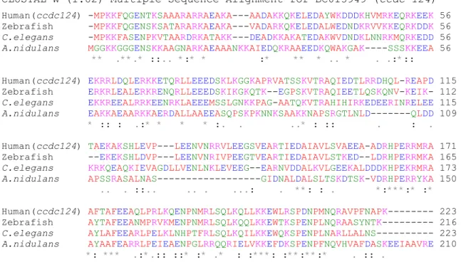
This gene is described as coiled-coiled domain containing 124 (Ccdc-124) in Genome Browser database at the University of California Santa Cruz. Ccdc-124 is conserved in all eukaryotes from the lower eukaryote fungus Aspergillus nidulans to
Homo sapiens. This gene has 75% identity scores in mammals, 50% identity in
nematodes and insects and 30-40% identity in plants and ascomycetes. Its protein has no domain similarities with any known protein. The gene contains 4 coding exons and it is predicted to encode a 223 amino acid protein in human. Molecular weight and isoelectric point of the protein were calculated as 25835.2 Da and 9.54, respectively via using ProtParam tool. It consists of 43 negatively charged, 51
27
positively charged residues, which means, 42% of the protein consists of charged residues (Fig.3.1).
CLUSTAL W (1.82) Multiple Sequence Alignment for BC013949 (ccdc-124)
Human(ccdc124) -MPKKFQGENTKSAAARARRAEAKA---AADAKKQKELEDAYWKDDDKHVMRKEQRKEEK 56 Zebrafish -MPKKFQGENSKSATARARKAEAKA---VADARKQKELEDALWEDNDKRVVKKEQRKDDK 56
C.elegans -MPKKFASENPKVTAARDRKATAKK---DEADKKAKATEDAKWVDNDKLNNRKMQRKEDD 56
A.nidulans MGGKKGGGENSKKAAGNARKAEAAANKKAIEDQKRAAEEDKQWAKGAK----SSSKKEEA 56 ** .**.* ::.. *:* * :* ** * .. * . .:*::
Human(ccdc124) EKRRLDQLERKKETQRLLEEEDSKLKGGKAPRVATSSKVTRAQIEDTLRRDHQL-REAPD 115 Zebrafish ERKRLEALERKRENQRLLEEEDSKIKGKQTK--EGPSKVTRAQIEETLQSKQNV-KEIK- 112
C.elegans EKKREEALRRKEENRKLAEEEMSSLGNKKPAG-AATQKVTRAHIHIRKEDEERINRELEE 115
A.nidulans EAKKAEAARKKAERDALLAAEEASQPSKPKNNKSAAKKNAPSRGTLNLD---QLDD 109 * :: : .:* * * * :. . ..* : :: . : .
Human(ccdc124) TAEKAKSHLEVP---LEENVNRRVLEEGSVEARTIEDAIAVLSVAEEA-ADRHPERRMRA 171 Zebrafish --EKEKSHLDVP---LEENVNRIVPEEGTVEARTIEDAIAVLSTKED--LDRHPERRMKA 165
C.elegans KRKQEAQKIEVAGDLLVENLNKLEVEEG--EARNVDDALKVLGEEKALDDDKHPEKRMRA 173
A.nidulans APSSRASALNAS---GIDNALDALSLTSKDTSK-VDRHPERRYKA 150 .. . ::.. .. . ...: . **: : . *:***:* :*
Human(ccdc124) AFTAFEEAQLPRLKQENPNMRLSQLKQLLKKEWLRSPDNPMNQRAVPFNAPK--- 223 Zebrafish AYTAFEEANMPRVKMENPNMRLSQLKQQLKKEWTKSPENPLNQRAASYNTK--- 216
C.elegans AYLAFEEARLPELKLNHPTFRLSQLKQILKKEWQKSPENPLNARLLALNS--- 223
A.nidulans AYAAFEARRLPEIEAENPGLRRQQRIELVKKEFDKSPENPFNQVHVAFDASKEEIAAVRE 210 *: *** .:*.:: ::* :* .* : :***: :**:**:* . :: .
Figure 3.1: High conservation of ccdc-124 gene protein sequence in eukaryotes. Protein sequences
of ccdc124 from different eukaryotes (human, zebrafish, C. Elegans and A. nidulans) were aligned by using Clustal-W multiple sequence analysis program. In the figure, green shows hydroxyl and amino groups containing amino acids; red shows, little hydrophobic amino acids; blue shows acidic amino acids; pink shows alkaline amino acids. 42% of the human CCDC-124 consists of positively charged (K, R, H, D, E) amino acids.
3.1.2 Expressional Analysis of Ccdc 124
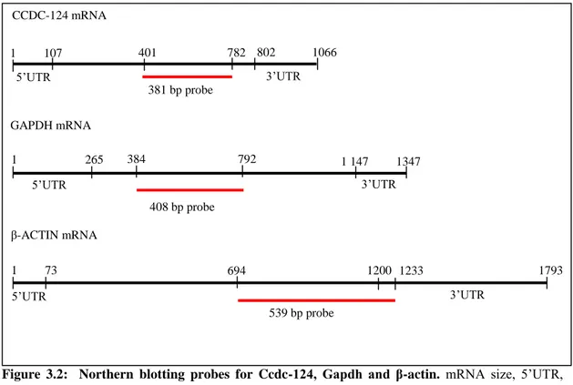
Northern blot analysis of Ccdc-124 was carried on a human tissue RNA transferred membrane (Human Blot 1, Ambion) and biotin labeled probes (Fig.3.2 and Fig.3.3).
28
Figure 3.2: Northern blotting probes for Ccdc-124, Gapdh and β-actin. mRNA size, 5‟UTR,
3‟UTR and probe size & location are indicated for each gene.
According to the results of Northern Blot analysis, Ccdc-124 is ubiquitously expressed in all human tissues tested.
Figure 3.3: Human tissue expression analysis of CCDC-124 by Northern blotting. Probes were
labeled via biotin and hybridized to Human Blot 1 membrane (see M.M.). Blots were exposed to X-ray films. β-actin and Gapdh were used as internal controls.
CCDC-124 mRNA GAPDH mRNA β-ACTIN mRNA 1 107 802 1066 1 265 1 147 1347 1 73 1200 1793 401 782 384 792 694 1233 381 bp probe 408 bp probe 539 bp probe 5‟UTR 3‟UTR 5‟UTR 3‟UTR 5‟UTR 3‟UTR