IDENTIFICATION OF THE INTERACTING DOMAIN OF p53
FAMILY MEMBERS WITH p33
ING1PROTEIN
A THESIS SUBMITTED TO
THE DEPARTMENT OF MOLECULAR BIOLOGY AND GENETICS
AND
THE INSTITUTE OF ENGINEERING AND SCIENCE OF
BILKENT UNIVERSITY
IN PARTIAL FULFILLMENT OF THE REQUIREMENTS
FOR THE DEGREE OF MASTER OF SCIENCE
By
DENİZ DİNÇEL
January, 2002
I certify that I have read this thesis and that in my opinion it is fully
adequate, in scope and in quality, as a thesis for the degree of Master of
Science.
_____________________________
Assist. Prof. Rengül Çetin-Atalay
I certify that I have read this thesis and that in my opinion it is fully
adequate, in scope and in quality, as a thesis for the degree of Master of
Science.
_____________________________
Prof. Mehmet Öztürk
I certify that I have read this thesis and that in my opinion it is fully
adequate, in scope and in quality, as a thesis for the degree of Master of
Science.
_____________________________
Assoc. Prof. Pervin Dinçer
Approved for the Institute of Engineering and Science
_____________________________
Prof. Mehmet Baray,
Director of Institute of Engineering
and Science
To My Family…
For letting me free to go after my dreams
ABSTRACT
IDENTIFICATION OF THE INTERACTING DOMAIN OF p53
FAMILY PROTEINS WITH p33
ING1DENİZ DİNÇEL
M.S. in Molecular Biology and Genetics Supervisor: Assist. Prof. Rengül Çetin-Atalay
January 2002, 106 Pages
p53 is a tumor suppressor gene, which is mutated, in about 50% of human cancers. The product of p53 gene encodes a sequence specific transcription factor. The genes transactivated by p53 code for proteins that are implicated in the negative regulation of cell proliferation and DNA damage repair. Two proteins, p63 and p73 members of p53 family, show striking homology to p53. p53 protein interacts with several viral and cellular proteins and these interactions are important in the regulation and dysregulation of the functions of p53. Another gene named, ING1 was identified as a candidate tumor suppressor gene due to its functions in apoptosis and cell cycle arrest. p24ING1, one of the protein product of ING1, was shown to enhance the growth suppressor functions of p53. Furthermore a physical association between p53 and p33ING1, another ING1 transcript, proteins has been detected by immunoprecipitation. In this study, we investigated the physical interaction between p53 family proteins and p33ING1 using in vitro techniques in order to determine the region(s) of p53 family proteins and p33ING1 that enabled this interaction. As a preliminary step for the study, the wild-type p53 cDNA and its several deletion mutant constructs were used in GST pulldown assays and Far Western assays with purified GST-p33 protein to map the interacting region on p53 protein. New deletion mutant constructs of p53 protein were created and cloned into expression vectors for the detailed analysis of the interacting domain of p53 protein. Also the other members of p53 protein family, p63 and p73 were examined in vitro for interaction with p33ING1. Deletion mutants of these proteins were created and cloned into expression vectors for protein-protein interaction assays. The results of this study shows that p53 protein interacts with p33ING1 and suggests that oligomerization domain of p53 protein is needed for this interaction. In addition, for the first time, it was showed that p63 and p73 proteins interact with p33ING1 and in p63 the C-terminus region is the primary determinant region, involved in these interactions with p33ING1.
Keywords: p33ING1, p53, p63, p73, tumor suppressor protein, protein-protein interaction, GST pulldown, Far Western Blotting
ÖZET
p53 PROTEİN AİLESİNİN p33
ING1PROTEİN’İ İLE İLİŞKİYE
GİREN BÖLGESİNİN BELİRLENMESİ
DENİZ DİNÇEL
Yüksek Lisans Tezi, Moleküler Biyoloji ve Genetik Tez yöneticisi: Yard. Doç. Dr. Rengül Çetin-Atalay
Ocak 2002, 106 Sayfa
p53, insan kanserlerinin yaklaşık %50’sinde mutasyona uğrayan bir tümör baskılayıcı gendir. p53 gen ürünü, DNA dizisi özgünlüğü olan bir transkripsiyon faktörüdür. p53 tarafından transkripsiyonu aktive edilen genlerin protein ürünleri, hücre bölünmesini önleyici yönde görev yapan kontrol elemanları ya da DNA tamir mekanizması elemanlarıdır. İki yeni protein, p63 ve p73 p53 protein ailesinin üyeleri, p53 proteinine hem gen seviyesinde hem de protein seviyesinde büyük benzerlik göstermektedir.p53 proteini bir çok hücresel ve viral protein ile etkileşime girer ve bu etkileşimler p53’ün işlevlerinin düzenlenmesinde ya da düzensizliğinde önemli yer tutar. ING1 (“Inhibitor of Growth 1”:Büyüme İnhibitörü 1) adlı yeni bir gen, apoptoz ve hücre döngüsün durdurulmasında ki rolleri dolayısıyla tümör baskılayıcı gen adayı olarak tanımlanmıştır. ING1’ın protein ürünlerinden biri olan p24ING1’ ın p53’ün büyüme baskılayıcı işlevlerini arttırdığı gözlemlenmiştir. Dahası, p53 ile p33ING1, diğer bir ING1 proteın ürünü, arasında fiziksel bir etkileşim, immunopresipitasyon yöntemiyle belirlenmiştir. Bu çalışmada, p53 protein ailesinin üyelerinin, p33ING1 ile etkileşen bölgelerini belirlemek amacıyla bu proteinler arasındaki fiziksel etkileşimi
in vitro yöntemlerle inceledik. İlk adım olarak, normal p53 cDNA’sı ve birçok
değişik p53 delesyon mutantı cDNA’ları, p53’ün p33ING1 ile etkileşen bölgesini belirlemek amacıyla ‘GST pulldown’ sisteminde ve ‘Far Western Blotting’ sisteminde kullanılmıştır. Bu çalışmalardan elde edilen sonuçlara bağlı olarak iki yeni p53 delesyon mutantı yaratılmış ve protein-protein etkileşiminin belirlenmesinde kullanılmıştır. p53 protein ailesinin diğer iki üyesi olan p63 ve p73 proteinlerinin p33ING1 proteini ile etkileşimi ve etkileşim bölgeleri C-terminali delesyon mutantları yaratılarak incelenmiştir. Bu çalışmanın sonuçları p53 proteinin, p33ING1 ile oligomerizasyon bölgesi aracılığı ile etkileşime girdiğine işaret etmektedir. Buna ek olarak, p63 ve p73 proteinlerinin, p33ING1 proteini ile etkileşime girdiği ve bu etkileşimin p63 proteininin C-terminalinden olduğu gösterilmiştir.
Anahtar Kelimeler: p33ING1, p53, p63, p73, tümör baskılayıcı, protein-protein etkileşimi, GST-pulldown, Far Western Blotting
ACKNOWLEDGEMENT
Thank You….
Dr. Rengül Çetin-Atalay, my supervisor, for all that I have learnt from you and for your patience
Dr. Mehmet Öztürk, for the knowledge and experience you have shared and the support you have given
Dr. Chris Lomer, for letting me know you Elif, for all the colors, you brought into my life
Bilhaj & Muna, my first friends at Bilkent, for these delicious lunchs Esin & Hüseyin, for being my supportive unit at all times, I love you!!!
Burcu, as the circumstances we shared together, made us come closer, for your friendship and time you spent with me
Tolga, the first person I called whenever I am in trouble, for making days standable and enjoyable!!! for me
Nuri, for patience you have shown towards my never-ending talks both at lab and on phone, in addition to your continues help during laborious cloning procedures
Çağla and Özgür, friends in need are friends indeed, for standing by me during years
Seda & Soner for the good time we had together
Hani for his friendship and help, whenever I looked at this thesis, I will remember you.
Tuba Gülbağcı for her warmth and understanding
Emek, for helps in the computer work
Tülay, Abdullah Bey, Sevim Hanım, Füsun, Ayşegül, Ömer, Ali and the others for the efforts you have spent to make things run smoothly
TABLE OF CONTENTS
ABSTRACT... III ÖZET... IV ACKNOWLEDGEMENT ...V TABLE OF CONTENTS ... VI LIST OF FIGURES ...X LIST OF TABLES ... XII ABBREVIATIONS ...XIII ABBREVIATIONS ...XIIICHAPTER 1. INTRODUCTION... 1
1.1 General Introduction ... 1
1.2 p53 Tumor Suppressor Gene and Protein Product ... 2
1.2.1 Background ... 2
1.2.2 Cellular Functions of p53 protein... 3
1.2.3 Structure of p53 protein ... 5
1.2.4 Regulation of p53 Activity... 7
1.2.5 Downstream Events ... 11
1.2.6 The p53 Family ... 13
1.3 ING1 Gene and its Protein product p33 ... 19
1.3.1 Structure of ING1 gene ... 22
1.3.2 ING1 Gene products in chromatin remodeling complexes ... 24
1.3.3 Cancer Development and p33... 25
CHAPTER 2: MATERIALS AND METHODS... 28 2.1 Materials ... 28 2.1.1 Chemicals ... 28 2.1.2 Bacterial Strains ... 28 2.1.3 Enzymes... 28 2.1.4 Oligonucleotides ... 28 2.1.5 Cloning Vectors ... 30 2.1.6 Antibodies ... 31
2.1.7 Commercially Available Kits ... 32
2.1.8 Apparatus ... 33
2.1.9 Materials for Autoradiograghy ... 33
2.1.10 DNA and Protein Size Markers ... 34
2.2 Solutions and Media ... 34
2.2.1 Agarose Gel Electrophoresis Solutions ... 34
2.2.2 Solutions for Plasmid DNA Isolation (MiniPrep) ... 34
2.2.3 Solutions for Bacterial Transformation ... 34
2.2.4 Microbiological media and antibiotics ... 35
2.2.5 Polyacrylamide Gel Electrophoresis Solutions ... 35
2.2.6 Far Western Buffers ... 36
2.2.7 Solutions for Trichloroacetic acid Precipitation... 37
2.3 Methods... 38
2.3.1 PCR amplification of cDNA ... 38
2.3.4 Methods for Cloning and Subcloning of Genes... 39
2.3.4.1 PolyA Extension Reaction... 39
2.3.4.2 Insertion of Products of PolyA Extention Reaction into pGEM-T easy vector... 39
2.3.4.3 Restriction Enzyme Digestion of DNA ... 40
2.3.4.4 DNA Ligation Reactions ... 40
2.3.4.5 Transformation of E.coli ... 40
2.3.4.6 Plasmid DNA Preparation ... 41
2.3.5 Quantification of Nucleic Acids... 42
2.3.6 Growth and Storage of Bacterial Strains ... 43
2.3.7 SDS Polyacrylamide Gel Electrophoresis ... 43
2.3.8 In vitro Transcription-Translation (IVTT) Reactions ... 44
2.3.9 GST Pulldown Assays ... 45
2.3.10 Far Western Blotting ... 47
CHAPTER 3: RESULTS ... 49
3.1 Examination of Deletion Mutant Constructs of p53... 49
3.2 Confirmation of Previously Obtained Results ... 51
3.3 Specific Monoclonal Antibodies to p53 and p33ING1 can not Inhibit the Interaction ... 54
3.4 Demonstration of p53 and p33ING1 Interaction by Far Western Analysis ... 55
3.5 Subcloning of p53 cDNA and p53 Deletion Mutant Constructs ... 57
3.6 Further Detailed Mapping of the Interacting domain of p53 with p33ING1 by In Vitro Techniques ... 59
3.7 Demonstration of Interaction between p33ING1 and other p53 Family
Members ... 61
3.8 Cloning of C-terminally deleted forms of p63, and p73... 62
3.9 Subcloning of C-terminally deleted forms of p63 and p73... 64
3.10 Production of Radioactively Labeled Deletion Mutant p63∆C ... 65
3.11 Examination of Interaction between p63∆C and p33ING1 in vitro ... 66
3.12 Cloning and the Subcloning of the deletion mutant of p33ING1... 67
DISCUSSION ... 72
REFERENCES ... 76
APPENDICES... 88
Appendix A: ... 88
LIST OF FIGURES
Figure 1.1: Upstream elements activating the p53... 4
Figure 1.2: Domain structure of p53 , together with mutational hotspots and interacting proteins... 6
Figure 1.3: Summary of posttranscriptional modifications and domains of p53... 9
Figure 1.4 Major downstream targets of p53 ... 12
Figure 1.5: Exon-intron organization of p53 family members ... 14
Figure 1.6: Homology between the p53 family members ... 15
Figure 1.7: Genomic structure of human ING1 ... 24
Figure 2.1 Vector map of pGEMT-easy ... 30
Figure 2.2: Vector Map of pBlueScript II KS- ... 31
Figure 2.3: Vector Map of pcDNA3... 32
Figure 3.1: Restriction enzyme digestion analysis of MiniPreps of deletion mutant constructs. ... 51
Figure 3.2: SDS-PAGE analysis of IVTT reactions of deletion mutant constructs of p53: ... 51
Figure 3.3: GST pulldown assay of wt p53 with GSTp33... 52
Figure 3.4: GST pulldown with p53 deletion constructs. ... 53
Figure 3.5: GST pulldown assay with p53 and p33ING1 antibodies. ... 55
Figure 3.6: Optimization of Far Western assay ... 56
Figure 3.7: Far Western analysis of wt p53 and C-terminally deleted mutant of p53.56 Figure 3.8: AGE analysis of MiniPrep of pBksF6R6 and pBksFuR6 constructs... 58
Figure 3.9: GST pulldown with p53 deletion constructs ... 59
Figure 3.10: Far Western with p53 deletion constructs ... 60
Figure 3.11: Far Western analysis of wt p63 and wt p73. ... 61
Figure 3.12. AGE of p63∆C, p73∆C PCR-products... 63
Figure 3.13: AGE analysis of MiniPrep of pGEMTp63∆C... 64
Figure 3.14: AGE analysis of MiniPrep of pGEMTp73∆C constructs. ... 64
Figure 3.15: AGE analysis of MiniPrep of pcDNA3p63∆C (a) and pcDNA3p73∆C (b) constructs... 65
Figure 3.16: SDS-PAGE analysis of IVTT reactions of deletion mutant construct of p63... 66
Figure 3.18: GST pulldown with p63 deletion construct... 67
Figure 3.19: AGE of p33ING1∆C PCR-product ... 68
Figure 3.20: AGE analysis of MiniPrep of pGEMT-p33ING1∆C constructs... 69
Figure 3.21: AGE analysis of MiniPrep of pBks p33ING1∆C constructs ... 70
LIST OF TABLES
Table 1.1 Representative examples of proteins interacting with p53. ... 10 Table 3.1: Different deletion products of p53 and their interaction status with p33ING1 ... 50
ABBREVIATIONS
A600 absorbance at 600 nanometer wavelength aa amino acid
AGE agarose gel electrophoresis APS ammonium persulfate
bp base pair
BSA bovine serum albumin cDNA complementary DNA
cm centimeter
ddH2O deionized, distilled water DEPC diethylpyrocarbonate
DMEM Dulbecco's Modified Eagle Medium DMSO Dimethylsulfoxid
DNA deoxyribonucleic acid dNTP deoxynucleotide triphosphate DTT Dithiothreitol
EDTA ethylenediaminetetra-acetic acid FCS fetal calf serum
g gram Gn Guanidium
GST Glutathione S-Transferase hr hour
IPTG isopropylthio-beta-D-galactoside IVTT in vitro transcription and translation
kb kilobase kD kilodalton LB Luria-Bertani medium M molar mA miliamper ME mercaptoethanol min minute
MidiPrep medium-scale isolation of plasmid DNA by alkaline lysis method MiniPrep small-scale isolation of plasmid DNA by alkaline lysis method ml milliliter
nm nanometer
PAGE polyacrylamide gel electrophoresis PBS phosphate buffered saline
pBsk pBluescript II KS- phagemid vector PCR polymerase chain reaction
rpm revolution per minute RNA ribonucleic acid RNase ribonuclease RT room-temparature
SDS sodium dodecyl sulphate
sec second
TAE tris/acetic acid/EDTA buffer
TEMED N,N,N,N-tetramethyl-1,2 diaminoethane Tris 2-amino-2-[hydroxymethyl]- 1,3 propandiol U unit
V volt
v/v volume for volume
wt wild type
w/v weight for volume w/w weight for weight
CHAPTER 1. INTRODUCTION
1.1 General Introduction
In an adult, the rates at which new cells in a tissue are formed and old cells die are in balance, producing a steady state in which the tissue does not increase in size. If the mechanisms regulating cell division are not functioning properly, the affected cells may produce new cells faster than old cells are removed, forming a growing mass of tissue known as a tumor. If the tumor cells grow into the surrounding tissues, disrupting their functions, and spread to other regions of the body, the tumor is said to be malignant tumor. Malignant tumors are composed of cancer cells, that have lost the ability to respond the normal mechanisms that regulate cell growth (Hanahan & Weinberg 2000).
Two classes of genes are mutated in cancer formation. The first class of genes (gatekeepers) comprising tumor suppressor genes, directly controls cellular proliferation. They can do this either by controlling the rate of cell birth or the rate of cell death. The second class of genes (caretaker genes) does not directly control cell growth, but instead controls the rate of mutation. Cells with defective mutator genes acquire mutations in all genes including oncogenes and tumor suppressor genes at an elevated rate. This higher rate leads to accelerated tumorigenesis (for review see Vogelstein and Kinzler, 1998).
Tumor suppressor gene is a genetic element whose loss or inactivation allows a cell to display one or another phenotype of neoplastic growth deregulation. The
products of the tumor suppressor genes are required for normal cell function, and they antagonize the proliferative actions of oncogenes to prevent tumorigenesis. Tumor suppressor mutations act as a recessive trait in the formation of tumors. Tumor suppressor genes constitute a very heterologous class of genes in terms of functions of their protein products. They can be transcription factors (e.g. p53, WT-1), cyclin dependent kinase inhibitors (e.g. p16) or receptor tyrosine kinases (e.g. RET) (for review see Haber, 1998).
1.2 p53 Tumor Suppressor Gene and Protein Product 1.2.1 Background
p53 protein is known as guardian of the genome, functional inactivation of p53 by gene mutation and deletion, protein degradation or viral oncogene binding renders a mammalian cell susceptible to oncogenic stimuli and environmental factors that promote growth deregulation and malignant progression. More than half of all human cancers shows either absence of p53 protein or mutations in this gene (for reviews, see Wang & Harris, 1997; Adams & Kaelin, 1998; Ko & Prives, 1996) . p53 is a nuclear phosphoprotein. It was originally discovered in SV40-transformed cells, where it is associated with T antigen (Lane et al., 1979; Linzer et al., 1979). A large increase in the amount of p53 protein is found in many transformed cells or lines derived from tumors. In early experiments, the introduction of cloned p53 was found to immortalize cells. These experiments caused p53 to be classified as an oncogene, However subsequent studies revealed that p53 cDNAs used in these studies were mutant and wild type counterparts were shown to suppress the growth of transformed cells in vitro and tumorigenic potential of these cells in animals (Finlay et al., 1989). Mutations of p53
alleles were implicated in many human and animal tumors. Indeed, members of Li-Fraumeni cancer-prone families were shown to carry germ-line p53 mutations (Malkin
et al., 1990). The role of p53 in cancer was more directly accessed by the observation
that p53 knock out mice developed tumors at high frequencies (Donehower et al., 1992). Therefore, p53 was re-classified as a tumor suppressor gene.
1.2.2 Cellular Functions of p53 protein
The human p53 is located in the short arm of chromosome 17, 17p13.1. The gene has 11 exons and spans 20kb of genomic DNA. Many organisms have p53 homologues, such as monkey, chicken, rat, frog, fruit fly and worm (Derry B. W., et al., 2001; Jin S., et al., 2000; Soussi T., et al.,1988; Rigaudy P., et al.,1989). The general organization of the p53 protein is well conserved during evolution.
p53 integrates signals from the cell’s internal and external environment to respond to inappropriate growth promoting or growth inhibiting conditions. The tumor suppressor activity of p53 stems from its ability to both inhibit the mitotic cell cycle and promote apoptosis. The functional character of the p53 protein was determined by experiments showing that p53 contains a strong transcriptional activation domain within its amino terminus and that it is a sequence specific DNA-binding protein (El-Deiry et
al., 1992). Although the p53 protein acts as a transcriptional activator of genes
containing p53 binding sites, it is also capable of inhibiting transcription from many genes lacking p53 binding sites. Several oncogenic DNA viruses express viral gene products that associate with and inhibit the trans-activation function of p53, notably SV40 large antigen, the adenovirus E1B 55-kD protein, and E6 protein of oncogenic forms of human papillomavirus (HPV E6) (for review, see Prives & Ko, 1996).
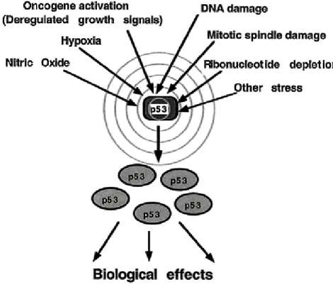
Figure 1.1: Upstream elements activating the p53 ( From Oren 1999).
p53 plays multiple roles in cells. Expression of high levels of wild-type p53 has two outcomes: cell cycle arrest, or apoptosis. In unstressed cells, p53 appears to be present at low levels and exist in a latent inactive form that requires modification to become active. The types of modification that p53 is subject to seem to be stress-, species- and cell-type-specific. Levels and/or activity of p53 increase in response to DNA damaging agents (Maltzman and Czyzyk, 1984; Kastan et al., 1991), decreased oxygen (Graeber et al., 1994), oncogenic stimuli (Debbas and White, 1993), cell adhesion (Nigro et al., 1997), altered ribonucleotide pools (Linke et al., 1996), and redox stress (Hainaut et al., 1993) (Figure 1.1). The importance of p53 and modifications that affect its functions are not limited to malignant disease. The activity of p53 can increase in normal tissues when undergoing pathophysiological changes that
result in oxidative or redox stress, such as ischemia, reperfusion injury of the brain, heart, and other tissues.
1.2.3 Structure of p53 protein
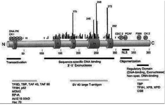
The human p53 protein is a single polypeptide of 393 amino acids. It can be divided into different domains (for review, see Ko & Prives, 1996): at the N-terminus, a transactivation domain (residues 1 to 43) and proline-rich domain (residues 62 to 91); in the middle core, a DNA-binding domain (residues 100 to 300); and at the C-terminus, a tetramerization domain (residues 326 to 354) and a regulatory region (363 to 393) (Figure1.2).
p53 binds in vitro to several proteins through its activation domain. The amino terminus of p53 interacts with many general transcription factors such as the TATA box-binding protein (TBP) component of general transcription factor TFIID, several TBP-associated factors (TAFs), including Drosophila melanogaster 40 and TAF-60 (Thut et al., 1995), the human TAF-31 (Lu and Levine, 1995) and TFIIH (Xiao et
al., 1994). p53 transcriptional activation is negatively regulated by the adenovirus
E1B-55Kd protein (Kao et al., 1990) and human Mdm2 protein (Haupt et al., 1997). In both cases, p53 amino acid residues 22 and 23 play a key role in the binding of p53 to E1B-55Kd or Mdm2 protein. Thus the negative regulators of p53-mediated transcription target some of the same p53 amino acids critical to positive regulation of transcriptional activation. This evidence showed that p53 uses a hydrophobic interface in its N terminal domain to interact with the transcriptional machinery of the cell and its negative regulators. Also this domain is the target of phosphorylation by various kinases (for review, see Levine, 1997).
Proline rich region spans the amino acids 62-91. There are five tandemly repeated proline rich motifs in the form of PXXP (where P is proline and X is any amino acid) and these motifs were shown to bind SH3-containing proteins. This region is implicated in both growth arrest and apoptosis (Venot et al., 1998).
The sequence-specific DNA-binding domain of p53 is localized between amino acid residues 100 and 300. It is a protease resistant and independently folded domain containing a Zinc ion that is required for its sequence-specific DNA-binding activity.
Figure 1.2: Domain structure of p53 , together with mutational hotspots and interacting proteins.
Dark Grey Boxes I-V: regions conserved among species; at the N-terminus: Transactivation domain;
Next to it proline rich domain; In the middle of the protein: DNA binding domain ; At the C-terminus: there is both oligomrization and regulatory domain (Adapted from Ko & Prives 1996).
The p53 protein binds to four repeats of a consensus DNA sequence 5’-PuPuPuC(A/T)-3’, and this sequence is repeated in two pairs each arranged as inverted repeats (El-Deiry et al., 1992). More than 90% of the missene mutations in the p53 reside in this sequence-specific DNA-binding domain, and these mutations fall into two
classes: mutations which result in defective contacts with the DNA and loss of the ability of p53 to act as a transcription factor, or mutations which disrupt the three dimensional (3D) conformation of the protein (for review, see Levine, 1997).
The native p53 protein functions as a tetramer, and amino acid residues 326-354 are required for this oligomerization of the protein. The structure of this domain contains a dimer of a dimer with two β sheets and two α helices. This tetramerization domain is linked to the sequence specific DNA-binding domain by a flexible linker of 37 residues (Lee et al., 1994; Clore et al.,1995; Jeffery et al.,1995).
The C-terminus 26 amino acids form an open, protease sensitive, domain composed of nine basic amino acid residues that bind to DNA and RNA (Lee et al., 1995). There is a considerable evidence showing that the p53 protein requires a structural change to activate it for sequence-specific binding to DNA. This non-DNA binding form can be regulated by basic C-terminus domain. Deletion of this domain, phosphorylation of certain residues by kinases, or by the binding of certain specific antibodies all activate site-specific DNA binding by the central domain (Hupp and Lane, 1994). Short (20-39 nucleotide) single strands of DNA interacting with this C-terminus domain can also activate specific p53 DNA binding. The C-C-terminus domain helps to catalyze the reassociation of single-stranded DNA or RNA to double strands (Lee et al.,1995). It also binds preferentially to DNA ends and to internal deletion loops in DNA as generated by replication errors.
1.2.4 Regulation of p53 Activity
Because p53 is such a critical cellular protein, multiple mechanisms have evolved to regulate its activity. These regulatory mechanisms exist both tightly and
rapidly control the activities of p53 and to provide alternative regulatory mechanisms for different cell types and different physiological stimuli. Normally, in a cell, the p53 protein is kept at a low concentration by its relatively short half-life. In addition to its low protein concentration, in some cells p53 probably also exists in a latent form (Kastan et al.,1991). To activate the protein, p53 must receive a signal or alternation. Several stressful situations, such as DNA damage, γ irradiation, presence of DNA repair intermediates after ultraviolet irradiation or chemical damage to DNA, can activate p53. This results in a rapid increase in the level of p53 in the cell and activation of a p53 as a transcription factor. Although levels of p53 mRNA does not change in response to stimuli, the levels of p53 protein increase rapidly (Kastan et al., 1991). The half-life of p53 increases several folds after DNA damage. Enhanced translation of p53 mRNA also contributes to this induction. Therefore the regulation is mainly posttranslational. This type of regulation is necessary for very rapid and efficient control of the p53 levels in a cell. In addition to changes in the level of p53, several different modifications are implicated in the activation of p53 protein, such as phosphorylations, dephosphorylations, acetylations, and binding to certain proteins (for review see Meek 1994; Chernov et al., 1998). Both posttranslational modifications and alternations in p53 binding proteins appear to be major contributors to the modulation of p53 activity. Carboxy terminal phosphorylation and incubation with a Carboxy terminal binding anti-p53 antibody markedly enhanced sequence-specific DNA binding of anti-p53 (Hupp et al., 1993). This evidence showed that Carboxy terminus domain of p53 is the regulatory region of the protein. Mdm2 protein as a major intracellular regulator of ubiquitin-mediated degradation of p53 protein (Haupt et al., 1997; Kubbutat et al., 1997). Mdm2 protein can inhibit p53 function by binding to and inhibiting transactivation by p53.
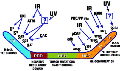
The existence of these various regions of p53 provides a way by which a primary p53 regulatory mechanism could be by specific post-translational modifications or one or more of these domains, or modulation of proteins that bind to these domains. The cell uses different functions and proteins to recognize different classes of DNA damage and different systems of enzymes to repair them. For example, ATM kinase activity is enhanced following ionizing radiation (IR) and this protein is shown to phosphorylate Serine-15 of p53. ATM was also implicated in a radiation-induced dephosphorylation event. IR causes a loss of a phosphate group from Serine 376 in the carboxy terminus of p53 in an ATM dependent manner and this dephosphorylation event results in a creation of a binding for 14-3-3 proteins and enhancement of sequence-specific DNA binding (Waterman et al., 1998) (Table 1.1).
Figure 1.3: Summary of posttranscriptional modifications and domains of p53. (From Giaccia &Kastan, 1998).
Ultra violet (UV) irradiation is also a potent inducer of p53 protein; however, clear differences exist in p53 induction following UV radiation versus IR. UV signaling to p53 does not depend on ATM kinase but lysine residues in the N terminal domain of p53 are acetylated by PCAF and p300/CBP (Lill et al., 1997; for review see Giaccia &
Kastan 1998) (Figure 1.3) . In addition to DNA damage, hypoxia is able to stimulate p53 levels and activate p53 protein in a different way from UV radiation and IR. It was suggested that hypoxia increases p53 protein levels via induction of another protein, HIF-1β ( An et al., 1998).
Yet a third signal to activate p53 is sent when ribonucleoside triphosphate pools fall below a critical threshold. Normal nucleoside triphosphate pools are necessary to support DNA replication and progression through the cell cycle (Linke et al., 1996). As seen in the examples, activation of p53 is complex and tightly regulated process.
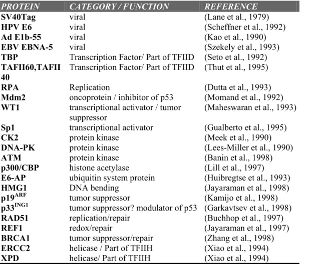
Table 1.1 Representative examples of proteins interacting with p53.
PROTEIN CATEGORY / FUNCTION REFERENCE
SV40Tag viral (Lane et al., 1979)
HPV E6 viral (Scheffner et al., 1992)
Ad E1b-55 viral (Kao et al., 1990)
EBV EBNA-5 viral (Szekely et al., 1993)
TBP Transcription Factor/ Part of TFIID (Seto et al., 1992)
TAFII60,TAFII 40
Transcription Factor/ Part of TFIID (Thut et al., 1995)
RPA Replication (Dutta et al., 1993)
Mdm2 oncoprotein / inhibitor of p53 (Momand et al., 1992)
WT1 transcriptional activator / tumor suppressor
(Maheswaran et al., 1993)
Sp1 transcriptional activator (Gualberto et al., 1995)
CK2 protein kinase (Meek et al., 1990)
DNA-PK protein kinase (Lees-Miller et al., 1990)
ATM protein kinase (Banin et al., 1998)
p300/CBP histone acetylase (Lill et al., 1997)
E6-AP ubiquitin system protein (Huibregtse et al., 1993)
HMG1 DNA bending (Jayaraman et al., 1998)
p19ARF tumor suppressor (Kamijo et al., 1998)
p33ING1 tumor suppressor? modulator of p53 (Garkavtsev et al., 1998)
RAD51 replication/repair (Buchhop et al., 1997)
REF1 redox/repair (Jayaraman et al., 1997)
BRCA1 tumor suppressor/repair (Zhang et al., 1998)
ERCC2 helicase / Part of TFIIH (Xiao et al., 1994)
XPD helicase/ Part of TFIIH (Xiao et al., 1994)
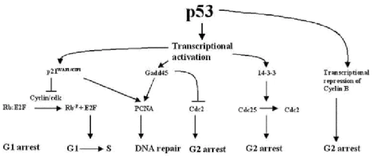
1.2.5 Downstream Events
The transcriptional activating function of p53 is a major component of its biological effects. By binding to specific sites within their promoters, p53 activates the transcription of various genes including, p21WAF1, GADD45, mdm2, bax, thrombospondin 1, cyclin G, and insulin-like growth factor binding protein 3 (IGF-BP3) (Figure1.4) (for review see Somasundaram & El-Deiry 2000).
The p53-dependent G1 arrest occurs largely through the transactivation of p21WAF11 (el-Deiry et al., 1993). p21WAF1 is a member of small cyclin-dependent kinase inhibitor (CKI) family. It binds to a number of cyclin and cyclin-dependent kinase (cdk) complexes, by this way inhibits the transition to S-phase. p21WAF1 also binds to proliferating cell nuclear antigen (PCNA), and inhibits the role of PCNA as a DNA polymerase processivity factor in DNA replication (Waga et al., 1994).
More recently, p53 has been implicated in a G2/M phase checkpoint. When mitotic spindle inhibitors are added to cells with wild-type p53, the cells are blocked in G2. In the absence of wild-type p53, these cells reinitiate DNA synthesis. These data suggest that p53 may be part of a G2/M checkpoint, preventing premature entry into another S phase. In addition p53 appears to be an integral part of the process that regulates the number of centrosomes in a cell (Cross et al.,1995; Fukasawa et al., 1996).
Another way for eliminating abnormal cells is apoptosis. p53 mediates apoptosis in several cell types in response to several stimuli, including DNA damage, adenovirus E1A expression, myc expression or withdrawal of growth factors. p53 may use
transcriptional activation or direct protein signaling (protein-protein interactions) (Hupp
et al., 1995) or both to initiate apoptosis.
Figure 1.4 Major downstream targets of p53 ( From Somasundaram & El-Deiry 2000).
Two of the genes that are regulated by p53 could influence the decision to commit to an apoptotic pathway: bax and IGF-BP3 (Miyashita and Reed, 1995; Buckbinder et al., 1995). Bax is a tumor suppressor gene whose protein product interacts with bcl-2 protein. Bcl-2 is an anti-apoptotic factor and increased bax expression block the functioning of this protein. A second p53 regulated gene product that could affect growth regulation is the IGF-BP3. IGF-BP3 protein blocks the IGF mitotic signaling pathway by binding to IGF and preventing its interaction with its receptor. Thus, the blocking of IGF activity could enhance apoptosis or lower the mitogenic response of cells.
Number of factors, like extend of DNA damage, certain oncogenic disruptions, availability of survival or growth factors, and cell type, affect the decision of a cell to enter a p53-mediated cell cycle arrest or apoptotic pathway.
1.2.6 The p53 Family
For a long time, p53 was thought to be alone, but two mammalian proteins, p73 and p63, with significant sequence homology to p53 have recently been identified. Its first relative, TP73, which encodes p73, was identified by chance in 1997 (Kaghad et
al., 1997), and its second, TP63 (which encodes p63 and is also known as KET, P51,
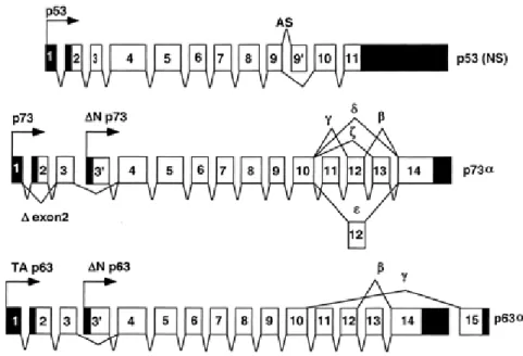
P40, chronic ulcerative stomatitis protein (CUSP) and P73L), was identified independently by several groups (Yang et al., 1998; Trink et al., 1998; Osada et al., 1998; Augustin et al., 1998; Senoo et al., 1998; Lee et al., 1999). There are several common features among the p53 gene family members. (1) They all contain very large introns, (2) exon1 is always non-coding, (3) the exon/intron organization of all the family members is highly similar (Figure 1.5).
Both p63 and p73 give rise to many differently spliced mRNAs, which are translated into several different proteins. Most of the splicing occurs at the 3’ end and creates proteins that have different C-termini. There are at least six alternatively spliced forms of human p73: p73α, p73β, p73γ, p73δ, p73ξ, p73ε (Kaghad et al., 1997; De Laurenzi et al., 1998) in addition to these COOH-termini splice forms, two additional forms, ∆Np73α, and ∆Np73β, result form the use of an alternative promoter located in intron 3 (Yang et al., 1998). The TP73 gene contains 14 exons and is located at chromosome 1p36.33. The TP63 gene is located at chromosome 3q27-29 and contains 15 exons (Yang et al., 1998; Osada et al., 1998) and can be transcribed from two different promoters, which are located upstream of exon1 and within intron 3 (Yang et
al., 1998). Recognized variants of TP63 include the α, β, and γ forms of p63. Furthermore p63 utilizes a cryptic promoter located in intron 3 to generate additional
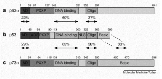
transcripts called ∆Np63α, ∆Np63β, ∆Np63γ (Figure 1.5). The three members of the p53 family share very significant homology both at the genomic and at the protein level. Each contains a transcriptional activation domain (TAD), a DNA-binding domain (DBD) and an oligomerization domain (OD). In addition p63 and p73, but not p53, contain long C-termini (for review see Levrero et al., 2000).
Figure 1.5: Exon-intron organization of p53 family members, showing different transcripts.
The highest level of homology is reached in the DBD (63% identity between p53 and p73, and 60% identity between p53 and p63), which suggests that the three proteins can bind to the same DNA sequences and transactivate the same promoters. The vast majority of missense mutations of p53 found in human tumors are clustered in the sequence specific DNA-binding domain. In addition, p73 and p63 are 29% and 22% identical to the region corresponding to the N-terminus transactivation domain of p53 respectively. Residues in the C-terminus region of p73 and p63 are 38% and 37% identical to the corresponding C-terminus oligomerization domain of p53 (for review see Chen 1999). The nuclear import and nuclear export signals lie within the
oligomerization domain and are conserved among p53 from different species as well as in p63 and p73. The sequences of p63 and p73 diverge from p53 mostly in their C-termini. Furthermore different p63 and p73 C-termini are generated as a result of alternative mRNA splicing. The determination of the three dimensional solution structure of the p73 C-terminus has shown that this region contains a sterile alpha motif (SAM), which is a protein-protein interaction domain (Chi et al., 1999). The SAM domain is a globular domain composed of four α-helices and a small 310-helix. SAM domains are protein-protein interaction modules found in proteins involved in developmental regulation. This domain is also present in p63 C terminus. Although SAM domains are known to participate in homotypic or heterotypic interactions, the p73 and p63 domains bind neither to themselves nor to each other. Nevertheless, it is possible that these domains interact either with a distantly related SAM domain protein or perhaps with a non-SAM containing protein (Figure 1.6) ( for review see Irvin & Kaelin 2001; Kaelin 1999).
Figure 1.6: Homology between the p53 family members. AD, activation domain, PXXP proline rich
p73 was initially proposed as a possible tumor suppressor gene because it is related to p53, it maps to chromosome 1p36.33, a region frequently deleted in neuroblastoma and other human cancers and it has been found to be monoallelically expressed owing to genomic imprinting (Kaghad et al., 1997). However the status of p73 as a tumor suppressor gene has been challenged by recent observations. It was found that p73 can be biallelically expressed in both normal or tumor tissues and cell lines, including neuroblastoma and mutation of the p73 gene occurs infrequently in human cancers (Ichimiya et al., 1999; Han et al., 1999).
It is not also certain whether p63 is a tumor suppressor gene. p63 is located at chromosome 3q27-29, a region that is not a common site of loss of heterozygosity in human cancers. p63 gene was found to be mutated infrequently in both human tumor tissues and cancer cell lines (Osada et al., 1998). Although p63 might have functions similar to those of p53 in cell-cycle arrest and apoptosis, ∆Np63, which lacks an activation domain, inhibits the activity of both p53 and p63, thereby exhibiting oncogenic functions (Yang et al., 1998).
While p53 is ubiquitously expressed, p73 and p63 are restricted to certain tissues. In the developing mice, p73 is detected in the epidermis, sinuses, inner ear, and brain while p63 is expressed in proliferating basal cells of the epidermis, cervix and prostate. In the p63- deficient mice, the apical ectodermal ridge essential to limb development is defective, and the mice have truncated limbs. In addition, p63 knockout mice have no hair follicles, no teeth, no mammary, lachrymal or salivary glands (Mills
et al., 1999; Yang et al., 1999). Thus the phenotype of p63-deficient mice suggests that
mutations of p63 have been detected in children affected by ectrodactyly ectodermal dysplasia and facial clefts (Celli et al., 1999). Ectodermal dysplasia is manifested by changes in skin, hair, nails, teethlacrimal duct, and urogenital tract. Similar to p63-deficient mice, p73-p63-deficient mice exhibit severe defects, including hydrocephalus, hippocampal dysgenesis, chronic infections and inflammation, and abnormalities in the pheromone sensory pathway. However they do not develop any spontaneous tumors (Yang et al., 2000). According to these findings, both p63 and p73 play very important roles in ectodermal differentiation and neurogenesis respectively. However these findings do not exclude the roles of p63 and p73 as tumor suppressors. Therefore the functions of p53 family may overlap in some tissues because of the requirement for simultaneous activity of p53, p63 and p73 at specific stages of development.
Although there is a high degree of sequence similarity between p53 and p73, neither SV40 TAg, E1B55k, nor E6 oncoproteins bind to p73 (Marin et al., 1998; Higashino et al., 1998; Roth et al., 1998). However the possibility exists that they can be bound and inactivated by other viral proteins. Only one viral oncoprotein, adenovirus E4orf6, was capable of associating with p73 and inhibiting the activity of p73 in one experimental protocol (Steegenga et al., 1999), but not in another (Roth et al., 1998). Thus further studies are required to address this issue. Whether these viral oncoproteins can regulate p63 remains to be determined.
Like p53, p73 and p63 can induce cell-cycle arrest and apoptosis (Kaghad et al., 1997; Yang et al., 1998; Osada et al., 1998; Jost et al., 1997; Zhu et al., 1998). Loss of p73 transcriptional activity abrogates its activity in cell-cycle arrest and apoptosis (Kaghad et al., 1997; Jost et al., 1997; Zhu et al., 1998). It is known that p53
can induce p21WAF1 transcription. Although p73 can induce p21WAF1, the level of cellular p21WAF1 induced by p73 is several times lower than that induced by p53. Two other p53 targets GADD45 and B-cell translocation gene 2 antiproliferative are only weakly activated by p73.
Like p53, p73 can induce G2/M arrest, p73 is capable of inducing 14-3-3σ (Zhu
et al., 1998). However, p73 induces several fold higher levels of 14-3-3σ gene product than does p53. It remains to be found whether p63 can induce 14-3-3σ and cell-cycle arrest at G2/M.
Although both p53 and p73 can induce apoptosis (Jost et al., 1997; Zhu et al., 1998), the signaling pathways may be different because of the different abilities of p73 to activate some p53 target genes. While bax and several redox-related genes (PIG2, PIG3, PIG6 and PIG11) are possibly involved in mediating p53-dependent apoptosis (Miyashita et al., 1994; Polyak et al., 1997), they were not significantly induced by p73 (Zhu et al., 1998). Because p73 transcriptional activity is required for inducing apoptosis (Jost et al., 1997; Zhu et al., 1998), it is possible that p73 can activate a different subset of cellular genes for mediating apoptosis.
Mdm2, an oncogene that negatively regulates p53, and is also induced by p53 (Wu et al., 1993), is weakly induced by p73β and p73α. The interaction with Mdm2 leads to the inactivation of transcription and apoptosis functions of p73-β and p73-α (Zeng et al., 1999) but does not result in the rapid degradation of p73 protein (Zeng et
al., 1999; Dobbelstein et al., 1999). Disruption of Mdm2-p73 interaction is needed for
Accumulation of p73 in response to certain DNA-damaging stimuli relies on the activity of the c-Abl tyrosine kinase, which prolongs the half-life of p73 (Gong et al., 1999). The accumulation of p53 and p73 in response to certain stimuli occurs through independent pathways. The accumulation of p53 is regulated by the inhibition of its degradation, but the accumulation of p73 requires the activity of the stabilizing protein (c-Abl) ( Yuan et al., 1999; for review see Levrero et al.,2000).
Although ultraviolet light, actinomycin D, and methylmethane sulfonate (MMS) do not induce the accumulation of p73, cisplatin, which crosslinks DNA (Gong
et al., 1999), and taxol, an agent that stabilizes microtubules and prevents completion of
mitosis, induce p73 accumulation and increase its half-life. Later it was shown that cisplatin and ionizing irradiation could regulate p73 through protein accumulation or tyrosine phosphorylation, respectively (Gong et al., 1999; Agami et al., 1999; Yuan et
al., 1999). These post-translational modifications of p73 occur through its physical
interaction with c-Abl kinase. According to these findings, regulation of p73 in response to several types of DNA damage is a complex phenomenon that may be mediated by the recruitment of the different upstream proteins.
1.3 ING1 Gene and its Protein product p33
A novel gene called “Inhibitor of Growth 1, ING1” encoding a 33 kDa protein was identified by subtractive hybridization (Garkavstev et al., 1996) (see Appendix A for more info.). By using indirect immunofluorescence, it is shown that the p33ING1 protein is located in the nucleus, with its proposed role as a growth regulator. Also a genomic probe to human ING1 localized to chromosome 13 at q33 → q34 by fluorescence in situ hybridization (ISH). This area is close to known sites of genomic
alteration in several human cancers: primary gastric cancer, haematologic neoplasms, and head and neck squamous cell carcinomas (Zeremski et al., 1997).
All the initial studies of ING1 protein was done by using p33ING1, but later the authors reported that the constructs used in these studies were actually the p24ING1, one of the three products of human ING1 gene, due to a cloning defect (Garkavstev et al., 1999) (see detailes later).
First studies showed that, p33ING1 (p24ING1), the protein product of ING1 gene, regulate the growth negatively. Overexpression of transfected ING1 constructs efficiently decreased S-phase fraction by blocking the entry of cells into S-phase. Chronic expression of antisense constructs resulted in tumor induction in vivo and in foci formation and ability to grow in soft agar in vitro. Further studies suggested that p33ING1 may be involved in cell cycle control like many other tumor suppressor genes. Consistent with it playing a growth inhibitory role, the RNA and protein levels of ING1(p24ING1) increased eight- to ten-fold in senescent cells compared to young proliferation-competent human diploid fibroblasts. Additionally, chronic expression of antisense ING1 (p24ING1) RNA resulted in extension of the proliferative life span of normal human diploid fibroblasts (Garkavstev et al., 1997a).
The ING1 gene also appears to be involved in the modulation of some cellular forms of apoptosis including the developmentally programmed certain multipotent embryonic cells. Overexpression of the p33ING1 (p24ING1) protein conferred sensitivity to apoptosis in different cell models, whereas decreasing ING1 (p24ING1) expression using an antisense construct protected cells from apoptosis. These properties of p33ING1 (p24ING1) are very similar to some tumor suppressor genes, such as p53. Also it was shown that, in response to UV exposure that causes DNA damage, p33ING1b translocates
to the nucleolus. Two stretches of four amino acids act as a nucleolar targeting sequence and mutants of the nucleolar targeting sequence are less effective in inducing apoptosis in primary human diploid fibroblasts (Scott et al., 2001). Another study reported that p33ING1, is induced at both mRNA and protein levels in melonama cell line after UV irradiation and p33ING1 protein enhances nucleotide excision repair of both UV-damaged genomic DNA and exogenous plasmid DNA supporting that p33ING1 is a tumor suppressor (Cheung et al., 2001).
ING1 gene products contain a zinc finger motif. Zinc finger motifs comprise
several structural subfamilies and most of them are thought to participate in recognition of macromolecules, such as DNA, RNA and protein. The carboxyl-terminal 70 amino acid residues of ING1 contain the Cys4-His-Cys3 sequence of a Plant Homeo Domain (PHD) finger domain. PHD finger domains have been found in many different proteins, including transcription factors and other proteins implicated in chromatin mediated transcriptional regulation (Aasland et al., 1995).
A p33ING1 homolog was identified by a search of a database of randomly sequenced human cDNAs (see appendix B for details). The new gene was called as ING1L (for ING1-like protein). The full-length ING1L cDNA contained an open reading frame of 840bp, encoding a deduced protein of 280 amino acids with a predicted molecular weight of 32.8kDa. ING1L gene is mapped to 4q35.1 by ISH and radiation hybrid mapping techniques. Comparison of the deduced amino acid sequence of ING1L with p33ING1, the gene products showed 58.9% identity. A PHD-type zinc finger motif was present in the carboxyl terminal halves of both ING1L and p33ING1. Northern blot analyses revealed that ING1L is ubiquitously expressed in various human
tissues and there is a relative increase in the mRNA levels of ING1L in colon cancer tissues compared to normal counterparts (Shimada et al., 1998).
Association of ING1 gene products with growth suppression, replicative senescence, anchorage dependence, and apoptosis raised the question of its relationship with p53. In 1998, Garkavtsev and his collogues reported that p33ING1 (p24ING1) directly cooperates with p53 in growth regulation by modulating the ability of p53 to act as a transcriptional activator. Reduction of ING1 (p24ING1) expression inhibits the growth suppressive activity of p53, suggesting that p33ING1 (p24ING1) is essential for p53 function. The mechanism of the cooperation between p33ING1 (p24ING1) and p53 involves physical interaction between these two proteins, which form a complex detectable by immunoprecipitation. Furthermore, it was shown that activation of transcription from the p21WAF1 promoter, a key mechanism of p53 mediated control, depends on the expression of p33ING1 (p24ING1) (Garkavstev et al., 1998). These data place p33ING1 into a family of p53-interacting proteins, including Mdm2, Ref-1 and p300 which modulate p53 activity through physical interaction. The involvement of p33ING1 (p24ing1) in this signaling pathway indicates that ING1 may be a potential tumor suppressor gene, the loss or inactivation of which may contribute to alter cell growth control, resistance to apoptosis or establishment of the immortal phenotype in tumors retaining wild type p53.
1.3.1 Structure of ING1 gene
In 1999, Zeremski and his collaborators reported the detailed structural and expression studies of mouse ortholog of the human ING1, ing1, gene. Mouse homolog of ING1, ing1, is transcribed from at least three differently regulated promoters, and
resulting transcripts encode at least two different proteins. All of the transcripts share a common region encoded by a common exon but differ in their 5’-exons. Two of the three alternative exons do not contain protein-coding sequences, while the third one does. Therefore one of the ing1 transcripts encodes a 37-kDa protein (p37ING1), while two others are translated into a shorter protein 24 kDa that surprisingly runs as if it was 31 kDa (p31 ING1). p37ING1 is ubiquitously expressed in thymus, spleen, liver, lung, brain, heart, and testis in mouse but p31 ING1 levels vary among organs and cells. There is a link between the proliferation rate of the cell and ing1 expression both at the protein and mRNA levels. The most important and interesting finding of this work is the two products of ing1 have opposite effects on p53-dependent transcription regulation; one, p31ING1, acts as a p53 cooperator, while the other, p37ING1, acts as a p53 suppressor. p53 is found in a complex with the long but not with the short product of ing1 and overexpression of the longer product inhibits accumulation of p53 protein after DNA damage. This may show a new genetic mechanism of promoting cancer that involves an imbalance between the two products of one gene. Overexpression of the longer product of ING1 could be the mechanism of attenuation of p53 activity in tumors that do not require mutations in p53 itself (Zeremski et al., 1999).
Alignment of the predicted amino acid sequences with that of human p33ING1 revealed 89% similarity between the mouse and human proteins (Zeremski et al., 1999).
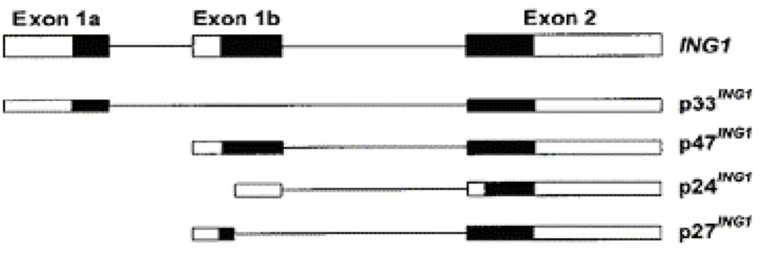
Human ING1 gene has three exons, from which four mRNA variants are transcribed from three different promoter regions (Figure 1.7) (Gunduz et al., 2000; Cheung & Li 2001).
Figure 1.7: Genomic structure of human ING1 and its alternatively spliced mRNA variants. p47ING1, p32ING1, p27ING1, and p24ING1 .(■) Coding sequence in exon. (□) Noncoding sequence in exon.
1.3.2 ING1 Gene products in chromatin remodeling complexes
Recently, the functions of three yeast proteins Yng1, Yng2, and Pho23, which have strong sequence identity in their C-termini PHD finger domains with the human ING1 gene, have been investigated. These three yeast proteins have a role in chromatin remodeling and transcriptional regulation. Yng2 associates with Tra1, a yeast homolog of TRRAP, which associates with PCAF histone acetyl transferase (HAT) complex. TRRAP was identified as a cofactor that interacts with c-Myc and E2F-1 and is required for transformation by c-Myc and E1A (McMahon et al., 1998). It has also been shown that Yng1, Yng2 and Pho23 are complexed with HAT activities. HAT activities are often found in large, modular, multiprotein complexes containing known transcriptional regulators. Yng1, Yng2, and Pho23 associate with HAT activities with preferences for different histones, suggesting that they associated with different HAT complexes. The homology between these yeast proteins and p33ING1 (p24ING1) suggests that these proteins are functionally related. ING1 also associates with HAT activity when it is expressed in yeast. The structure and function of ING1 and Yng2 particularly well
conserved suggesting that human ING1 may be associated with HAT activity and TRRAP, the mammalian homolog of Tra1, in mammals (Loewith et al., 2000). A model in which ING1 regulates HAT activity would be consistent with its putative tumor-suppressor role.
Another study showed that alternative transcripts of human p33ING1 differentially associate with mSin3 transcriptional corepressor complex and have HDAC1-dependent transcriptional corepressor activity in reporter gene assays. While p33ING1b is associated with the Sin3/HDAC1 mediated transcriptional repression, p24ING1c does not seem to interact with the Sin3/HDAC1 complexes (Skowyra et
al.,2000). These findings suggest that this interaction is through N-terminal region of
p33ING1 protein because the three alternative transcripts vary in their N-terminal region.
1.3.3 Cancer Development and p33
ING1 gene emerged as a candidate tumor suppressor gene because: 1) it is located on 13q, a frequent deletion target in many tumor types, including head and neck cancer; 2) it was found to be genetically altered in one neuroblastoma cell line, and decreased rates of protein expression have been detected in several breast cancer cell lines; 3) it interacts with the p53 protein in vitro; and 4) the biological effects of the p33ING1 and p53 proteins may be interrelated, since the ING1 gene is implicated in p53 mediated cell growth inhibition. For these reasons scientists started to investigate p33ING1 mutations, deletions and rearrangements in different cancer type ( for review see; Cheung & Li 2001).
Recently, it has been reported that the ING1 gene is mutated very rarely in primary breast and ovarian cancers and breast cancer cell lines. Also expression of
ING1 gene is reduced in high proportion of breast tumors (44%) and in all breast cancer cell lines. Furthermore it has been suggested that ING1 expression levels correlate strongly with metastasis, with tumors showing reduced levels of ING1 being metastatic at a frequency at least six fold greater than these showing increased levels of ING1 (Garkavstev et al., 1999).
However molecular analysis of the ING1 in human head and neck tumors with 13q deletions, showed no somatic mutation in any of the tumors or cell lines (Sanchez-Cespedes et al., 1999). Recently another study suggested that of 34 informative cases of head and neck squamous cell carcinoma, 68% of tumors showed loss of heterozygosity at chromosome 13q33-34, where the ING1 gene is located. Three missense mutations and three silent changes were detected in the ING1 gene in some tumors with allelic loss at the 13q33-34 region. These missense mutations were found within the PHD finger domain and nuclear localization motif in ING1 protein. These changes may effect the PHD finger and break the three-dimensional structure of ING1 protein leading, the loss of the function (Gunduz et al., 2000). In 31 informative esophageal squamous cell cancer cases, nearly 60% of tumors showed allelic loss at chromosome 13q33-34 and four tumor specific missense mutations detected within the PHD-finger domain and nucleolar localization motif of the ING1 (Chen et al., 2001).
The p33ING1 status in colorectal carcinomas showed that allelic deletion of, or mutations within, the ING1 gene do not appear to occur during colorectal carcinogenesis ( Sarela et al., 1999).
1.4 Aim and Scope of the Project
From the evidences presented above, it is obvious that understanding the signaling pathways of p53 family proteins and the proteins involved in these pathways is crucial to solve the cancer puzzle.
This study aims at the identification of the interacting domain of p53 family proteins with p33ING1 protein. For this purpose, mapping of the interacting regions of p53 family proteins with p33ING1 protein has been investigated, by using in vitro methods namely GST pulldown and Far Western Blotting. For this purpose different deletion mutants of p53 family proteins were created and cloned into expression vectors and used in vitro protein-protein interaction assays.
Such a study is thought to highlight the significant points in the interactions among mentioned proteins.
CHAPTER 2: MATERIALS AND METHODS
2.1 Materials 2.1.1 Chemicals
The laboratory chemicals were analytical grade and supplied from Sigma Biosciences Chemical Company Ltd. (St. Louis, MO, U.S.A) or from Carlo-Erba (Milano, Italy).
2.1.2 Bacterial Strains
The bacterial strain used in this work had the following genotypes:
E. coli, DH5α: F-, (f80dÊ(lacZ)M15), recA1, endA1, gyrA96, thi1,hsdR17,(r-km-k),
supE44,relA1, deoR, Ê(lacZYA-ar gF)U169
2.1.3 Enzymes
Restriction endonucleases were purchased either from MBI FERMENTAS Inc. (NY, U.S.A) ( SalI, XbaI, XhoI , BamHI, NotI, ApaI, EcoRI, NheI, KpnI) or from Appligene-Oncor (Illkirch, France) (HindIII, SacI). Cloned Pfu Polymesase was purchased from Stratagene (CA, U.S.A). T4 Ligase was purchased from MBI FERMENTAS Inc. (NY, U.S.A).
2.1.4 Oligonucleotides
The oligonucleotide primers used for the PCR amplification of p33ING1,p63, p73 and for C-terminal deleted forms of these genes were synthesized in the Beckman Oligo 1000M DNA synthesizer (Beckman Instruments Inc. CA. U.S.A) at the Bilkent
University, Faculty of Science, Department of Molecular Biology and Genetics, (Ankara, Turkey).
The primers for p33 are:
Forward: 5’- AGACGTCGACAAATGTTGAGTCCTGCCAACG -3’ SalI Reverse: 5’- AGACAAGCTTCTACCTGTTGTAAGCCTCTC -3’ HindIII Reverse: 5’-ATATAAGCTTATTAGGGGACGAATCTCGCTC-3’ HindIII The primers for p63 are:
Forward: 5’-GATAGGATCCATGTCCCAGAGCACACAG-3’
BamHI
Reverse: 5’-CGCGCTCGAGTCACTTTGTACTGTCCGAAAC-3’
XhoI The primers for p73 are:
Forward: 5’-TATAGGATCCATGGCCCAGTCCACCGCC-3’
BamHI
Reverse: 5’-CGCGCTCGAGTCACTTGGCGGAGCTCTCGTT-3’
XhoI
Prior to synthesis, the primers were optimized for their annealing temperature, %GC content, hairpin and dimer formation potential with the help of Primer Designer Version 2.0 computer program (Scientific & Educational Software,1990,1991). It was confirmed by the Clone Manager Version 4.0 computer program (Scientific & Educational Software) that p33ING1 cDNA contained no apparent SalI, HindIII, KpnI
and EcoRV restriction enzyme recognition sequences, p63 cDNA and p73 cDNA contained no apparent BamHI and XhoI restriction enzyme recognition sequences .
2.1.5 Cloning Vectors
PGEM-T Easy is a convenient vector for the cloning of PCR products. This
type of vectors have 3’-terminal thymidine at both ends. These single 3’ overhangs at the insertion site greatly improve the efficiency of ligation of a PCR product into a plasmids by preventing recircularization of the vector and providing a compatible overhang for PCR products generated by certain thermostable polymerases, like Taq DNA Polymerase (Figure 2.1).
Figure 2.1 Vector map of pGEMT-easy
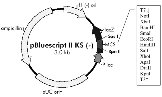
pBluescript II KS- (GenBank #X52326) is a phagemid that permits expression of cloned genes in bacterial systems or in vitro transcription-translation (IVTT) reactions in eukaryotic systems. The vector contains T3 and T7 viral promoters that
span the multiple cloning site and are oppositely oriented. These promoters enable eukaroytic expression of the cloned gene in the presence of respective viral RNA poymerases (Figure 2.2).
Figure 2.2: Vector Map of pBlueScript II KS-
pcDNA 3 is a mammalian expression vector. The vector contains both T7 and
Sp6 viral promoters, spanning the multiple cloning site. These promoters and CMV promoter permits the expression of this plasmid both IVTT reactions and mammalian systems respectively (Figure 2.3).
2.1.6 Antibodies
The primary antibodies used in GST pulldown assays are as follows: monoclonal anti p33 antibodies (3G6, 20H6, 15B9, 9H9), produced by Rengül Çetin-Atalay, monoclonal mouse-anti-p53 antibodies (7D3: specific to amino acids 211-220 ;
T7 ↓ NotI XbaI BamHI SmaI EcoRI HindIII SalI XhoI ApaI DraII KpnI T3↑
HR221: specific to amino acids 371-380; 9E4 specific to amino acids 281-290) kindly provided by Dr. Esma Yolcu (Yolcu et al.,2001).
Figure 2.3: Vector Map of pcDNA3
2.1.7 Commercially Available Kits
QIAquick PCR Purification Kit (by Qiagen (Chatsworth, CA, U.S.A)) is used
for cleaning nucleic acids from contaminants.
QIAEXII (by Qiagen (Chatsworth, CA, U.S.A)) is for extraction of DNA bands
from agarose gels.
QIAGEN Plasmid Purification Kit (by Qiagen (Chatsworth, CA, U.S.A)) is
utilized to obtain plasmid DNA at higher amounts and concentrations with higher purity than those achieved by MiniPrep.
pGEM-T and pGEM-T Easy Vector Systems is purchased from Promega
(Madison, WI, USA) for the easy cloning of the genes in vectors.
TNT Coupled Reticulocyte Lysate System is purchased from Promega (Madison,
WI, USA) for the single step in vitro transcription-translation (IVTT) of the cloned genes in an eukaryotic system.
Amplify Solution from Amersham (Uppsala, Sweden) is a flourimetric method
to enhance the visualization of the signals generated by radioisotopes on polyacrylamide gels.
2.1.8 Apparatus
Vertical mini gel apparatus for polyacrylamide gel electrophoresis and the power supply are products of E-C Apparatus Corp. (Florida, U.S.A). Horizontal midi gel apparatus used for agarose gel electrophoresis is purchased from Stratagene. Thermal cycler for PCR is a product of Perkin Elmer (CA, USA). Slab Gel Dryer is from Savant Instruments Inc.(N.Y.,USA). Semi-dry transfer unit for Far Western Blotting and GelDoc2000 Image analyzer for agarose gels are purchased from Bio Rad Laboratories (CA, U.S.A).
2.1.9 Materials for Autoradiograghy
Radiolabelled methionine (35S-Methionine) and light-proof film cassette (Hypercassette) and film developing unit are products of Amersham (Uppsala, Sweden). Medical X-ray films are purchased from Fuji (Tokyo, Japan).
2.1.10 DNA and Protein Size Markers
As a DNA size marker, 1 kb DNA Ladder from MBI FERMENTAS Inc. (NY, U.S.A) is used.SDS-PAGE protein size markers are either from Pharmacia (Uppsala, Sweden) or Bio Rad Laboratories (CA, U.S.A) (prestained markers).
2.2 Solutions and Media
2.2.1 Agarose Gel Electrophoresis Solutions
Tris-acetic acid-EDTA (TAE) 40mM Tris-acetate
1mM EDTA
Ethidium bromide: 10 mg/ml in water (stock solution),
30 ng/ml (working solution)
1x Gel loading buffer: 0.25% bromophenol blue, 0.25% xylene cyanol, 50% glycerol, 1mM EDTA
2.2.2 Solutions for Plasmid DNA Isolation (MiniPrep)
Solution I 50 mM Glucose, 25 mM, Tris.Cl, pH 8.0,
10M EDTA. Sterilize in autoclave.
Solution II 0.2 N NaOH, 1% (wt/vol) SDS
Solution III 3 M Potassium acetate, pH 4.8
11.5% (v/v) glacial acetic acid
Phenol/ Chloroform 100% equilibrated phenol / 100% chloroform
2.2.3 Solutions for Bacterial Transformation
CaCl2 50 mM in double distilled water,
Transformation Buffer 10 mM PIPES, 55 mM MnCl2, 15 mM CaCl2, 250 mM KCl, Filter Sterilized and stored and 4˚C.
2.2.4 Microbiological media and antibiotics
Luria-Bertani medium (LB) Per liter: 10 g nutrient broth, 5 g bacto-
yeast extract, 8 g NaCl, 0.52g Tris Base
For LB-agar plates, add 15 g/L bacto agar.
Sterilized by autoclave.
SOB Per liter: 2% tryptone, 0.5% yeast extract, 10 mM
NaCl, 2.5 mM KCl, Autoclaved to sterilize. Then 20 mM MgSO4 and 10 mM MgCl2.
SOC Per liter: SOB + 20 mM Glucose from filter
sterilized 1 M Glucose stock solution in ddH2O.
DMSO Commercially available, 100%
Ampicillin working solution: 100 µg/ml or 50 µg/ml stock solution :100 mg/ml in ddH2O (stored at -20°C)
2.2.5 Polyacrylamide Gel Electrophoresis Solutions
Resolving (Lower) Buffer (2X) 375mM TrisHCl, 0.2% SDS, pH 8.9
Stacking (Upper) Buffer (2X): 250mM TrisHCl, 0.2% SDS, pH6.8
Sample Dye (2X) 50mM TrisHCl pH6.8, 1% SDS, 2mM EDTA, 1% 2-ME, 0.02% Bromophenol Blue,10% glycerol Acrylamide-Bisacrylamide mix 24% acrylamide, 0.64% bisacrylamide (stock)
Amonium persulphate (APS) 10% (w/v) ammonium persulphate in ddH2O
Coomassie Staining Solution Solution1: 10% Acetic Acid
Solution2: 0.04% Coomassie G Blue, 0.5% CuSO4 in 27% ethanol
Mix Solution1 and 2 at a ratio of 1:1
Destain Solution 14% ethanol, 7% acetic acid in ddH2O
2.2.6 Far Western Buffers
Denaturation Buffer: Hepes (pH:7.8) 25mM NaCl 25mM MgCl2 5mM GnHCl 6M DTT 1mM
Renaturation Buffer: Hepes (pH:7.8) 25mM NaCl 25mM MgCl2 5mM GnHCl 0.187M DTT 1mM
Saturation Buffer: Hepes (pH:7.8) 25mM NaCl 25mM
MgCl2 5mM DTT 1mM NP-40 0.05% Milk Powder 1%
IVTT Buffer: NaCl 0.1M Tris HCl (pH: 7.6) 20mM DTT 1mM EDTA 1mM Glycerol 10% Milk Powder 1%
2.2.7 Solutions for Trichloroacetic acid Precipitation