1Department of Anesthesiology and Reanimation, Baskent University Faculty of Medicine, Istanbul, Turkey 2Department of General Surgery, Baskent University Faculty of Medicine, Istanbul, Turkey
Submitted: 26.08.2013 Accepted after revision: 01.09.2014
Correspondence: Dr. Özlem Çınar. Başkent Üniversitesi Tıp Fakültesi, Anesteziyoloji ve Reanimasyon Anabilim Dalı, Istanbul, Turkey. Tel: +90 - 216 - 554 15 00 e-mail: [email protected]
© 2016 Turkish Society of Algology
JANUARY 2016 42
Özet
Bu olguda, 77 yaşında ciddi koroner arter hastalığı öyküsü olan, ultrason eşliğinde sürekli paravertebral blok tekniğiyle radikal mastektomi ve aksiller lenf nodu diseksiyonu uygulanan bir hasta sunuldu. Genellikle aksiller diseksiyon ve radikal mastektomi genel anestezi altında uygulanan bir cerrahi prosedür olup endotrakeal entübasyon gerektirir. Koroner arter hastalığı ve uyku apne sendromu olan hastalarda genel anestezi yüksek risklidir. Ultrason eşliğinde sürekli paravertebral blok tekniği basit ve güvenli bir anestezi alternatifi olduğu kadar minimal yan etkiyle ameliyat sonrası analjezi de sağlamaktadır.
Anahtar Kelimeler: Koroner arter hastalığı; mastektomi; ultrason eşliğinde sürekli paravertebral blok.
Summary
The case of a 77-year-old patient with severe coronary heart disease who underwent radical mastectomy with axillary lymph node dissection by ultrasound-guided continuous paravertebral block (CPVB) is described in the present report. Radical mas-tectomy with axillary dissection is a surgical procedure that necessitates endotracheal intubation and is usually performed under general anesthesia, which carries heightened risk for patients with coronary heart disease (CHD) and sleep apnea syn-drome (SAS). Ultrasound-guided CPVB is a simple and safe alternative technique that allows for the use of anesthesia and postoperative analgesia with minimal side effects.
Keywords: Coronary heart disease; mastectomy; ultrasound-guided continuous paravertebral block.
Introduction
We report here the use of ultrasound-guided con-tinuous paravertebral block (CPVB) as a primary anes-thetic technique in a patient with severe CHD and SAS undergoing radical mastectomy with axillary lymph node dissection for infiltrating ductal carcinoma. Thoracic paravertebral block (TPVB) results in an ip-silateral somatic motor and sensory nerve block of multiple contiguous thoracic dermatomes above and below the site of injection.[1,2] Location of the
paraver-tebral space may be technically difficult, as location of the transverse process by blind needle placement is necessary and its overall failure rate is more than
10%.[3] Failure to identify the transverse process,
re-sults in several needle redirections which not only cause pain and discomfort but also increase the po-tential risk of pneumothorax. The use of ultrasound offers the capability to place a catheter in the para-vertebral space with real-time image guidance. So-nographic measurements obtained using ultrasound scanning of vertebral transverse processes and pari-etal pleura can give an accurate measurement of the depth from the skin to the paravertebral space.[4]
Radical mastectomy with axillary dissection is a surgi-cal procedure that is usually performed under general anesthesia and necessitates endotracheal intubation.
Ultrasound guided continuous paravertebral block in a patient
with coronary heart disease and sleep apnea syndrome
Koroner arter hastalığı ve uyku apne sendromu olan bir hastada
ultrason eşliğinde sürekli paravertebral blok
Bilgiser ESEn,1 Hüseyin Yüce Bircan,2 Özlem ÇInar,1 ayda TürkÖz1 Agri 2016;28(1):42–45
doi: 10.5505/agri.2014.82474
c a S E r E P O r T
Here we report a patient affected by coronary heart disease and sleep apne syndrome presenting with re-spiratory distress scheduled for radical mastectomy, in whom anesthesia was managed succesfully using only sedation with thoracic paravertebral block.
case report
A 77 years old, 69 kg, 155 cm woman, with American Society of Anesthesiology (ASA) physical status III was scheduled for right radical mastectomy with axillary lymph node dissection for infiltrating ductal carcino-ma. She was suffering from severe CHD and SAS. She had left ventricular aneurysm and her left ventricular ejection fraction (LVEF) was %25. Since the patient pre-sented a higher risk of complications for endotracheal intubation, we have decided to perform the proce-dure with CPVB. After discussing with the patient and her family, written informed consent was obtained. One hour before surgery, midazolam 5 mg was ad-ministered orally as premedication. Routine moni-toring (pulse oximeter, noninvasive blood pressure cuff, and electrocardiogram) were applied, and in-travenous access was established. Patient was intra-venously administered 50 µgr fentanyl as needed for block procedural sedation/analgesia. The initial blood pressure, heart rate and peripheral oxygen sat-uration were 160/80 mmHg, 80 beats/min and 94%, respectively. Paravertebral injection was performed with the patient in the sitting position. Sonosite Ti-tan (SonoSite, Bothell, Washington, USA) 7–13 MHz linear transducer was applied longitudinally to the paravertebral area.
The skin and subcutaneous tissue were anesthetized with 5 ml lidocaine (10 mg/ml). An 18G Tuohy needle was inserted perpendicularly at T7 to hit the trans-verse process via an out-of-plane approach (Figure 1). The needle was then directed over the top of the bony structure. The deviation of the needle from the perpendicular line was kept at 15º. The paraverte-bral space was identified using loss of resistance to normal saline injection without ultrasound. After negative aspiration, test dose with 3 ml 1% lidocaine with epinephrine 1:200.000 was administered incre-mentally with ultrasound guidance. Catheter place-ment was performed by an attending anesthesiolo-gist experienced in regional anesthetic techniques. 18 gauge, 10 cm Tuohy needle access followed by
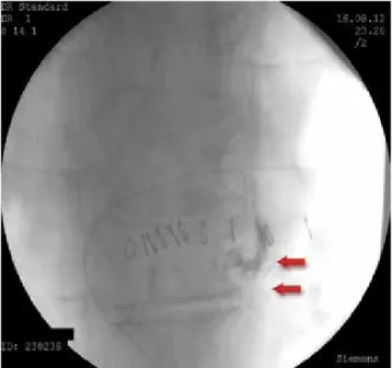
20 gauge open tip, single orifice epidural catheter advanced 5 cm into the paravertebral space (Figure 2). During catheter placement, a surgical block was instituted with 0.3 ml/kg, 0.5% levobupivacaine. On-set of sensory loss occured 12 minutes after injection with surgical anesthesia ensuing 25 minutes after in-jection. Prior to incision, blood presure decreased to 110/55 mmHg. Intraoperative sedation was provided with IV 1 mg/hours infusion of midazolam. Anesthe-sia depth was monitored with Index of Conscious-ness (IOC) (Morpheus Medical, Barcelona, Spain) dur-ing the operation, and IOC values were within 60–70.
JANUARY 2016 43
Ultrasound guided continuous paravertebral block in a patient with coronary heart disease and sleep apnea syndrome
Figure 1. Ultrasound-guided paravertebral block in an ‘out-of-plane’ technique.
Figure 2. Distribution of the radioopaque that is given through the paravertebral catheter.
Surgery lasted 135 minutes and the patient remained comfortable during the procedure (Figure 3).
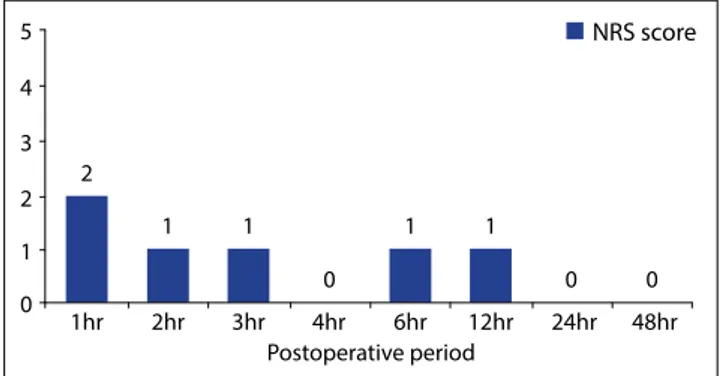
Radical mastectomy was completed without any complication and the patient was transferred to the post anesthesia care unit (PACU). In the PACU, 5–10 ml/h 0.25% levobupivacaine infusion begun through the paravertebral catheter and continued for 48 hours. Pain scores; at rest, on movement and during coughing were evaluated for 48 hrs in the postoper-ative period using a numeric rating scale (NRS) rang-ing from 0 (no pain) to 10 (worst imaginable pain) (Figure 4). Nausea and vomiting were seen neither in PACU nor in ward. During her hospitalization, there was no need for opioid medication and the patient was discharged on the third postoperative day.
Discussion
The main finding of this case report was that ade-quate intraoperative anesthesia and postoperative analgesia can be achieved by ultrasound guided CPVB, in a patient with CHD and SAS, undergoing radical mastectomy.
Regional anesthesia using paravertebral block is an al-ternative to general anesthesia for breast cancer sur-gery.[5] The technique provided good operative
anes-thesia for major chest wall procedures as well as good postoperative analgesia.[6] Benefits include, reduction
in postoperative nausea and vomiting and potential for ambulatory discharge. Infiltration of the paraver-tebral space results in unilateral sensory, motor and sympathetic blockade. So, this technique could facili-tate maintenance of the normal haemodynamic sta-tus.[7] PVB can provide profound, long-lasting sensory
differentiation. As a result, greater attenuation of the surgical stress response may be transformed into re-duced inotropic stimulation of the heart.
Additionally, unlike general anesthesia, PVB ensures superior postoperative analgesia, shortens recov-ery and hospitalization time, decreases analgesic requirement. Also, the use of PVB in patients under-going ambulatory breast cancer surgery has a cost-saving potential.[1]
This case demonstrates that, older patients with multiple comorbidities can undergo radical mastec-tomy using CPVB with the avoidance of general
an-esthesia. First of all, opioids were not used and blood pressure and heart rate were stable during the op-eration. Secondly, pain scores at rest, on movement and during coughing did not increase above 1 dur-ing the postoperative 48 hours. That’s why, this tech-nique should be considered as a potential anesthetic management in such patients.
The use of paravertebral catheters in the management of thoracic pain after surgery or trauma has been well described.[8,9] Percutaneous paravertebral catheters
have traditionally been placed using the method de-scribed by Eason and Wyatt[3] using loss of resistance
to air or saline with needle and syringe. Although acci-dental pleural puncture is rare,[10] paravertebral block
is still considered potentially dangerous by many anesthetist.[5] Using ultrasound to visualize anatomical
landmarks, it will be possible to improve the safety of the block. Ultrasound imaging not only helps to deter-mine needle insertion sites, but also provides informa-tion about the depth to the paravertebral space.[11]
Ul-trasound guided CPVB is a simple and safe alternative technique that offers anesthesia as well as postopera-tive analgesia with minimal side-effects.
JANUARY 2016 44 PAINA RI 180 160 140 120 100 80 60 40 20 0 Befor e PVB
After PVBPVB 5minPVB 10minPVB 15minIncisionPostop 1hrPostop 2hrPostop 3hrPostop 4hrPostop 6hr Post op 12hr Post op 24hr Post op 48hr SAP DAP * * * * * * *
Figure 3. Systolic and diastolic blood pressure values.*P<0.05 in
the groups.
Figure 4. Postoperative pain scores. 5 4 3 2 1 1hr 2 1 1 0 0 0 1 1 2hr 3hr 4hr 6hr 12hr 24hr 48hr 0 NRS score Postoperative period
Conflict-of-interest issues regarding the author-ship or article: None declared.
Peer-rewiew: Externally peer-reviewed.
references
1. Karmakar MK. Thoracic paravertebral block. Anesthesiol-ogy 2001;95(3):771–80.
2. O Riain SC, Donnell BO, Cuffe T, Harmon DC, Fraher JP, Shorten G. Thoracic paravertebral block using real-time ultrasound guidance. Anesth Analg 2010;110(1):248–51. 3. Eason MJ, Wyatt R. Paravertebral thoracic block-a
reap-praisal. Anaesthesia 1979;34(7):638–42.
4. Pusch F, Wildling E, Klimscha W, Weinstabl C. Sonographic measurement of needle insertion depth in paravertebral blocks in women. Br J Anaesth 2000;85(6):841–3.
5. Richardson J, Sabanathan S. Thoracic paravertebral anal-gesia. Acta Anaesthesiol Scand 1995;39(8):1005–15.
6. Buckenmaier CC 3rd, Klein SM, Nielsen KC, Steele SM. Con-tinuous paravertebral catheter and outpatient infusion for breast surgery. Anesth Analg 2003;97(3):715–7.
7. Karmakar MK. Thoracic paravertebral block. Anesthesiol-ogy 2001;95(3):771–80.
8. Dhole S, Mehta Y, Saxena H, Juneja R, Trehan N. Compari-son of continuous thoracic epidural and paravertebral blocks for postoperative analgesia after minimally invasive direct coronary artery bypass surgery. J Cardiothorac Vasc Anesth 2001;15(3):288–92.
9. Paniagua P, Català E, Villar Landeira JM. Successful man-agement of pleuritic pain with thoracic paravertebral block. Reg Anesth Pain Med 2000;25(6):651–3.
10. Lönnqvist PA, MacKenzie J, Soni AK, Conacher ID. Paraver-tebral blockade. Failure rate and complications. Anaesthe-sia 1995;50(9):813–5.
11. Hara K, Sakura S, Nomura T, Saito Y. Ultrasound guided thoracic paravertebral block in breast surgery. Anaesthesia 2009;64(2):223–5.
JANUARY 2016 45