Radioprotective effect of montelukast sodium against hepatic
radioiodine (
131I) toxicity: A histopathological investigation in the rat
model
Hasan Ikbal ATILGAN1, Nihat YUMUŞAK2, Murat SADIC3, Salih Sinan GULTEKIN4,
Gokhan KOCA3, Sinem OZYURT5, Koray DEMIREL3, Meliha KORKMAZ3
1Ministry of Health Kahramanmaras Necip Fazil City Hospital, Division of Nuclear Medicine, Kahramanmaraş; 2Harran University,
Faculty of Veterinary Medicine, Department of Pathology, Şanlıurfa; 3Ministry of Health Ankara Training and Research Hospital,
Department of Nuclear Medicine, Ankara; 4Ministry of Health Dışkapı Yıldırım Beyazıt Training and Research Hospital, Division of
Nuclear Medicine, Ankara; 5Ministry of Health Sami Ulus Children Hospital, Division of Nuclear Mecicine, Ankara, Turkey.
Summary: Radioactive iodine (131I) is a known radionuclide which is used both for diagnostic and therapeutic purposes in the
treatment of hyperthyroidism and thyroid cancer. Montelukast sodium (Cysteinyl leukotriene receptor-1 antagonist) is also a well-known antioxidant drug. This study aimed to evaluate the histopathological changes in rat livers at the third month following 131I
treatment and the radioprotective effect of Montelukast sodium (ML) against 131I-related liver damage. Thirty female Wistar Albino
rats were randomly divided into three groups as control group (n=10), untreated rats; second (RAI) group (n=10), oral radioiodine (111 MBq) administrated rats, and third (ML) group (n=10), oral radioiodine and ML administrated rats. In the third group, ML administration was started 3 days before and ended 10 days after RAI administration. In the third month of radioiodine (RAI) administration, the animals were decapitated and the livers were removed for histopathological examination. The histopathologic data were evaluated comparatively by using Mann Whitney U and Fisher’s Exact Tests Differences were determined in all the parameters in terms of intensity between the control and 131I groups, which showed the harmful effects of 131I. In the comparison of
the 131I and ML groups, hyperemia was determined respectively 80% to 40%, the presence of inflammatory cells 70% to 60% and
capsule thickening 70% to 40%. Montelukast sodium was observed to have a protective effect especially on hyperemia and capsule thickening. According to the study results, radioactive iodine (131I) treatment seems to cause morphological damage to the rat liver,
and Montelukast sodium effectively protects the liver against damage. Key words: Iodine-131, histopathology, liver, montelukast sodium, rat.
Karaciğerde radyoiodin (131İ) toksisitesine karşı montelukast sodyumun radyoprotektif etkisi: Rat
modelinde histopatolojik incelemeler
Özet: Radyoaktif iyot (131I) hipertiroidizm ve tiroid kanserlerinin tanı ve tedavisi amacıyla kullanılan bir radyonükliddir.
Montelukast sodyum (Cysteinyl leukotriene receptor-1 inhibitörü) ise antioksidan olarak bilinen bir ilaçtır. Bu çalışma ile rat karaciğerinde radyoiodinden (RAI) kaynaklanan radyasyon hasarına karşı Montelukast sodyumun (ML) radyoprotektif etkisinin histopatolojik olarak değerlendirilmesi amaçlandı. Çalışmada 30 adet dişi Wistar Albino rat kullanıldı ve bunlar rastgele üç gruba ayrıldı; birinci grubu hiçbir işleme tabi tutulmayan kontrol grubu (n=10), ikinci (RAI) grubu, (n=10), oral olarak RAI (111 MBq) uygulanan ratlar, üçüncü (ML) grubunu ise (n=10) oral olarak RAI ve ML uygulanan grup oluşturdu. Üçüncü çalışma grubunda ML uygulaması RAI uygulamasından 3 gün önce başlatıldı ve 10 gün sonra bitirildi. Üçüncü ay sonunda nekropsileri yapılan ratların karaciğer doku örnekleri rutin doku takibine alınarak histopatolojik olarak incelendi. Radyoiodin ve ML grubunda tüm histopatolojik bulgular Mann Whitney U testi ve Fisher’in ekzak test ile karşılaştırılarak değerlendirildi. Bütün parametreler açısından, RAI’un zararlı etkisini gösterecek şekilde, kontrol ve RAI grubu arasında insidans açısından farklılık vardı. Radyoiodin ve ML grubunun karşılaştırmasında, parametrelerin insidansı hiperemi açısından sırasıyla %80’e %40, inflamatuar hücre varlığı %70’e %60, kapsül kalınlaşması %70’e %40 idi. Montelukastın özellikle hiperemi ve kapsül kalınlaşması açısından koruyucu etkisi olduğu görüldü. Montelukast sodyumun RAI tedavisi sonrası karaciğerde özellikle hiperemi ve kapsül kalınlaşması üzerine parsiyel radyoprotektif etkisi olduğu belirlendi.
Anahtar sözcükler: İodine-131, histopatoloji, karaciğer, montelukast sodyum, rat.
Introduction
Radioactive iodine (131I) therapy is widely used in the treatment of hyperthyroidism due to Graves’ disease
or active nodules and ablation, recurrences or metastases of differentiated thyroid cancer in cats, dogs and humans (3, 14, 18, 24). 131I therapy has been used for the ablation
of well-differentiated thyroid carcinoma for more than six decades (21). This therapy is a rapid, stress-free, highly effective and simple for cats, dogs and people with hyperthyroidism (14, 25). Nonetheless, treatment with 131I dose requires nuclear medicine equipment, special licensing and hospitalization facilities (25). Another disadvantage of this therapy is the multiple doses required to maintain therapeutic levels (18).
The dose of administered 131I activity changes according to the thyroid disease, age, gender and clinical experience. High doses of 131I are used to ablate remmant thyroid tissue after thyroidectomy, and 131I avid metastases of differentiated thyroid cancer. However, some cats, dogs and humans may develop side effects such as, dysphagia, vomiting, thyroiditis, hypothyroidism and renal disease during treatment at higher doses 131I (3, 11, 24, 25).
Both beta minus, which is used for the treatment, and gamma rays, which are used for diagnosis, are emitted by 131I (26). Beta rays especially cause harmful effects. After oral ingestion of 131I, it is absorbed via the stomach and intestines, then passes to the liver and systemic circulation via the portal vein. Retention of iodide in the thyroid depends on the metabolic activity of the thyrocytes and it is assumed that 30% of iodine is trapped by the thyroid (32). However, the thyroid gland in a variety of nonthyroidal tissue has weak iodide uptake (8, 10). Diffuse hepatic uptake may be seen due to residual or metastatic thyroid tissues, but factors other than functional thyroid tissue may also cause diffuse hepatic uptake (22). There have been several reports on the effects of drugs such as pilocarpine (28), amifostine (7, 9), vitamin C (19) and vitamin E (1, 6) for the prevention of 131I-related tissue damage. There has been only one study in literature about the protective effect of Cysteinyl leukotriene receptor-1 (montelukast sodium). The leukotriene concentrations in lacrimal or any other radioidine-treated tissue are not present in the literature, it is possible that ML, as a leukotriene antagonist, has an additional protective action by inhibiting possible early neutrophilic inflammation. ML probably prevents inflammation by blocking the cysteinyl leukotriene receptor type 1, which is found in neutrophils. ML has been shown to down-regulate the human monocyte chemotaxins and to inhibit production of tumor necrosis factor-alpha (TNF- α) and monocytechemotactic protein-1 (protein-17, 3protein-1). In another study, it has been reported that ML depressed the TNF- α response in sepsis-induced ileal and hepatic injury in rats (17, 27). Koca et al. (17) recently reported that montelukast effectively protects against 131I induced damage to lacrimal glands. The aim of this study was to investigate 131I-induced liver changes and the radioprotective effects of montelukast sodium (ML) in rat livers.
Material and Methods
Animals: This study was conducted with the
approval of the Local Ethics Committee of Animal Experiments at Ankara Training and Research Hospital (03.05.2012/0008-099). All procedures were carried out in compliance with the relevant national laws relating to the conduct of animal experimentation.
This study was conducted on 30 female Wistar Albino rats aged 4-6 months and weighing 225-275 gr. The rats were acclimatised for at least 1 week before the study and were housed for three months under standard laboratory conditions at a constant temperature of 21±2°C and relative humidity of 65-70% with a 12-hour light and dark cycle in polypropylene cages using disposable absorbent cloths under sterile paddy husks to avoid contamination from radioactive urine. The animals were fed with standard chow and water ad libitum throughout the three months of the experiment.
Experimental design: The animals were randomly
assigned to one of three groups of 10 rats in each. The first group was the control group, which received no 131I therapy or ML. The rats in the second group (131I group, n=10) were treated with oral 131I (111 MBq). The rats in the third group (ML group, n=10) were treated with intraperitoneal (ip) ML as a radioprotective agent. Radioiodine (MON-IYOT-131 Eczacıbaşı/Monrol Nükleer Ürünler Sanayi ve Ticaret Anonim Şirketi Gebze, Kocaeli, Türkiye) was applied at 111 MBq (3 mCi) after replacing nasogastric sondage to the rats in Groups 2 and 3. Montelukast sodium (Singulair, Merck Sharp & Dohme, Istanbul, Turkey) was started three days before the 131I administration and continued for 10 days at a dosage of 10 mg/kg/day orally in Group 3. In the third month of 131I administration, the animals were anesthetized with 50 mg/kg, i.p. Propofol (Abbott Laboratuvarı Anonim Sirketi, Istanbul, Turkey) and then decapitated. The liver was removed from each animal (Figure 1).
Histopathological evaluation: Tissue samples were
fixed in 10% buffered formaldehyde, subjected to the routine pathology processes, and embedded in parafin. Tissue sections with 5 μm thickness were stained with the Haematoxylin-Eosin (HE) and examined with a light microscope. Pathological samples were evaluated for hyperemia, steatosis, cellular changes, bile duct proliferation, fibrosis, venous lesions, capsule thickening and granuloma formation and were classified into four grades as Grade 0: absent, Grade 1: mild, Grade 2: severe, Grade 3: very severe. The presence of inflammatory cells was classified into four grades as absent, few, severe and very severe.
Data analysis: Data analysis was performed using
Statistical Package for Social Sciences for Windows software (SPSS version 17.0, SPSS Inc. Chicago, USA).
All of the variables were categorical. Descriptive statistics were expressed as numbers and frequency (%). Comparisons between two groups were carried out with the Mann Whitney U test and Fisher’s exact test for all histopathological parameters. A p value of <0.05 was considered statistically significant.
Results
In macroscopic appearance, on the surface of liver diffuse and different shape grayish granulomas were seen (Figure 1). In microscopical examination; a difference was determined in terms of intensity between the control and 131I groups for all the parameters. The comparative results were as follows: fibrosis (10% to 50%), granuloma formation (0% to 80%), cellular changes (10% to 60%), hyperemia (20% to 80%), presence of inflammatory cells (10% to 70%), capsule thickening (0% to 70%), steatosis (30% to 60%), biliary duct proliferation (0% to 50%) and venous lesions (0% to 40%) (Figure 2A-D). There was no significant difference
Figure 1. Multifocal granulomas on the liver. Şekil 1. Karaciğerde multifokal granulomlar.
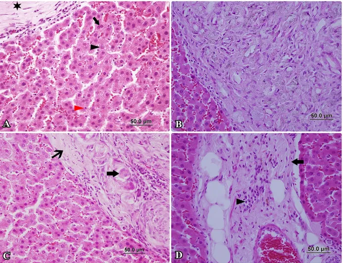
Figure 2. Group of radioiodine. A- Granuloma pattern (star), large nucleated cells (black arrowhead), small nucleated cells (red arrowhead), dual-core pattern (arrow), HxE, x400. B- Large granuloma pattern, HxE, x400. C- Fibrose capsula arround of granuloma (thin arrow), multinucleated giant cells (arrow), HxE, x400. D- Perivascular fibrosis (arrow) and perivascular infiltration (arrowhead), HxE, x400.
Şekil 2. Radyoiyot grubu. A- Granulom yapısı (yıldız), makronükleus (siyah okbaşı), mikronükleus (kırmızı okbaşı), çift çekirdekli hücre (ok), HxE, x400. B- Çevresinden sınırlı büyük granulom, HxE, x400. C- Granulom çevresini saran fibröz kapsül (ince ok), çok çekirdekli dev hücre (ok), HxE, x400. D- Perivasküler fibrozis (ok) ve yangısal hücre infiltrasyonu (okbaşı), HxE, x400.
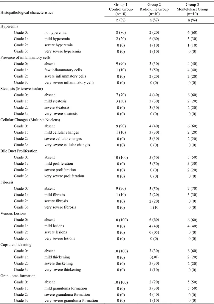
Table 1: Grading of histopathological characteristics among of the groups. Tablo 1: Gruplar arasındaki histopatolojik bulguların derecelendirilmesi.
Histopathological characteristics Group 1 Control Group (n=10) Group 2 Radioidine Group (n=10) Group 3 Montelukast Group (n=10) n (%) n (%) n (%) Hyperemia Grade 0: no hyperemia 8 (80) 2 (20) 6 (60)
Grade 1: mild hyperemia 2 (20) 6 (60) 3 (30)
Grade 2: severe hyperemia 0 (0) 1 (10) 1 (10)
Grade 3: very severe hyperemia 0 (0) 1 (10) 0 (0)
Presence of inflammatory cells
Grade 0: absent 9 (90) 3 (30) 4 (40)
Grade 1: few inflammatory cells 1 (10) 5 (50) 4 (40)
Grade 2: severe inflammatory cells 0 (0) 2 (20) 2 (20)
Grade 3: very severe inflammatory cells 0 (0) 0 (0) 0 (0) Steatosis (Microvesicular)
Grade 0: absent 7 (70) 4 (40) 6 (60)
Grade 1: mild steatosis 3 (30) 3 (30) 2 (20)
Grade 2: severe steatosis 0 (0) 3 (30) 2 (20)
Grade 3: very severe steatosis 0 (0) 0 (0) 0 (0)
Cellular Changes (Multiple Nucleus)
Grade 0: absent 9 (90) 4 (40) 6 (60)
Grade 1: mild cellular changes 1 (10) 3 (30) 2 (20)
Grade 2: severe cellular changes 0 (0) 3 (30) 2 (20)
Grade 3: very severe cellular changes 0 (0) 0 (0) 0 (0)
Bile Duct Proliferation
Grade 0: absent 10 (100) 5 (50) 5 (50)
Grade 1: mild proliferation 0 (0) 5 (50) 3 (30)
Grade 2: severe proliferation 0 (0) 0 (0) 2 (20)
Grade 3: very severe proliferation 0 (0) 0 (0) 0 (0)
Fibrosis
Grade 0: absent 9 (90) 5 (50) 7 (70)
Grade 1: mild fibrosis 1 (10) 2 (20) 3 (30)
Grade 2: severe fibrosis 0 (0) 2 (20) 0 (0)
Grade 3: very severe fibrosis 0 (0) 1 (10 0 (0)
Venous Lesions
Grade 0: absent 10 (100) 6 (60) 6 (60)
Grade 1: mild lesions 0 (0) 4 (40) 4 (40)
Grade 2: severe lesions 0 (0) 0 (05) 0 (0)
Grade 3: very severe lesions 0 (0) 0 (0) 0 (0)
Capsule thickening
Grade 0: absent 10 (100) 3 (30) 6 (60)
Grade 1: mild thickening 0 (0) 3(30) 2 (20)
Grade 2: severe thickening 0 (0) 3 (30) 2 (20)
Grade 3: very severe thickening 0 (0) 1 (10) 0 (0)
Granuloma formation
Grade 0: absent 10 (100) 2 (20) 5 (50)
Grade 1: mild granuloma formation 0 (0) 3 (30) 5 (50)
Grade 2: severe granuloma formation 0 (0) 4 (40) 0 (0)
between the control and ML groups for fibrosis (10% to 20%), hyperemia (20% to 40%) and steatosis (30% to 40%), whereas the difference was more than double for granuloma formation (0% to 50%), cellular changes (10% to 40%), presence of inflammatory cells (10% to 60%), capsule thickening (0% to 40%), biliary duct proliferation (0% to 50%) and venous lesions (0% to 40%) (Table 1). There was a statistically significant difference in hyperemia, the presence of inflammatory cells, capsule thickening, biliary duct proliferation and granuloma formation between the 131I and control groups (p<0.05) (Figure 3A-D). Biliary duct proliferation and granuloma formation was statistically significantly different between control and ML groups (p<0.05).
These results indicated that ML has a protective effect on hyperemia, the presence of inflammatory cells and capsule thickening. In the comparison of the 131I and ML groups for the intensity of the parameters, distributions were determined as 80% to 40% for hyperemia, 70% to %60 for the presence of inflammatory cells and 70% to 40% for capsule thickening, respectively.
Discussion and Conclusion
Large scale and whole body ionizing radiation causes multiple organ dysfunctions by the reactive oxygen species that cause an increase in cellular oxidative stress. This induces the damage to DNA, lipids, proteins and membranes (13). Ionizing radiation causes
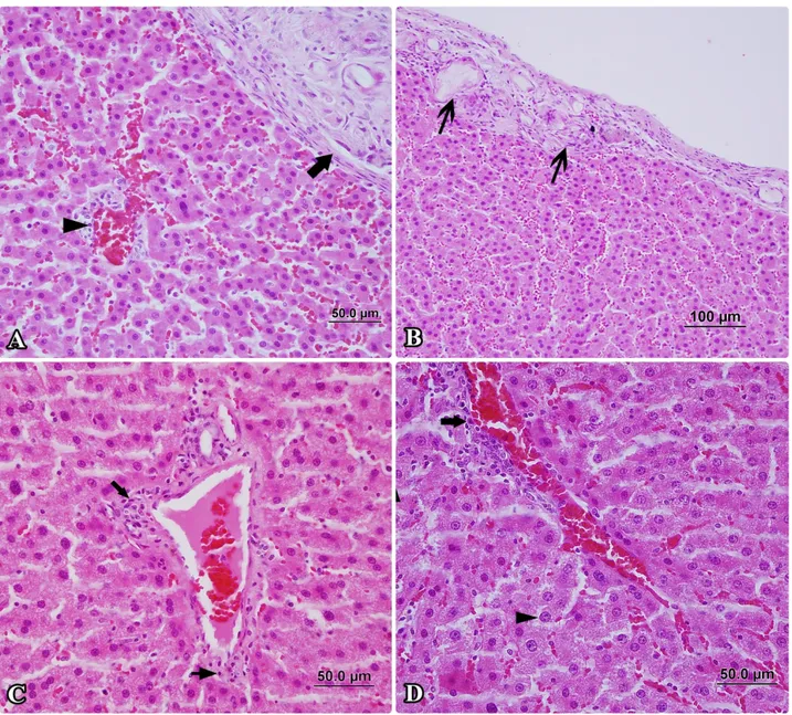
Figure 3. The pathological changes are more less prominent in the montelukast group than in the radioiodine group. A- Decreased granuloma pattern (arrow), perivascular infiltration (arrowhead), HxE, x400. B- Decreased granuloma pattern (arrows), HxE, x200. C- Mild perivascular fibrosis (arrow) and infiltration (thin arrow), HxE, x400. D- Mild perivascular infiltration (arrow) and large nucleated cell (arrowhead), HxE, x400.
Şekil 3. Radyoiyot grubuna göre daha hafif patalojik bulgular görülen montelukast sodium grubu. A- Gerilemiş granulom (ok), perivasküler hücre infiltrasyonu (okbaşı), HxE, x400. B- Gerilemiş granulomlar (oklar), HxE, x200. C- Hafif perivasküler fibrosis (ok) ve infiltrasyon (ince ok), HxE, x400. D- Hafif perivasküler hücre infiltrasyonu (ok) ve makronükleus (okbaşı), HxE, x400.
an imbalance between the pro-oxidants and antioxidants (15). Antioxidant treatment may delay and prevent the damage of ionizing radiation (13). DNA synthesis decreases after radiation. The level of the decrease depends on the size of the dose and the stage of the regeneration. A smaller dose of radiation during the relatively sensitive presynthetic interval is enough to reduce DNA synthesis (2). Koca et al. (16) described RAI-induced changes in rat lacrimal glands during the third month. RAI treatment leads to a decrease in peripheral basophilia and scanty cytoplasm, acinar atrophy and fibrosis, ductal abnormality and lipofuscin accumulation. In the current study, radiation caused cellular changes, hyperemia, fibrosis, granuloma formation, the presence of inflammatory cells, capsule thickening, steatosis, biliary duct proliferation and venous lesions in the liver as harmful effects.
The liver has many vital functions such as synthesis, storage, secretion, transformation, breakdown and detoxification. The protection of the liver should be an important issue for patients undergoing ionizing radiation(29). The greatest cause of cellular damage is lipid peroxidation (LPO). An increase in radiation dose increases LPO in rat liver mitochondri and microsomes (30). Occasionally, foci of inflammatory cells and microvascular steatosis are seen in rat livers which have received RT (12). Many kinds of synthetic or natural drugs have been used, such as antioxidants, cytoprotective agents, angiotensin converting enzyme inhibitors, angiotensin-II type-1 receptor antegonists, immunmodulators, prostaglangins, and vitamins A, C, E to diminish the damage of the radiation (4). However, none of these drugs have the properties of an ideal radioprotective agent. Therefore, in this study, montelukast sodium was investigated as an alternative agent for the protection of the liver from radiation.
Koca et al. (17) recently showed that RAI treatment causes morphological damage to rat lacrimal glands, and montelukast sodium effectively protects lacrimal glands against the damage of the treatment. Acar et al. (1) described RAI-induced changes in rat lacrimal glands after oral RAI administration and the effect of vitamin E in preventing these probable changes. According to the results of the current study, histopathological examinations revealed that ML protects rat liver against radioiodine related liver damage.
Beytur et al. showed the protective effect of ML against the side effects of cisplatin including testicular, spermatological, and hormonal damage in rats (5). Mohamadin et al. studied the protective effect of ML on the Escherichia coli lipopolysaccharides (LPS)-induced oxidative stress in rat livers. Montelukast sodium suppressed the release of inflammatory cytokines and enhanced the enzymatic antioxidant activities (20). Ozkan et al. reported the anti-inflammatory and
antioxidant protective effects of ML in liver injury due to ischemia-reperfusion (23). In this study, the plasma cells were significantly lower in the ML group than in the RAI group in the liver tissue, while the groups were similar regarding the polymorphonuclear cells and lymphocytes in liver. Koca et al. used ML for the protection of lacrimal (17) and salivary glands (16) from radiation damage. In both studies ML was found to be effective against radiation damage. According to the results of the current study, histopathological examinations revealed that ML protects rat liver against radioiodine-related liver damage. The presence of hyperemia and capsule thickening was significantly less frequent in the liver of ML-protected rats.
Radiation affects the liver and may be important especially in the patients with liver disease. To the best of our knowledge, this is the first study to evaluate the radioprotective effects of ML on the liver. In this study, there was a difference between the 131I group and the ML group in respect of hyperemia and capsule thickening and little difference was determined between the groups compared to previous studies with ML for the other parameters especially the presence of inflammatory cells, steatosis, cellular changes, bile duct proliferation, fibrosis, venous lesions, thickening of the capsule and granuloma formation. In conclusion, Montelukast sodium seems to have a partially radioprotective effect on the liver after 131I therapy, especially in respect of hyperemia and capsule thickening.
References
1. Acar U, Atilgan HI, Acar DE, Akaya ZY, Yumusak N, Korkmaz M, Koca G (2013): The effect of short-term
vitamin E against radioiodine-induced early lacrimal gland damage. Ann Nucl Med, DOI
10.1007/s12149-013-0763-z.
2. Albert MD, Bucher NL (1960): Latent injury and repair
in rat liver induced to regenerate at intervals after x-radiation. Cancer Res, 20: 1514-1522.
3. Alexander C, Bader JB, Schaefer A, Finke C, Kirsch CM (1998): Intermediate and long-term side effects of
high-dose radioiodine therapy for thyroid carcinoma. J
Nucl Med, 39: 1551-1554.
4. Arora R, Gupta D, Chawla R, Sagar R, Sharma A, Kumar R, Prasad J, Singh S, Samanta N, Sharma RK (2005): Radioprotection by plant products: present status
and future prospects. Phytother Res, 19: 1-22.
5. Beytur A, Ciftci O, Oguz F, Oguzturk H, Yilmaz F (2012): Montelukast attenuates side effects of cisplatin
including testicular, spermatological, and hormonal damage in male rats. Cancer Chemother Pharmacol, 69:
207-213.
6. Bhartiya US, Raut YS, Joseph LJ, Hawaldar RW, Rao BS (2008): Evaluation of the radioprotective effect of
turmeric extract and vitamin E in mice exposed to therapeutic dose of radioiodine. Indian J Clin Biochem,
7. Bohuslavizki KH, Klutmann S, Jenicke L, Brenner W, Feyerabend B, Henze E, Clausen M (1999):
Radioprotection of salivary glands by S-2-(3aminopropylamino)-ethylphosphorothioic (amifostine) obtained in a rabbit animal model. Int J Radiat Oncol Biol
Phys, 45: 181-186.
8. Brown-Grant K (1961): Extrathyroidal iodide concentrating mechanisms. Physiol Rev, 41: 189-213.
9. Capizzi RL, Oster W (2000): Chemoprotective and
radioprotective effects of amifostine: an update of clinical trials. Int J Hematol, 72: 425-435.
10. Carrasco N (1993): Iodide transport in the thyroid gland. Biochim Biophys Acta, 1154: 65-82.
11. Goolden AWG, Mallard JR. Farran HE (1957):
Radiation sialitis following radioiodine therapy. Br J
Radiol, 30: 210-212.
12. Guha C, Sharma A, Gupta S, Alfieri A, Gorla GR, Gagandeep S, Sokhi R, Roy-Chowdhury N, Tanaka KE, Vikram B, Roy-Chowdhury J (1999): Amelioration
of radiation-induced liver damage in partially hepatectomized rats by hepatocyte transplantation. Cancer
Res, 59: 5871-5874.
13. Gultekin FA, Bakkal BH, Guven B, Tasdoven I, Bektas S, Can M, Comert M (2013): Effects of ozone oxidative
preconditioning on radiation-induced organ damage in rats. J Radiat Res, 54: 36-44.
14. Hibbert A, Gruffydd-Jones T, Barrett EL, Day MJ, Harvey AM (2009): Feline thyroid carcinoma: diagnosis
and response to high-dose radioactive iodine treatment.
Journal of Feline Medicine and Surgery, 11: 116-124. 15. Koc M, Taysi S, Buyukokuroglu ME, Bakan N (2003):
Melatonin protects rat liver against irradiation-induced oxidative injury. J Radiat Res, 44: 211-215.
16. Koca G, Gültekin SS, Han U, Kuru S, Demirel K, Korkmaz M (2013): The efficacy of montelukast as a
protective agent against 131I-induced salivary gland damage in rats: scintigraphic and histopathological findings. Nucl Med Commun, 34: 507-517.
17. Koca G, Yalniz-Akkaya Z, Gultekin SS, Yumusak N, Demirel K, Korkmaz M, Atilgan HI, Altinparmak UE, Onal B, Ornek F (2013): Radioprotective effect of
montelukast sodium in rat lacrimal glands after radioiodine treatment. Rev Esp Med Nucl Imagen Mol,
32: 294-300.
18. Liptak JM (2007): Canine Thyroid Carcinoma. Clin Tech Small Anim Pract, 22: 75-81.
19. Liu B, Kuang A, Huang R, Zhao Z, Zeng Y, Wang J, Tian R (2010): Influence of vitamin C on salivary
absorbed dose of 131I in thyroid cancer patients: a prospective, randomized, single-blind, controlled trial. J
Nucl Med, 51: 618-623.
20. Mohamadin AM, Elberry AA, Elkablawy MA, Gawad HS, Al-Abbasi FA (2011): Montelukast, a leukotriene
receptor antagonist abrogates lipopolysaccharide-induced toxicity and oxidative stress in rat liver. Pathophysiology,
18: 235-242.
21. Nascimento AC, Lipsztein JL, Corbo R, Rebelo AM (2010): 131I Biokinetics and cytogenetic dose estimates in ablation treatment of thyroid carcinoma. Health Phys, 99:
457-463.
22. Omur O, Akgun A, Ozcan Z, Sen C, Ozkılıç H (2009):
Clinical implications of diffuse hepatic uptake observed in postablative and post-therapeutic I-131 scans. Clin Nucl
Med, 34: 11-14.
23. Ozkan E, Yardimci S, Dulundu E, Topaloğlu U, Sehirli O, Ercan F, Velioglu-Ogunc A, Sener G (2010):
Protective potential of montelukast against hepatic ischemia/reperfusion injury in rats. J Surg Res, 159:
588-594.
24. Peremans K, Vandermeulen E, Van Hoek I, Daminet S, Vermeire S, Bacher K (2008): Interference of iohexol
with radioiodine thyroid uptake in the hyperthyroid cat.
Journal of Feline Medicine and Surgery, 10: 460-465. 25. Peterson ME (2006): Radioiodine Treatment of
Hyperthyroidism. Clin Tech Small Anim Pract, 21: 34-39.
26. Riesco-Eizaguirre G, Santisteban P (2006): A
perspective view of sodium iodide symporter research and its clinical implications. Eur J Endocrinol, 155: 495-512.
27. Sener G, Sehirli O, Cetinel S, Ercan F, Yuksel M, Gedik N, et al. (2005): Amelioration of sepsis-induced
hepatic and ileal injury in rats by the leukotriene receptor blocker montelukast. Prostaglandins Leukot Essent Fatty
Acids, 73: 453-62.
28. Silberstein EB (2008): Reducing the incidence of
131I-induced sialadenitis: the role of pilocarpine. J Nucl Med,
49: 546-549.
29. Srinivasan M, Sudheer AR, Pillai KR, Kumar PR, Sudhakaran PR, Menon VP (2006): Influence of ferulic
acid on gamma-radiation induced DNA damage, lipid peroxidation and antioxidant status in primary culture of isolated rat hepatocytes. Toxicology, 228: 249-258.
30. Srinivasan M, Sudheer AR, Pillai KR, Kumar PR, Sudhakaran PR, Menon VP (2007): Modulatory effects
of curcumin on γ-radiation-induced cellular damage in primary culture of isolated rat hepatocytes. Environ
Toxicol Pharmacol, 24: 98-105.
31. Takasaki J, Kamohara M, Matsumoto M, Saito T, Sugimoto T, Ohishi T, et al. (2000): The molecular
characterization and tissue distribution of the human cysteinyl leukotriene CysLT(2) receptor. Biochem Biophys
Res Commun, 274: 316-22.
32. Wyszomirska A (2012): Iodine-131 for therapy of thyroid diseases. Physical and biological basis. Nucl Med Rev Cent East Eur, 15: 120-123.
Geliş tarihi: 20.01.2014 / Kabul tarihi: 22.05.2014
Address for correspondence:
Dr. Nihat Yumuşak University of Harran, Faculty of Veterinary Medicine Department of Pathology, Eyyubiye-Şanlıurfa/Turkey