Review / Derleme
Tularemia: A re-emerging disease
Aysegul Ulu KILIC, Mehmet DOGANAY
Department of Infectious Diseases, Faculty of Medicine, Erciyes University, Kayseri, Turkey.
Summary: Tularemia is a bacterial zoonotic disease. The etiologic agent is Francisella tularensis which is a gram negative
coccobacillus and has an exceedingly low infectious dose. Natural infections with F. tularensis have been reported in a range of vertebrates including mammals, birds, amphibians and fish. Tularemia occurs in humans only in the northern hemisphere and most frequently in Scandinavian countries, northern America, Japan and Russia. It is also an epidemic disease in some part of Turkey. The infection is transmitted to humans by arthropod bites (ticks, flies, mosquitoes), by direct contact with infected animals, by contact with infected tissues or fluids of infectious animals, by ingestion of contaminated water or food or by inhalation of infective aerosols. Clinical forms are ulceroglandular or glandular, oculoglandular, oropharyngeal, respiratory and typhoidal forms. The diagnosis is based on the isolation of F.tularensis and/or positive serology. In the therapy, the first choice is an aminoglycosides (streptomycin or gentamicin) and alternative choice is ciprofloxacin or doxycycline. Duration of therapy is suggested as 10-14 days. A live attenuated vaccine is only in use some parts of the former Soviet Union but no licensed vaccine available.
Key words: Francisella tularensis, ulceroglandular, oropharyngeal, streptomycin.
Tularemi: Yeniden ortaya çıkan bir hastalık
Özet: Tularemi bakteriyel zoonotik bir hastalıktır. Etyolojik ajan son derece düşük enfektif doza sahip, gram negatif,
kokobasil olan Francisella tularensis’tir. Bakteri; memeliler, kuşlar, balıklar, karada ve suda yaşayan hayvanları da içeren birçok omurgalıda bildirilmiştir. Sadece kuzey yarımkürede yaşayanlarda, daha çok İskandinav ülkeleri, Kuzey Amerika, Japonya’da ve Rusya’da görülmektedir. Ayrıca son yıllarda Türkiye’nin bazı bölgelerinde salgınlar şeklinde ortaya çıkmaktadır. Enfeksiyon insanlara böcek ısırıkları (kene, sinek, sivrisinek), enfekte hayvanlara doğrudan temas, enfekte hayvanların dokuları veya vücut sıvılarına temas, kirli su ve yiyeceklerin yenmesi ve enfekte aerozollerin solunması yoluyla bulaşmaktadır. Klinik formları ülseroglandüler veya glandüler, oküloglandüler, orofaringeal, solunum ve tifoidal formlardır. Tanı için Francisella tularensis’in izolasyonu ve/veya pozitif seroloji esas alınmaktadır. Tedavide ilk seçenek aminoglikozidlerdir (gentamisin veya streptomisin), alternatifler ise siprofloksasin veya doksisiklindir. Tedavi süresi 10-14 gündür. Sadece eski Sovyetler Birliği’nin bazı bölgelerinde kullanılan bir zayıflatılmış canlı aşısı kullanılmasına rağmen lisanslı aşısı bulunmamaktadır.
Anahtar sözcükler: Francisella tularensis, ülseroglandüler, orofaringeal, streptomisin.
Tularemia (also referred to as Deerfly Fever or Rabbit Fever) is a zoonosis caused by Francisella
tularensis, a small gram negative, facultative,
intracellular bacterium. The bacterium is first isolated by Mc Coy and Chapin in Tulare country, California in 1911. The causative agent isolated from rodents named as Bacterium tularensis. The genus was named as
Francisella tularensis in honor of American
bacteriologist Edward Francis, who defined and characterized the organism, and the disease associated with it (18). Tularemia affects more than 250 species including wild and domestic mammals, birds, reptiles, fish, and humans (23). The disease has been known to occur only in the northern hemisphere predominately between 30 and 71 degrees north latitude and mostly has been reported from Scandinavia, northern America and
parts of Eurasia (21, 24). Tularemia is a known historical disease in Turkey seen before 1956. The latest outbreak has been occurred at the Marmara region at 1988 and later spread all of Turkey. Currently, tularemia is an epidemic and a re-emerging disease in Turkey.
Etiology and Pathogenesis: F. tularensis is a
relatively small (0.2 to 0.7-1 μm), nonmotile, non-spore-forming, aerobic, gram negative coccobacillus. Four subspecies of the bacteria identified according to virulence testing, biochemical characteristics (citrulline ureidase activity, acid production from glycerol) and epidemiological features: subsp. tularensis (also known as biovar type A), subsp. palaearctica (also known as biovar type B or holarctica), mediasiatica and novicida. Type A and Type B biovars are particularly associated with human disease (1, 7, 15). The organism is fastidious
and slow-growing bacteria as it requires iron, cysteine or cystine for optimal growth. Smooth, greenish-white colonies 2 to 4 millimeters in length grow on cysteine heart agar medium 24 to 72 hours after incubation. It is a facultative intracellular pathogen, replicating mainly in macrophages. The virulence mechanisms for F.
tularensis have not been well characterized. Until now,
no toxins have been described in F. tularensis (9). It thought to encode a thin and carbohydrate-rich capsule (24). This capsule is considered as a virulence factor as it protects the bacteria from the bactericidal components of serum and phagocytosis. Recently, presence of pili (type 4) have been reported which may contribute to the virulence of the organism (8). LPS is another surface structural component produced by F. tularensis, particularly by the more virulent subspecies (24). F.
tularensis evades macrophage-killing by utilizing genes
encoding a pathogenicity island, iglABCD. The iglABCD operon is responsible for intracellular growth by preventing phagosome from becoming acidified (6). After gaining access to macrophages, bacteria use these cells as a niche for replication and dissemination to other organs within the host.
The main portal of entry for human infection is through the skin or mucous membrane in ulceroglandular form of infection. This may occur through the bite of a contaminated arthropod or by way of unapparent abrasions. Within 48-72 hr after cutaneous inoculation,
F.tularensis multiplies locally, producing an erythematous,
tender, or pruritic papule appearing at the portal of entry. This papule may enlarge and form an ulcer with a black base. Macrophages and circulating mononuclear phagocytes ingest and harbor the organism and carry from the local lesion to the regional lymph nodes. Once
F. tularensis reaches the lymph nodes, the organism may
multiply, form granulomas and cause enlarged, tender
nodes that may suppurate. Abscess formation is typical for involved lymph nodes. Bacteremia may also be present, and although any organ of the body may be involved, the reticuloendothelial system is the most commonly affected (2, 7, 15, 19, 20). Approximately 2 weeks after infection, specific T lymphocytes are activated, and macrophages ingest and kill the organism. From the lymph nodes, the organisms can spread via the lymphatic system to other organs and tissues, including lungs, liver, spleen, kidneys, and the central nerve system (2, 19). As few as 10-50 organisms may cause disease if inhaled or injected into the skin, whereas more than 108 organisms are required when introduced orally (3).
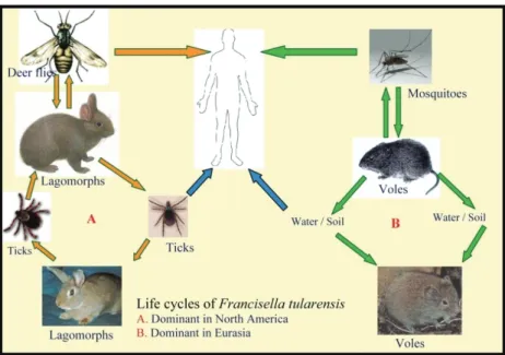
Epidemiology: Tularemia is usually a disease of
animals. The bacteria are commonly found in various terrestrial and aquatic mammals (rabbits, hares, muskrats, beavers and voles), some domesticated animals (dogs, cats, hamsters) and exotic animals (monkeys, prairie dogs) (7). Infection can be acquired by humans by various modes: i) bites by infected arthropods, ii) handling of infected animal tissues or fluids, direct contact with or ingestion of contaminated water, food, or soil, iii) inhalation of infected aerosols (including laboratory exposure) (20). Person-to-person transmission is rare or nonexistent. In addition, humans could be exposed as a result of bioterrorism (3). Figure 1 summarizes the life cycles of F. tularensis in nature. F.
tularensis is a hardy bacterium capable of surviving for
weeks at low temperatures in water, moist soil, hay, straw, or decaying animal carcasses (20, 21). F.
tularensis tularensis (Type A) is found in lagomorphs
(rabbits and similar animals) in North America and it is highly virulent in humans. Type A is usually transmitted to humans by contact with infected animals or is bitten by insects that have fed on an infected animal. Ticks
(Dermacentor variabilis, Amblyomma americana), deer
Figure 1: Life cycles of Francisella tularensis in nature (Original).
flies (Chrysops discalis), and mosquitoes have been shown to transmit tularemia between animals and humans (2, 7, 15, 20).
Two primary disease manifestations, ulceroglandular and glandular, can arise from the bite of an infected vector (22). Conjunctival inoculation may result in infection of the eye with preauricular lymphadenopathy (oculoglandular form). Inhalation or ingestion of F.
tularensis can also result in infection. F. tularensis is an
aerosolizable intracellular bacterium and one of the most infectious pathogens known. It is considered to be a serious potential biological threat agent capable of causing a debilitating or fatal disease with low doses (17). All ages are susceptible to the infection and recovery is followed by permanent immunity. F.
tularensis palaearctica (Type B) occurs mainly in
aquatic rodents (beavers, muskrats) in North America and in hares and small rodents in northern Eurasia. It is less virulent for humans causing a milder infection. Type B is mainly associated with water and animals living near water (15). The dominant mode of transmission is consumption of contaminated spring water in Turkey, and to a lesser extent in Russia and the Balkan countries (5, 10, 11, 13).
The majority of infections in humans and animals are caused by Type A and B. Human disease is rarely associated with the subspecies mediasiatica and novicida (20). Cases of ulceroglandular tularemia that occur in summer are usually associated with arthropod bites, while winter exposures are commonly associated with hunter contact of rabbit carcasses. Outbreaks during the fall and winter may also seen associated with contaminated water and typically result in oropharyngeal form (12, 16). Tularemia can be transmitted to humans through the skin when handling infected animals or contact with tissues, fluids, feces or saliva of infected animals. This can occur when hunting, skinning or butchering infected animals especially rabbits. Bare-handed contact should be avoided when handling any sick or dead animals (2, 3). Inhalation of contaminated agricultural dusts and aerosols are occasional sources for infection in humans. Handling diagnostic samples poses a significant risk for infection via the respiratory route and laboratory personnel should be alerted when tularemia is suspected. Infection through inhalation generally leads to respiratory or the typhoidal form of tularemia (2, 3).
The epidemics seen in Turkey have been caused by
F.tularensis subsp. holarctica (15). Outbreaks linked to
ingestion of contaminated natural spring waters have been reported in Black Sea, Marmara and Thrace and western regions of Turkey in the early 21st century (5, 10, 11, 13, 30). A waterborne outbreak of F.tularensis
subsp. holartica were found to be associated with
consuming contaminated and unchlorinated water in Central Anatolia emerged between 2009 and 2011 (26).
Clinical Manifestations and Diagnosis: The
symptoms of tularemia are varied and depend upon where the organism enters the body. Symptoms usually begin 3-5 days (range of 1-14 days) after being exposed to the bacteria. Patients with tularemia often develop a sudden influenza-like illness, characterized by fever/chills, body aches, nausea, headache and fatigue. Patients also may complain of abdominal pain, emesis, or diarrhea. The type of disease is often named for the most prominent symptoms. There are six main clinical presentations in tularemia: ulceroglandular, glandular, oculoglandular, oropharyngeal, pneumonic and typhoidal (Table1). Dermatological manifestations including erythema nodosum, erythema multiforme, diffuse maculopapular rash, acneiform and vesicular lessions reported as many as 20% of patients with tularemia (2, 4, 20, 25). Secondary skin lesions of infection are observed most frequently in women than men and eruptions completely resolve after treatment (25). Fulminant manifestations of tularemia are reported including meningitis, abscesses and endocarditis (2, 29). Although the ulceroglandular form is the most frequent throughout the endemic areas of the world, oropharyngeal tularemia with tonsillopharyngitis and swollen cervical lymph nodes is the most common form of the disease in Turkey (15).
Diagnosis: The choice of specimen for diagnostic
testing is dependent on the form of clinical illness. The following specimens are acceptable for the various forms of illness as specified: whole blood, serum, respiratory secretions, swabs of visible lesions, aspirates from lymph nodes or lesions, tissue biopsies, autopsy materials (lymph node, lung, liver, spleen, cerebrospinal fluid, and bone marrow) (2, 20). Definite diagnosis of tularemia requires isolation of the causal agent. Culture provides a conclusive diagnosis of infection and an invaluable resource for molecular epidemiology, subtyping and discovery of novel species and subspecies. Whenever possible, culture should be attempted. F. tularensis grows well on several types of cysteine/cystine-supplemented agar (2, 20). The diagnosis of tularemia is mainly based on serological analysis, because isolation of the causative agent is time-consuming, extremely hazardous and requires biosafety level-3 containment in order to avoid risks of laboratory infection. Agglutination, either microagglutination or tube agglutination, is the standard serological test used for determining the presence of antibody to F. tularensis. More recently an ELISA (directed against LPS) combined with Western blot (against antigen extracted from whole killed cells) showed very good sensitivity and specificity for diagnosis of infection (2). PCR can be a valuable diagnostic tool when organisms are non-cultivable or when culture is not recommended due to biosafety concerns.
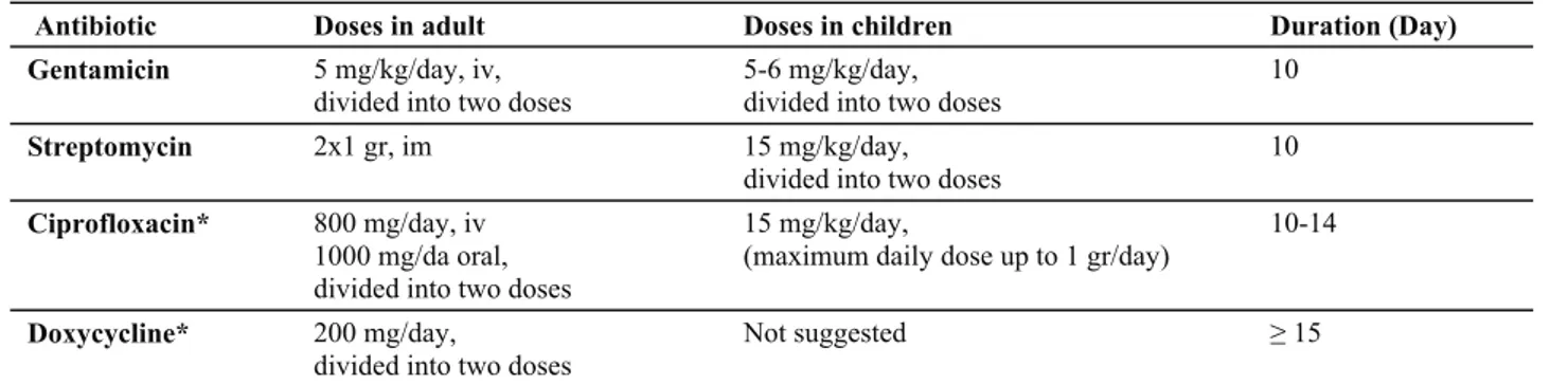
Treatment, prevention and control: The aminoglycosides (streptomycin and gentamicin) are bactericidal agent against F. tularensis. WHO recommends parenteral administration of an aminoglycoside as the first drug of choice for treatment in severe tularemia which requires hospitalization. Gentamicin is preferred at 5 mg/kg daily, divided into two doses and monitored by
assay of serum concentrations of the drug. If streptomycin is available, it is an alternative given by intramuscular injection 2 g daily, divided in two doses, for 10 days. In severe cases, the duration of therapy depends on clinical response, and may be 10 or more days (2, 7). Treatment failure and relapse are rarely seen due to their bactericidal effect. However, ototoxic and Table 1. Clinical manifestations of Tularemia*.
Tablo 1. Tularemi hastalığında klinik tablolar.
Ulceroglandular This form is usually caused by inoculation of the skin and is associated with ulcer at the site of infection and then spreads lymphatically, causing swollen, painful regional lymph nodes. Ulceroglandular form is contracted to humans through the bite of an infected tick.
Glandular F tularensis is considered to enter through an inapparent abrasion and then to spread lymphatically. This form has the same signs and symptoms of ulceroglandular tularemia, with no visible skin ulcers.
Oculoglandular F tularensis enters through the conjunctivae, humans contracted from either splashing of blood or rubbing of eyes after contact with contaminated tissue fluids. Patients with oculoglandular tularemia usually present with acute conjunctivitis, itching, lacrimation and pain. Clinical manifestations are usually unilateral and preauricular or cervical lymphadenitis may accompany with. Chemosis, periorbital edema, and small nodular or ulcerative lesions of the palpebral conjunctivae are also reported. If untreated, corneal ulceration may occur.
Oropharyngeal After consumption of contaminated food or water oropharyngeal or gastrointestinal tularemia may occur depending on the site of colonization. Patients with oropharyngeal tularemia usually report a fever, painful sore throat and swelling on the neck. Clinical presentation is typically enlargement of tonsils, formation of yellow-white pseudo membrane accompanied by swollen cervical lymph nodes. Due to a considerable delay in the start of effective treatment, lymph nodes continue to enlarge; an abscess formation followed by spontaneous suppuration may develop during the course of oropharyngeal tularemia. Suppurating lymph nodes were present in almost 20-40% of the cases in outbreaks in Turkey. Depending on the infecting dose, gastrointestinal tularemia ranges from mild to severe disease. Abdominal pain (due to mesenteric lymphadenopathy), nausea, vomiting, diarrhea, and, occasionally, frank gastrointestinal bleeding (caused by intestinal ulcerations) may be observed.
Pneumonic Disease involving the lungs is termed pneumonic disease. Primary tularemia pneumonia is uncommon and occurs after inhalation of the F tularensis.Rarely acquired naturally, pneumonic tularemia may develop in laboratory workers. Pneumonia develops after hematogenous spread in patients with ulceroglandular tularemia and typhoidal tularemia. Patients usually report a dry cough, dyspnea, and pleuritic-type chest pain. Chest radiography may reveal patchy ill-defined infiltrates in one or more lobes. Frank lobar pneumonia may also develop. Bilateral hilar adenopathy may be present. Bloody pleural effusions are characteristic and demonstrate a mononuclear cellular response. Adult respiratory distress syndrome (ARDS) develops in some patients.
Typhoidal Tularemia that predominately affects the bloodstream and body organs is referred to as typhoidal tularemia. It is more severe and probably represents F tularensis bacteremia. Patients usually present with fever, chills, myalgias, malaise, and weight loss. They often have pneumonia. Diagnosis is difficult because ulcers and lymphadenopathy are usually absent.
* Modified by the references of 2, 7, 14, 20, 27.
Table 2. Suggested antimicrobial treatment and duration in Tularemia*. Tablo 2. Tularemide önerilen antimikrobiyal tedavi ve süreleri.
Antibiotic Doses in adult Doses in children Duration (Day)
Gentamicin 5 mg/kg/day, iv, divided into two doses
5-6 mg/kg/day, divided into two doses
10
Streptomycin 2x1 gr, im 15 mg/kg/day,
divided into two doses 10 Ciprofloxacin* 800 mg/day, iv
1000 mg/da oral, divided into two doses
15 mg/kg/day,
(maximum daily dose up to 1 gr/day) 10-14 Doxycycline* 200 mg/day,
divided into two doses Not suggested ≥ 15
* Table is modified by the references of 2, 15, 20.
nephrotoxic side effects, and the absence of oral aminoglycoside formulations limit their use in clinical practice (15, 27). Fluoroquinolones are also bactericidal; they can be used orally, with high bioavailability. Good penetration into the cell, reaching sufficient concentrations in tissues, fewer toxic effects compared to aminoglycosides, and the absence of the need to monitor drug levels are among the several advantages of quinolones. Fluoroquinolones showed the lowest MIC values in a recent study and seem to have potential as an effective, oral, and first-line therapy for tularemia particularly in remote endemic areas or in outbreak situations with limited resources (27, 28). WHO recommends oral ciprofloxacin or doxycycline in less severe cases or in a mass casualty setting. Ciprofloxacin 800-1000 mg daily, divided into two doses, may be given intravenously or by oral administration. The duration of therapy should be at least 10-14 days (2). In the recent outbreak in Central Anatolia oral ciprofloxacin found to be as effective as aminoglycosides in 1000-1500mg daily dose offering an effective and convenient choice in epidemics of tularemia (27). An alternative is doxycycline, 200 mg daily, divided in two oral doses and, due to the bacteriostatic nature of the drug, given for at least 15 days. Table 2 summaries WHO recommendations for tularemia in adults and children. Potential side-effects have to be weighed against the benefits of treatment of a severe infection in pregnancy. Even though gentamicin and ciprofloxacin are not approved for administration in pregnancy by the Food and Drug Administration of the USA, their use in pregnancy in tularaemia has been recommended by a working group on civilian biodefence. Ciprofloxacin is an option and a brief course of gentamicin treatment is an alternative. Doses are the same as for non-pregnant subjects and the treatment period should be individualized (2).
Post-exposure prophylaxis: Antibiotic prophylaxis
is considered at the below situations (2): i) Accidental exposure of laboratory personnel: antibiotic treatment should be initiated within 24 h and a treatment period of 14 days is recommended with either ciprofloxacin 1000 mg daily divided in two doses, or oral doxycycline 200 mg daily, divided in two doses. ii) Exposure most likely did not occur (in the laboratory): an increased vigilance may be sufficient, including daily measurement of body temperature for 14 days and a readiness to treat if symptoms appear. iii) Incidental spread of F. tularensis by aerosol: potentially exposed persons should be instructed to be alert to the development of fever within 14 days of exposure, and treatment initiated if necessary according to schedule above.
Human-to-human transmission is not recorded. Since tularemia is not spread from person to person, the
patients with tularemia, is not required a strict isolation in hospital ward. A live attenuated vaccine is only in use some parts of the former Soviet Union but no licensed vaccine available (2, 19).
Acknowledgement
This article was presented at 1st National Symposium on Vectors and Vector-Borne with International Participation, 9-10 September, 2012, Avanos, Cappadocia, Nevsehir, Turkey.
References
1. Akalin H, Helvaci S, Gedikoğlu S (2009): Re-emergence of tularemia in Turkey. Int J Infect Dis, 13, 547-551. 2. Anonymous (2007): WHO Guidelines on Tularaemia,
World Health Organization. http://www.who.int/csr/ resources/publications/WHO_CDS_EPR_2007_7.pdf. Erişim Tarihi: 28.01.2013.
3. Anonymous (2009): Biosafety in Microbiological and Biomedical Laboratories. 5th Edition. Centers of Disease and Control (CDC). http://www.cdc.gov/biosafety/ publications/bmbl5/BMBL5_sect_VIII_a.pdf. Erişim Tarihi: 28.01.2013.
4. Byington CL, Bender JM, Ampofo K, Pavia AT, Korgenski K, Daly J, Christenson JC, Adderson E (2008): Tularemia with vesicular skin lesions may be mistaken for infection with herpes viruses. Clin Infect Dis, 47, e4-6.
5. Celebi G, Baruönü F, Ayoğlu F, Cinar F, Karadenizli A, Uğur MB, Gedikoğlu S (2006): Tularemia, a reemerging disease in Northwest Turkey: epidemiological investigation and evaluation of treatment responses. Jpn J Infect Dis, 59, 229-234.
6. Clemens DL, Lee BY, Horwitz MA (2004): Virulent and avirulent strains of Francisella tularensis prevent acidification and maturation of their phagosomes and escape into the cytoplasm in human macrophages. Infect Immun, 72, 3204-3217.
7. Ellis J, Oyston PCF, Gren M, Titball RW (2002): Tularemia. Clin Microbiol Rev, 15, 631-46.
8. Gil H, Benach JL, Thanassi DG (2004): Presence of pili on the surface of Francisella tularensis. Infect Immun, 72, 3042-3047.
9. Gil H, Platz GJ, Forestal CA, Monfett M, Bakshi CS, Sellati TJ, Furie MB, Benach JL, Thanassi DG (2006): Deletion of TolC orthologs in Francisella tularensis identifies role s in multidrug resistance and virulence. Proc Natl Acad Sci USA, 103, 12897-12902. 10. Gurcan S, Otkun MT, Otkun M, Arikan OK, Ozer B
(2004): An outbreak of tularemia in Western Black Sea region of Turkey. Yonsei Med J, 45, 17-22.
11. Helvaci S, Gedikoglu S, Akalin H, Oral HB (2000): Tularemia in Bursa, Turkey: 205 cases in ten years. Eur J Epidemiol, 16, 271-276.
12. Hopla CE (1974): The ecology of tularemia. Adv Vet Sci Comp Med, 18, 25-53.
13. Karadenizli A, Gurcan S, Kolayli F, Vahaboglu H (2005): Outbreak of tularemia in Golcuk, Turkey in 2005: report of 5 cases and an overview of the literature from Turkey. Scand J Infect Dis, 37, 712-716.
14. Kilic S (2010): Francisella tularensis ve Türkiye’de tularemi epidemiyolojisine genel bir bakış. Flora, 15, 37-58. 15. Kilic S, Yesilyurt M (2011): Tularemia: A General
Overview on Current Treatment Options. Klimik Journ, 24, 2-10.
16. Mörner T (1992): The ecology of tularemia. Rev Sci Tech Off Int Epiz, 11, 1123-1130.
17. Oyston PC, Sjostedt A, Titball RW (2004). Tularaemia: bioterrorism defence renews interest in Francisella tularensis. Nat Rev Microbiol, 2, 967-978.
18. Oyston PC (2008): Francisella tularensis: unravelling the secrets of an intracellular pathogen. J Med Microbiol, 57, 921-930.
19. Pechous RD, McCarthy TR, Zahrt TC (2009): Working toward the future: Insights into Francisella tularensis pathogenesis and vaccine development. Microbiol Mol Biol Rev, 73, 684-711.
20. Penn RL (2010): Francisella tularensis (Tularemia). 2927-2937. In: Mandell GL, Bennett JE, Dolin R (Eds). Mandell, Douglas, and Bennett’s Principles and Practice of Infectious Diseases. 7th ed. Philadelphia, PA, Churchill Livingstone.
21. Petersen JM, Schriefer ME (2005): Tularemia: emergence/re-emergence. Vet Res, 36, 455-467.
22. Petersen JM, Mead PS, Schriefer ME (2009): Francisella tularensis: an arthropod-borne pathogen. Vet Res, 40, 7.
23. Rhyan JC, Gahagan T, Fales WH (1990): Tularemia in a cat. J Vet Diagn Invest, 2, 239-241.
24. Tärnvik A, Berglund L (2003): Tularaemia. Eur Respir J, 21, 361-373.
25. Ulu Kilic A, Cicek Senturk G, Tutuncu EE, Kilic S, Altay FA, Gurbuz Y, Sencan I (2010): Two Tularemia Cases with Atypical Presentation. Klimik Journ, 23, 120-123.
26. Ulu Kilic A, Kilic S, Sencan I, Cicek Senturk G, Gurbuz Y, Tutuncu EE, Celebi B, Kiciman O, Ergonul O (2011): A water-borne tularemia outbreak caused by Francisella tularensis subspecies holarctica in Central Anatolia region. Mikrobiyol Bul, 45, 234-247. 27. Ulu Kilic A, Gulen G, Sezen F, Kilic S, Sencan I (2012):
Tularemia in Central Anatolia. Infection, (Epub ahead of print).
28. Ulu Kilic A, Kilic S, Celebi B, Sencan I (2013): Tigesiklinin Francisella tularensis alttip holarctica’ya in vitro etkinliği; doksisiklin, siprofloksasin ve aminogliko-zidlerle karşılaştırılması. Mikrobiyol Bul, 47, 189-191. 29. Van de Beek D, Steckelberg JM, Marshall WF,
Kijpittayarit S, Wijdicks EF (2007): Tularemia with brain abscesses. Neurology, 68, 531.
30. Willke A, Meric M, Grunow R, Sayan M, Finke EJ, Splettstösser W, Seibold E, Erdogan S, Ergonul O, Yumuk Z, Gedikoglu S (2009): An outbreak of oropharyngeal tularaemia linked to natural spring water. J Med Microbiol, 58, 112-116.
Geliş tarihi: 07.02.2013 / Kabul tarihi: 27.03.2013 Address for correspondence:
Professor Mehmet Doganay Department of Infectious Diseases, Faculty of Medicine,
Erciyes University, 38039, Kayseri, Turkey email: [email protected]