Spontaneous Regression of Optic Disc Pit Maculopathy in a Six-Year-Old Child
Tam metin
Şekil
Benzer Belgeler
High reflec- tance signal of the solid lesion was continuous with the retinal nerve fiber layer in the adjacent retina and as might be expected, it was not continuous with
Vitrectomy, argon laser, and gas tamponade for serous retinal detachment associ- ated with an optic disc pit: a case report. Hirakata A, Hida T, Wakabayashi T,
Retinal and glaucoma specialists can evaluate vascular circulation in every possible separate layer for the first time in ocular imagining history and acquire a new understanding
Reduced retinal nerve fiber layer and macular thickness in patients with multiple sclerosis with no history of optic neuritis identified by the use of spectral domain
[2] Brain abscess may develop as outcome of congenital heart disea- se, meningitis, mastoiditis, orbital cellulitis, intraoral infection, surgical intervention, or penetrating head
Clinical picture of children with brain abscess changes depending on size and location of abscess, presence of edema around abscess, and virulence of infectious agent, if
serous maculopathy associated with optic disc pit included observation, laser photocoagulation, pneumatic displacement, macular buckling and pars plana vitrectomy with
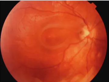
18,19 In our patient, one eye had fluid accumulation under the optic nerve head and an intrapapillary septum structure (Figure 2C). Although macular and optic nerve
