Submandibular Gland Surgery: Our Clinical Experience
Selim Sermed Erbek1, Alper Köycü2, Özgül Topal3, Hatice Seyra Erbek1, Levent Naci Özlüoğlu1 1Department of Otorhinolaryngology, Başkent University Ankara Hospital, Ankara, Turkey2Clinic of Otorhinolaryngology, Siirt State Hospital, Siirt, Turkey
3Department of Otorhinolaryngology, Başkent University Konya Hospital, Konya, Turkey Original Investigation
Address for Correspondence:
Alper Köycü,
E-mail: [email protected] Received Date: 27.12.2015 Accepted Date: 26.04.2016
© Copyright 2016 by Official Journal of the Turkish Society of Otorhinolaryngology and Head and Neck Surgery Available online at www.turkarchotorhinolaryngol.org DOI: 10.5152/tao.2016.1467
Abstract Objective: The aim of this study was to assess the
demographic findings and surgical results of patients who underwent submandibular gland excision at a tertiary care center.
Methods: The clinical characteristics and
histopatho-logical results of 45 patients who had undergone sub-mandibular gland excision between 1997 and 2014 were evaluated in detail.
Results: Twenty-eight (62.2%) and 17 (37.8%)
pa-tients presented with a complaint of a painful mass and painless mass, respectively. Histopathologic in-vestigation of the surgical specimens revealed sialoli-thiasis in 14 patients (31.1%), chronic sialadenitis in 16 (35.6%), benign tumor in 12 (26.7%), malignant tumor in two (4.4%), and mucocele extravasation in one. As complications, permanent paralysis of the
marginal mandibular branch of the facial nerve was seen in one patient (2.2%), temporary paralysis of the marginal mandibular branch of the facial nerve was seen in seven (15.6%), orocutaneous fistula was seen in one (2.2%), and temporary paralysis of the hypo-glossal nerve was seen in one (2.2%).
Conclusion: This study revealed that in patients
pre-senting with complaints of a submandibular gland mass, sialolithiasis, sialadenitis, and benign masses were the mostly diagnosed disorders. Transcervical submandibular gland excision is a satisfactory pro-cedure with low complication and recurrence rates when it is performed on selected patients and obeyed to surgical techniques.
Keywords: Salivary gland, submandibular gland,
surgery, complication
Introduction
Primary masses of the submandibular gland are extremely rare in comparison to the parotid. There-fore, the cause of a significant portion of surgeries of this gland is the stones in the salivary gland and the chronic infections of the salivary gland usually developing secondary to sialolithiasis. Although the incidence of sialolithiasis is reported as 1% in autopsy studies, symptomatic stones are clinical-ly much rarer (1). Of the symptomatic stone cas-es, 80-92% arises from the submandibular gland (1). A decrease in the production of saliva in the long term on the basis of chronic obstruction and chronic inflammation reflecting to the clinic as tightening and stiffening that give the impression of a focal mass in the gland with palpation may develop in these patients. No surgery is needed in these patients who are asymptomatic most of the time. However, recurrent episodes of acute inflam-mation require this treatment.
Only 10-15% of the neoplasms arising from sali-vary gland are seen in the submandibular gland (2). Of these masses, 50-57% are benign (pleomorphic adenoma most commonly) and 43-50% are malig-nant (adenoid cystic carcinoma most commonly) (3).
Transcervical submandibular gland excision has been used for many years in chronic sialadenitis, sialolithiasis and benign or malignant gland tu-mors for diagnosis or treatment. However, only limited numbers of reports related to long-term large series of cases of submandibular gland exci-sion are available in the literature (4, 5). The aim of this study is to share the symptoms, demographic features, clinical and radiological diagnostic meth-ods, surgical specimen histopathological findings, surgical complications and follow-up information of the patients who underwent submandibular gland excision in our clinic.
Methods
This study has been retrospectively conducted by using the file information belonging to 45 patients who underwent subman-dibular gland excision between the years of 1997 and 2014 in a tertiary hospital. The symptoms, demographic features, clinical and radiological diagnostic methods, histopathological findings of surgical specimen and surgical complications were obtained by retrospectively scanning the files of patients who underwent excision of the submandibular gland in our clinic. The study has been approved by the local Ethics Committee and conducted with the permission number KA14/108.
Results
In the study, the files of 54 patients who underwent submandib-ular salivary gland excision were initially scanned, but nine pa-tients were excluded because their pathology did not primarily originate from submandibular glands (four patients with lymph-adenitis, three patients with lipoma, two patients with metasta-ses) and 45 patients 21 of whom were women and 24 were male were included in the study. The average age was 44.8 (14-82). Right submandibular salivary gland excision was performed in 23 (51.1%) of the patients and left submandibular salivary gland excision in 22 (48.9%). The average follow-up duration is 38.4 months (3-60 months) (Table 1).
Twenty-eight (62.2%) of the patients applied with the com-plaint of swelling and 17 (37.8%) of them with the comcom-plaint of painless swelling. According to the findings obtained from the physical examination of submandibular triangle, there were palpable mass in 15 of the patients (33.3%), pain-sensitivity of glands on palpation in 21 (46.7%) of the patients, hard gland in 6 (13.3%) of the patients and palpable stone in 3 (6.7%) of the patients (Table 1).
Most frequently, ultrasonography (USG) was used as the diag-nostic method (31 patients, 68.8%). Four patients (8.8%) were diagnosed with computed tomography (CT) and three patients (6.6%) with magnetic resonance imaging (MRI). Both ultraso-nography (USG) and magnetic resonance imaging (MRI) were preferred as a diagnostic method in one patient (2.2%). While fine needle aspiration (FNA) was performed in six (13.3%) pa-tients who applied to the clinic with the complaint of painless swelling and who was considered to have tumor in preoperative evaluation, the accurate diagnosis could be made only in three (50%) of them.
In the histological examination of the surgical specimen, sialo-lithiasis was found in 14 (31.1%) patients, chronic sialadenitis in 16 (35.6%) patients, benign tumor in 12 (26.7%) patients, malignant tumor in two (4.4%) patients and extravasation mu-cocele in one (2.2%) patient (Table 2).
Pleomorphic adenoma was found the most often among the be-nign tumors at a ratio of 24.4% (11 patients) (Figure 1). The re-maining one patient (2.2%) had cavernous hemangioma arising from submandibular gland. The diagnoses of two patients with malignant tumor were lymphoma and adenoid cystic carcino-ma. When these diagnoses were compared with the preoperative FNA results, while accurate diagnosis was made in three pleo-morphic adenomas, the lymphoma case was found to be report-ed as lymphoid cells.
As postsurgical complications, it was observed that permanent paralysis of facial nerve marginal mandibular branch developed in one patient (2.2%), temporary paralysis of the marginal man-dibular branch in seven (15.6%) patients, orocutaneous fistula in one patient (2.2%) and hypoglossal nerve temporary paresis in one patient (2.2%). No additional treatment was applied to the patients with temporary paralysis of marginal mandibular branch of the facial nerve and a complete spontaneous remis-sion was observed in approximately 3 weeks. Reviremis-sion surgery was not implemented due to postsurgical infections, stones or recurrent mass.
Discussion
Submandibular gland disorders can be divided into two groups as neoplastic and non-neoplastic. Chronic sialadenitis devel-oping on the basis of sialolithiasis comes into prominence as the most common non-neoplastic pathology in submandibular gland excision. This rate is quite high as 44.8 – 73.5% in some studies in the literature (6, 7). It was found as 18.2% in the study of Dalgıç et al. (4) and as 35.6% in our series.
Neoplastic submandibular gland masses constitute 8-22% of all salivary gland tumors (8). Of these tumors, 50-57% are benign and the rest are malignant tumors (8). In particular, rare inci-dence of malignant tumors to this extent leads to the lack of large series of cases and thus, inability to reach a consensus on the size of the surgical and nonsurgical oncological treatment. Consistent with the literature, the diagnoses of benign tumors (26.7%) were made the most frequently and among those,
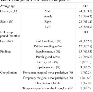
di-Table 1. Demographic characteristics of the patients
Average age 44.8 Gender, n (%) Male 24 (%53.3) Female 21 (%46.7) Side, n (%) Right 23 (%51.1) Left 22 (%48.9) Follow-up 38.4 period (months)
Complaint Painful swelling, n (%) 28 (%62.2)
Painless swelling, n (%) 17 (%37.8)
Findings Palpable mass, n (%) 15 (%33.3)
Painful gland, n (%) 21 (%46.7)
Firm gland, n (%) 6 (%13.3)
Palpable stone, n (%) 3 (%6.7)
Complication Permanent marginal nerve paralysis, n (%) 1 (%2.2)
Temporary marginal nerve paralysis, n (%) 7 (%15.6)
Orocutaneous fistula 1 (%2.2)
Temporary paralysis of the Hypoglossal N. 1 (%2.2)
agnoses of pleomorphic adenoma (11 patients, 24.4%) were the most frequent in our study. Our total rate of malignant tumors is very low (4.4%) and adenoid cystic carcinoma stands out as the primary malignant tumor. The patient with the diagnosis of adenoid cystic carcinoma was postoperatively followed for about 10 years and the patient received a repeated radiotherapy and chemotherapy during this time. The patient in whom wide-spread metastases in the neck and lungs were found died at the end of the 10th year. The ratio of being benign or malignant of the tumors was found to be 85.7% / 14.3% in our study.
The rate of salivary gland tumors in children is much lower than in adults. It is seen 10 times more often in parotid than in submandibular gland (9). Therefore, pediatric submandibular gland pleomorphic adenomas are sporadically reported rather as case reports. A 14-year-old girl patient has been diagnosed with pleomorphic adenoma in our study as well.
Ultrasonography is an important diagnostic tool in subman-dibular gland lesions. It is useful especially in submansubman-dibular gland superficial lesions (10). Papaspyrou et al. (6) reported the sensitivity and specificity of the USG as respectively 87% and
81.3% in the diagnosis of sialolithiasis. CT and MRI should be selected to investigate the situation of tumor spread, local inva-sion and perineural invainva-sion in the cases where malignant cells are seen in the disease or cytology extending to deep tissue (10). USG became the examination that the clinicians preferred most commonly in our series of patients with a ratio of 68.8%. There is still no consensus about the best biopsy method to per-form because the neoplasms of the submandibular gland are very rare. Olubaniyi et al. (11) reported that diagnosis could not be made in fine-needle aspiration biopsies at a rate of approxi-mately 73% in a study that they compared the biopsy methods in neoplastic pathologies of this area. In addition, because the fine needle biopsy will mostly remain insufficient in the distinc-tion of low grade lymphoma and reactive nodal hyperplasia, it creates excisional biopsy indication in most patients (12). For these reasons, both researchers suggest that “core needle” biopsy be performed in company with ultrasonography in terms of di-agnostic efficiency (11, 12). As eighteen gauge and smaller nee-dles are used, it is stated that there isn’t a defined tumor seeding. FNAB was used as a preoperative diagnostic tool in 13.3% of the patients in our clinic and a correct diagnosis was able to be reached in 50% of them. However, while the rate of correct diagnosis in FNAB was achieved only in pleomorphic adenoma, a correct diagnosis could not be made in other malignant pa-thologies. Excisional biopsy is the preferred method when the diagnosis can not be reached through the method of FNA in our clinic.
Temporary marginal mandibular nerve (MMN) damage after transcervical submandibular gland surgeries is reported as 36% (13) and the permanent damage as high a rate as 12% (14) in the literature. Beahm et al. (15) reported these rates between 7.7 and 36% in the study in which they conducted a literature review. Permanent marginal branch paralysis is 2.2% and temporary mar-ginal branch paralysis is 15.6% in our series. The patients with marginal branch paralysis were followed without applying any ad-ditional treatment in the postoperative period. While a complete recovery was observed in seven patients within three weeks on average, a permanent paralysis occurred in one patient. Hernando et al. (7) reported two permanent MMN paralyses in a series of
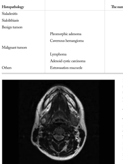
Figure 1. In the right submandibular gland, pleomorphic adenoma that gives hyperintense appearance in Magnetic Resonance Imaging
Table 2. Histopathological diagnoses of patients
Average age
Histopathology The number of patients (years) %
Sialadenitis 16 43.6 35.6% Sialolithiasis 14 48.8 31.1% Benign tumors 12 26.7% Pleomorphic adenoma 11 43 24.4% Cavernous hemangioma 1 36 2.2% Malignant tumors 2 4.4% Lymphoma 1 67 2.2%
Adenoid cystic carcinoma 1 39 2.2%
29 patients and while the diagnosis of one of these was malignant tumor of the gland, the other was reported to be sialadenitis. In our study, the patient with permanent MMN paralysis was diag-nosed with sialadenitis. Four of the seven patients with temporary paresis were diagnosed with sialadenitis, two patients with sialoli-thiasis and one patient with pleomorphic adenoma. The presence of temporary or permanent paralysis in our study increases with the inflammation, in other words with sialadenitis. There are three surgical maneuvers to reduce the risk of MMN damage in the transcervical surgical approach for the gland:
1. The technique that is performed with the gland identified lower than hyoid bone and in which the MMN is not sought. 2. The technique in which the MMN is found in the level
where it leaves the parotid tail, followed and protected. 3. The technique in which the facial veins are found at a lower
level and ligated, and the nerve is suspended by being ele-vated, thus MMN is kept out of the surgical area.
In our clinic, as Riffat et al. (16) suggests, after the neck incision 2 cm away from the mandibular corpus by bringing the patient’s neck into hyperextension, facial veins are ligated at the lower edge of the gland, elevated together with the gland fascia, and thus the MMN is protected. Besides, keeping the nerve in mind while using retractors during surgery, respectful approach to tis-sue and tistis-sue plans and behaving carefully while using bipolar cautery in hazardous areas during the operation keep the rate of complications at quite low levels. Transoral open and/or endo-scopic approaches, the use of which has been increasing recently reduce this risk significantly (15).
Open surgical approaches, endoscope assisted open surgical ap-proaches and total endoscopic apap-proaches are available in sub-mandibular gland surgery. Open surgical approaches are lateral transcervical, submental, retroauriculer and transoral ones. Semi endoscopic surgical procedures and total endoscopic surgical methods with a pouch created by giving carbon dioxide gas that are applicable in these regions are also available (15). These surgical procedures have different advantages and disadvantag-es when compared with each other. They are preferred and can be applied according to the surgeon’s experience and capability, accessibility to technology and the characteristics of the patient and disease.
Transoral approach first entered the literature in 1960, but gained popularity with the study that Hong et al. (17) conducted in 2000 (18). Hong et al. (17) reported the advantages of tran-soral approach to lateral transcervical surgery as the reduction in MMN paralysis and the risk of mucocele formation and lack of skin scars in their two series of 31 and 77 patients. However, in transoral surgery, they detected an abnormal sensation in the tongue at a rate of 74% and limitations in tongue movements at a rate of 70% in the early postoperative period. The limita-tion of the tongue movements declined to about 10% (17, 19) during the follow-up. Weber et al. (20) identified the abnormal sensation in the tongue at a rate of 43%, but they didn’t ob-serve any limitations in the tongue movements and wound scar.
They reported that they managed this by staying a little away from the lingual gingiva to make the incision and thus reduc-ing the tension. They also mentioned that abnormal sensation could be prevented by avoiding stretching the lingual nerve. A history of gland surgery and a small oral cavity were mentioned as relative contraindications for transoral surgery. In the study that Çukurova et al. (21) conducted, 10 patients who underwent transoral surgery and 10 patients who underwent transcervical surgery were compared. They found that transoral surgical ap-proach significantly shortened the duration of hospital stay in comparison to the transcervical approach. However, sialadenitis repeated due to the remaining glandular tissue in a patient who underwent transoral surgery (21).
Springborg et al. (5) reported postoperative infections at a rate of 9.4% in their series of 139 patients. They reported that they used preoperative antibiotic prophylaxis in order to prevent this complication. We don’t use preoperative prophylaxis in our clin-ic. However, prophylactic antibiotics are administered for 7 days postoperatively.
Among the surgical complications, hypoglossal nerve damage has been reported very rarely (0-1.4%) (5). This situation is caused by the nerve’s settlement partially sheltered by the pos-terior belly of the digastric muscle that is in the inferomedi-al of the lower 1/3 of the gland. In the patient with cavernous hemangioma who was operated in our clinic, paresis of the 12th nerve that lasted for 5 months postoperatively developed due to heavy bleeding during the surgery and frequent and uncon-trolled use of clamp/bipolar cauterization. In the follow-up, the patient recovered without sequelae.
Conclusion
The most common pathology in submandibular gland is the for-mation of stones in the gland or ducts. In particular, extremely rare incidence of malignant tumors leads to the lack of large se-ries of cases and thus, inability to reach a consensus on the type of the surgical and nonsurgical oncological treatment. Transcer-vical submandibular gland excision is an applicable surgery with acceptable rates of complications and sequelae. The risk for neu-rological complications which may occur due to the nerves close to surgical area can be reduced with approaches that are loyal to the surgical principles and respect to tissue planes.
Ethics Committee Approval: Ethics committee approval was received
for this study from the ethics committee of Başkent University Hospi-tal (KA 14-108).
Informed Consent: Written informed consent was obtained from
pa-tients or parents of the papa-tients who participated in this study.
Peer-review: Externally peer-reviewed.
Author Contributions: Concept - S.S.E., H.S.E.; Design - A.K.,
S.S.E., L.N.Ö.; Supervision - S.S.E., H.S.E., L.N.Ö.; Resources - A.K., Ö.T.; Materials - A.K., Ö.T.; Data Collection and/or Processing - A.K., Ö.T., H.S.E.; Analysis and/or Interpretation - A.K., H.S.E., L.N.Ö.; Literature Search - A.K., Ö.T.; Writing Manuscript - A.K., Ö.T.; Critical Review - S.S.E., L.N.Ö.
Conflict of Interest: No conflict of interest was declared by the
aut-hors.
Financial Disclosure: The authors declared that this study has received
no financial support.
References
1. Williams MF. Sialolithiasis. Otolaryngol Clin North Am 1999; 32: 819-34. [CrossRef]
2. Stenner M, Klussmann JP. Current update on established and novel biomarkers in salivary gland carcinoma pathology and the molecular pathways involved. Eur Arch Otorhinolaryngol 2009; 266: 333-41. [CrossRef]
3. Rinaldo A, Ferlito A, Pellitteri PK, Robbins KT, Shaha AR, Brad-ley PJ, et al. Management of malignant submandibular gland tu-mors. Acta Otolaryngol 2003; 123: 896-904. [CrossRef]
4. Dalgic A, Karakoc O, Karahatay S, Hidir Y, Gamsizkan M, Bir-kent H, et al. Submandibular triangle masses. J Craniofac Surg. 2013; 24: e529-31.
5. Springborg LK, Møller MN. Submandibular gland excision: long-term clinical outcome in 139 patients operated in a single institu-tion. Eur Arch Otorhinolaryngol 2013; 270: 1441-6. [CrossRef]
6. Papaspyrou G, Werner JA, Sesterhenn AM. Transcervical extir-pation of the submandibular gland: the University of Marburg experience. Eur Arch Otorhinolaryngol 2014; 271: 2009-12.
[CrossRef]
7. Hernando M, Echarri RM, Taha M, Fragueiro LM, Hernando A, Mayor GP. Surgical complications of submandibular gland excisi-on. Acta Otorrinolaringol Esp 2012; 63: 42-6. [CrossRef]
8. Spiro RH. Salivary neoplasms: overview of a 35-year experience with 2807 patients. Head Neck Surg 1986; 8: 177-84. [CrossRef]
9. Bentz BG, Hughes CA, Lüdemann JP, Maddalozzo J. Masses of the salivary gland region in children. Arch Otolaryngol Head Neck Surg 2000; 126: 1435-9. [CrossRef]
10. Lee YY, Wong KT, King AD, Ahuja AT. Imaging of salivary gland tumours. Eur J Radiol 2008; 66: 419-36. [CrossRef]
11. Olubaniyi BO, Chow V, Mandalia U, Haldar S, Gok G, Michl P, et al. Evaluation of biopsy methods in the diagnosis of submandi-bular space pathology. Int J Oral Maxillofac Surg 2014; 43: 281-5.
[CrossRef]
12. Sellon E, Moody A, Howlett D. Ultrasound guided core biopsy is the diagnostic tool of choice in salivary gland swellings. BMJ 2012; 345: e7782.
13. Smith WP, Peters WJ, Markus AF. Submandibular gland surgery: an audit of clinical findings, pathology and postoperative morbi-dity. Ann R Coll Surg Eng 1993; 75: 164-7.
14. De M, Kumar Singh P, Johnson AP. Morbidity associated with submandibular gland excision: a retrospective analysis. Internat J Head Neck Surg 2006; 1: 1.
15. Beahm DD, Peleaz L, Nuss DW, Schaitkin B, Sedlmayr JC, Ri-vera-Serrano CM, et al. Surgical approaches to the submandi-bular gland: A review of literature. Int J Surg 2009; 7: 503-9.
[CrossRef]
16. Riffat F, Buchanan MA, Mahrous AK, Fish BM, Jani P. Oncologi-cal safety of the Hayes-Martin manoeuvre in neck dissections for node-positive oropharyngeal squamous cell carcinoma. J Laryngol Otol 2012; 126: 1045-8. [CrossRef]
17. Hong KH, Kim YK, Intraoral removal of the submandibular gland: a new surgical approach. Otolaryngol Head Neck Surg 2000; 122: 798-802. [CrossRef]
18. Downton D, Qvist G. Intra-oral excision of the submandibular gland. Proc R Soc Med 1960; 53: 543-4.
19. Hong SM, Yang YS, Surgical results of the intraoral removal of the submandibular gland. Otolaryngol Head Neck Surg 2008; 139: 530-4. [CrossRef]
20. Weber SM, Wax MK, Kim JH, Transoral excision of the subman-dibular gland. Otolaryngol Head Neck Surg 2007; 137: 343-5.
[CrossRef]
21. Çukurova İ, Arslan İB, Bulgurcu S, Demirhan E. Transoral ver-sus transcervical approach to submandibular gland: techniques and outcomes. Kulak Burun Bogaz Ihtis Derg 2015; 25: 319-23.