Original Article
DOI: 10.5606/tftrd.2019.3296 ©Copyright 2019 by Turkish Society of Physical Medicine and Rehabilitation - Available online at www.turkishjournalpmr.com
The relationship of serum vitamin D receptor levels with disease activity
and clinical parameters in patients with ankylosing spondylitis
Received: June 24, 2018 Accepted: October 02, 2018 Published online: November 22, 2019
Corresponding author: Turgut Kültür, MD. Kırıkkale Üniversitesi Tıp Fakültesi, Fiziksel Tıp ve Rehabilitasyon Anabilim Dalı, 71450 Yahşihan, Kırıkkale, Türkiye. e-mail: [email protected]
Cite this article as:
Kültür T, Öztaş D, Keskin D, Keskin G, İnal A, Kara H. The relationship of serum vitamin D receptor levels with disease activity and clinical parameters in patients with ankylosing spondylitis. Turk J Phys Med Rehab 2019;65(4):389-393.
ABSTRACT
Objectives: The aim of this study was to investigate the relationship between serum vitamin D receptor (SVDR) levels and disease activity parameters in patients with ankylosing spondylitis (AS).
Patients and methods: Between July 2016 and January 2017, a total of 62 patients (51 males, 11 females; mean age 36.5±12.8 years; range, 23 to 49 years) with AS and 32 healthy volunteers (25 males, 7 females; mean age 41.57±13.6 years; range, 26 to 48 years) were included in the study. The SVDR levels were measured using the enzyme-linked immunosorbent assay. Erythrocyte sedimentation rate (ESR) and serum C-reactive protein (CRP) levels were recorded. The Bath Ankylosing Spondylitis Disease Activity Index (BASDAI) scores were used to assess disease activity.
Results: Although there was no significant difference between the patient and control groups (p=0.66), SVDR levels were significantly elevated in patients with active AS (BASDAI score ≥4) (p=0.01). The SVDR levels significantly increased in AS patients with peripheral joint involvement and enthesitis (p=0.01, p=0.05, respectively). The SVDR levels significantly elevated in patients treated with non-steroidal anti-inflammatory drugs, compared to those treated with biological agents and control group (p=0.01, p=0.03, respectively). The SVDR levels were positively correlated with the BASDAI, CRP and ESR in the patient group (p=0.01, r=0.751; p=0.01, r=0.75; p=0.01, r=0.81, respectively). Conclusion: Our study results suggest that serum SVDR levels are associated with the disease activity and clinical parameters in patients with AS. Based on these findings, SVDR level may be used as a marker of disease activity in AS.
Keywords: Ankylosing spondylitis, clinical and laboratory parameters, serum vitamin D receptor.
Turgut Kültür1, Dilek Öztaş2, Dilek Keskin1, Göksal Keskin3, Ali İnal4, Halil Kara5
1Department of Physical Medicine and Rehabilitation, Kırıkkale University Faculty of Medicine, Kırıkkale, Turkey 2Department of Public Health, Ankara Yıldırım Beyazıt University Faculty of Medicine, Ankara, Turkey 3Department of Immunology, Ankara University Faculty of Medicine, Ankara, Turkey
4Department of Immunology, Başkent University İstanbul Health Application and Research Hospital, İstanbul, Turkey 5Department of Pharmacology, Ankara Yıldırım Beyazıt University Faculty of Medicine, Ankara, Turkey
Ankylosing spondylitis (AS) is a chronic, systemic, inflammatory disease with an unknown etiology, which can primarily target sacroiliac, axial and peripheral joints, and occasionally some organs such
as eyes, heart, lungs, intestines, and kidneys.[1,2] In
general, acute phase responses such as erythrocyte sedimentation rate (ESR) and C-reactive protein (CRP) are used in the evaluation and follow-up of the disease. However, these parameters do not always correlate
with the disease activity.[1,2] Therefore, several studies
have investigated the association of tumor necrosis factor-alpha (TNF-α) and a number of inflammatory
cytokines with AS activity.[2]
Vitamin D is a hormone with immunosuppressive and immunoregulatory functions in addition to the classical effects on calcium-phosphorus metabolism. It acts by binding to a nuclear receptor called the vitamin D receptor (VDR) for activating or inhibiting the target
genes.[3-6] Rosca and Cheung[7] reported that although
vitamin D levels in AS patients were significantly lower, correction of these levels did not affect disease activity or function in AS patients. Although the proposed mechanisms have not been fully elucidated yet, it has been suggested that vitamin D deficiency and polymorphisms of VDR-encoded genes may play a role in the pathogenesis, disease activation, and
clinical features of AS.[3-7] Vitamin D regulates the immunological mediators, particularly via VDR. In addition, it has been proposed that VDR agonists can act as immunoreactive agents and modulators which may be used in the treatment of certain chronic
inflammatory autoimmune diseases.[3-7]
It is well-known that the effect of VDR levels on disease activation varies according to the
clinical condition of each individual patient.[8-10] In
the literature, there is no study examining serum vitamin D receptor (SVDR) levels in patients with AS. In the present study, therefore, we aimed to investigate the relationship between SVDR levels and disease activity parameters in patients with AS.
PATIENTS AND METHODS
This cross-sectional study included a total of 62 patients (51 males, 11 females; mean age 36.5±12.8 years; range, 23 to 49 years) diagnosed with AS according to the 1984 Modified New York Criteria in the Ankara University Faculty of Medicine Immunology Department between July 2016 and January 2017. The control group was comprised of a total of 32 age- and sex-matched healthy volunteers (25 males, 7 females; mean age 41.57±13.6 years; range, 26 to 48 years). Patients with acute or subacute viral/bacterial infections, pregnancy, cardiopulmonary disease, renal insufficiency, endocrine disorders such as hyperparathyroidism, malabsorption, recent history of surgical operation, systemic diseases (malignancy, diabetes mellitus, hypertension), prosthesis, mental retardation or severe emotional disturbance, smokers and alcohol consumers were excluded. A written informed consent was obtained from each participant. The study protocol was approved by the Clinical Research Ethics Committee of Ankara Yıldırım Beyazıt University Faculty of Medicine (13.07.2016/201). The study was conducted in accordance with the principles of the Declaration of Helsinki.
Data including demographic and clinical characteristics of patients and healthy controls were included. The presence of peripheral involvement and history of uveitis and enthesitis were obtained from the patient records. The medications of the patients were recorded (non-steroidal anti-inflammatory drugs [NSAIDs] and biological treatment).
The SVDR level (pg/mL) was measured using the enzyme-linked immunosorbent assay. The ESR (mm/h) was measured using the Westergren tube method. The CRP (mg/dL) was measured using the nephelometric method. The Bath Ankylosing Spondylitis Disease Activity Index (BASDAI) scores
were calculated to evaluate the disease activity.[11]
A BASDAI score of ≥4 indicates active AS.
Statistical analysis
The required sample size was calculated using a general power analysis program (G*power; version 3.1.9.2 software [Heinrich-Heine-Universität Düsseldorf, Düsseldorf, Germany]). The power of the study with 62 patients was calculated as 92.24% which was sufficient for a minimum of 80% power. Statistical analysis was performed using the SPSS version 16.0 software (SPSS Inc., Chicago, IL, USA). Descriptive data were expressed in mean ± standard deviation (SD) and median (min-max) or number and frequency. The Student’s t- and Mann-Whitney U tests were performed to assess group differences for continuous variables. Categorical variables were compared using the chi-square test. The relationship between two independent numerical variables was analyzed using the Spearman’s Rho correlation coefficient. A receiver operating characteristic (ROC) analysis was performed to determine the cut-off, sensitivity, and specificity values, and the calculation of validity and reliability analysis results for SVDR. A p value of <0.05 was considered statistically significant.
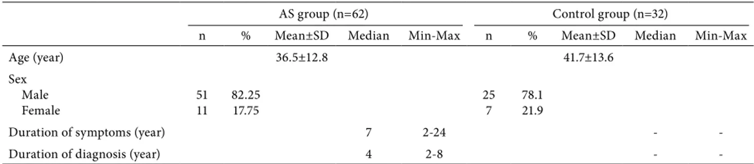
Table 1. Demographic characteristics of patient and control group
AS group (n=62) Control group (n=32)
n % Mean±SD Median Min-Max n % Mean±SD Median Min-Max
Age (year) 36.5±12.8 41.7±13.6
Sex Male
Female 5111 82.2517.75 257 78.121.9
Duration of symptoms (year) 7 2-24 -
-Duration of diagnosis (year) 4 2-8 -
RESULTS
The demographic and clinical characteristics of the patients and controls are shown in Table 1. There was no significant difference in the SVDR levels
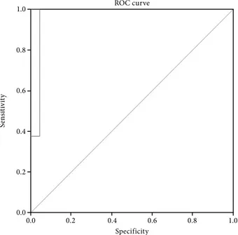
between the AS group and control group. However, the SVDR levels in the patients with active AS (BASDAI score ≥4) significantly increased, compared to both of the controls and inactive AS groups (p=0.01, p=0.01) respectively). The ROC analysis yielded a value of 0.972 (p=0.01) with a lower limit of 0.894 and upper limit of 0.997 (95% confidence interval [CI], 0.894-0.997). A SVDR value of >0.64 pg/mL had a very high sensitivity (100%) and specificity (95.5%) for the active patient group, and based on the ROC curve analysis, the most optimal cut-off value for active AS was 0.64 pg/mL with a negative predictive value of 100% and a positive predictive value of 97.5%. Figure 1 shows the ROC curve of the SVDR values for active AS patients. The SVDR levels significantly increased in the AS group with peripheral joint involvement and enthesitis (p=0.01, p=0.05, respectively), although there was no significant difference among the AS group with uveitis.
The SVDR levels in the AS group according to the BASDAI and clinical parameters are presented in Table 2. We also evaluated the relationship between the SVDR levels and treatment of AS by subgroup analysis. We found a significant difference in the SVDR levels in the patients receiving NSAID treatment compared to those using biological treatment (p=0.01). In addition, the SVDR levels of the patient group treated with NSAIDs were also significantly elevated, compared to the control group (p=0.03). Figure 1. Receiver operating characteristic curves of vitamin
D receptor values in differentiating patients with active ankylosing spondylitis from inactive active ankylosing spondylitis The blue curve represents the receiver operating characteristic curve, and the gray line represents the diagonal line used as a reference.
ROC: Receiver operating characteristic. 0.0 0.0 0.2 0.4 0.6 0.8 1.0 0.4 Specificity ROC curve Se ns iti vit y 0.8 0.2 0.6 1.0
Table 2. Comparison of serum vitamin D receptor levels according to demographic and clinical parameters in ankylosing spondylitis patients
n % Median Min-Max p SVDR levels (pg/mL) Sex Male Female 5111 82.317.7 - - 0.92 Involvement Axial Peripheral 4913 7921 - - 0.01** Enthesitis + - 1646 25.874.2 - - 0.04* Uveitis + - 1250 19.680.4 - - 0.20 BASDAI <4 >4 2141 33.965.1 6.563.04 0.24-3.804.78-7.12 0.01**
Min: Minimum; Max: Maximum; SVDR: Serum vitamin D receptor; BASDAI: Bath Ankylosing Spondylitis Disease Activity Index; (Active AS BASDAI >4); * p<0.05: Statistically significant; ** Mann Whitney U test.
Correlation analysis revealed that the SVDR levels were positively correlated with the BASDAI (r=0.75, p=0.01), CRP (r=0.75, p=0.01), and ESR (r=0.81, p=0.01). However, there was no correlation between the SVDR levels and disease duration in patients with AS (r=0.094, p=0.468).
DISCUSSION
In the present study, we investigated the relationship between SVDR levels and disease activity parameters in patients with AS. According to our findings, the SVDR levels were found to be elevated in patients with active AS, compared to patients with inactive AS. The SVDR levels were positively correlated with the BASDAI scores and systemic inflammatory markers such as CRP and ESR. In addition, we found a cut-off value for active disease with a very high sensitivity and specificity. These findings indicate that the SVDR level may be accepted as a new inflammatory marker and acute phase reactant to indicate clinical activity for AS. To the best of our knowledge, the present study is the first to evaluate VDR levels in patients with AS in the literature.
Interleukin (IL)-22, IL-17, TNF-α, IL-6, IL-10, toll-like receptor-4, and IL-23 are important in AS clinic and disease activation which are found to be increased and dysregulated in AS patients. The VDR is effective in the regulation of expression and function
of these mediators.[12-20] The VDR may act to regulate
nuclear factor-kappa B (NF-kB) activation which increases in AS and important for the expression of adhesion molecules and other pro-inflammatory
genes.[21,22] It is also effective on the regulation of
functions B cells, CD4, and CD8 T lymphocytes which play a critical role in AS clinic and disease activation, as well as reducing the activity of Wnt/β-catenin signaling pathway, which has important implications in cell adhesion and regulation of the functions in B,
CD4, and CD8 T lymphocytes.[12,21-26] In experimental
studies on mice, stimulation of the ß-catenin signal was shown to activate the Wnt pathway, leading to aberrant
chondrocyte maturation and joint ankyloses.[27] It was
also reported that VDR might induce apoptosis.[28,29]
The induced chondrocyte apoptosis in AS was thought
to play a role in the changes in bone and cartilage.[30]
Enthesopathy, according to some authors, is considered a completely pathological process; however, it is a
repair mechanism for others, indeed.[31] Pathogenesis
of AS is still not clear. According to the literature, autoimmune diseases may associate with microbiota which plays a role in the pathogenesis by leading
to dysfunction of VDR, due to increased release of antimicrobial peptides, and that the use of antibiotics may result in an increased VDR activation, thereby,
leading to an increased immunological response.[32]
In our study, the SVDR levels significantly increased in the AS group with peripheral joint involvement and enthesitis. However, further studies are needed to elucidate the immunomodulatory effect of SVDR in AS.
In the present study, the SVDR levels were also found to be elevated in patients receiving NSAIDs. Although SVDR levels in the AS patients were not significantly different from the controls, the BASDAI scores and the SVDR levels in the patients treated with NSAIDs significantly increased, compared to those using biological agents and control groups. These results may be associated with decreased SVDR levels due to suppression of disease activity with anti-TNF drugs. According to our results, SVDR may have a role as an inflammatory marker and, therefore, it may be suppressed by biological agents naturally. It was previously reported that VDR caused a significant
decrease in the TNF-α levels in mast cells.[10] Ziv et
al.[10] found that exposure of keratinocytes to TNF-α
caused a significant increase in VDR levels. Pojednic
et al.[33] also reported that VDR levels were correlated
with IL-6 gene expression and VDR activation could regulate intracellular inflammation. Thus, it can be assumed that SVDR plays a role in decreasing disease activity in AS, although further researches are needed to shed light into this subject.
The main limitations of the present study include the lack of vitamin D analysis and VDR gene polymorphism in the patients with AS.
In conclusion, we found elevated SVDR levels in patients with activated AS in our study. Based on these results, increased SVDR levels may be associated with peripheral joint involvement and the presence of enthesitis and it may be related to disease activity parameters in patients with AS.
Declaration of conflicting interests
The authors declared no conflicts of interest with respect to the authorship and/or publication of this article.
Funding
The authors received no financial support for the research and/or authorship of this article.
REFERENCES
1. Feldtkeller E, Erlendsson J. Definition of disease duration in ankylosing spondylitis. Rheumatol Int 2008;28:693-6.
2. Reveille JD. Biomarkers for diagnosis, monitoring of progression, and treatment responses in ankylosing spondylitis and axial spondyloarthritis. Clin Rheumatol 2015;34:1009-18.
3. Zhao S, Duffield SJ, Moots RJ, Goodson NJ. Systematic review of association between vitamin D levels and susceptibility and disease activity of ankylosing spondylitis. Rheumatology (Oxford) 2014;53:1595-603.
4. Wu S, Sun J. Vitamin D, vitamin D receptor, and macroautophagy in inflammation and infection. Discov Med 2011;11:325-35.
5. Adorini L, Penna G. Control of autoimmune diseases by the vitamin D endocrine system. Nat Clin Pract Rheumatol 2008;4:404-12.
6. Zhang P, Li Q, Qi J, Lv Q, Zheng X, Wu X, et al. Association between vitamin D receptor gene polymorphism and ankylosing spondylitis in Han Chinese. Int J Rheum Dis 2017;20:1510-6.
7. Rosca C, Cheung N. Does correction of vitamin d level influence the disease activity and functionality in ankylosing spondylitis patients? A longitudinal study. Rheumatology 2017;56(Suppl 2):209.
8. Muralidhar S, Nsengimana J, Pozniak J, O’Shea S, Laye J, Adams D, et al. Primary melanoma expression of the vitamin D receptor (VDR) is protective for melanoma survival and is associated with increased tumor immune response, decreased Wnt/B-catenin signaling and tumor proliferation. Proceedings of the American Association for Cancer Research Annual Meeting, April 14-18, 2018, Chicago, IL. Philadelphia (PA): AACR; Cancer Res 2018;78(13 Suppl):5205.
9. Singh RK, Turner R, Kim K, Sivagnanalingam U, Moore RG. Targeting vitamin D receptor (VDR)/immune checkpoint inhibitor receptor ligand PD-L1 axis for immunotherapy of ovarian cancer. Gynecologic Oncology 2018;149:48. 10. Ziv E, Koren R, Zahalka MA, Ravid A. TNF-α increases
the expression and activity of vitamin D receptor in keratinocytes: role of c-Jun N-terminal kinase. Dermatoendocrinol 2016;8:e1137399.
11. Akkoc Y, Karatepe AG, Akar S, Kirazli Y, Akkoc N. A Turkish version of the Bath Ankylosing Spondylitis Disease Activity Index: reliability and validity. Rheumatol Int 2005;25:280-4. 12. Yin K, Agrawal DK. Vitamin D and inflammatory diseases.
J Inflamm Res 2014;7:69-87.
13. Chang SH, Chung Y, Dong C. Vitamin D suppresses Th17 cytokine production by inducing C/EBP homologous protein (CHOP) expression. J Biol Chem 2010;285:38751-5.
14. Stubbs JR, Idiculla A, Slusser J, Menard R, Quarles LD. Cholecalciferol supplementation alters calcitriol-responsive monocyte proteins and decreases inflammatory cytokines in ESRD. J Am Soc Nephrol 2010;21:353-61.
15. Ramadan RA, Desouky LM, Moaaz M, Elnaggar MA, Selima M, Samir M, et al. Association of vitamin D receptor and toll like receptor genetic variants and haplotypes with colon cancer risk: A case control study in Egypt Meta Gene 2017;11:209-16.
16. James J, Weaver V, Cantorna MT. Control of Circulating IgE by the Vitamin D Receptor In Vivo Involves B Cell Intrinsic and Extrinsic Mechanisms. J Immunol 2017;198:1164-71.
17. Hoeck AD, Pall ML. Will vitamin D supplementation ameliorate diseases characterized by chronic inflammation and fatigue? Med Hypotheses 2011;76:208-13.
18. Smith JA. Update on ankylosing spondylitis: current concepts in pathogenesis. Curr Allergy Asthma Rep 2015;15:489. 19. Yan Y, Guo TM, Zhu C. Effects of nonsteroidal
anti-inflammatory drugs on serum proanti-inflammatory cytokines in the treatment of ankylosing spondylitis. Biochem Cell Biol 2018;96:450-6.
20. Ma D, Zhang RN, Wen Y, Yin WN, Bai D, Zheng GY, et al. 1, 25(OH)2D3-induced interaction of vitamin D receptor with p50 subunit of NF-κB suppresses the interaction between KLF5 and p50, contributing to inhibition of LPS-induced macrophage proliferation. Biochem Biophys Res Commun 2017;482:366-74.
21. Sun J, Kong J, Duan Y, Szeto FL, Liao A, Madara JL, et al. Increased NF-kappaB activity in fibroblasts lacking the vitamin D receptor. Am J Physiol Endocrinol Metab 2006;291:E315-22.
22. Cobas M, Wilson A, Ernst B, Mancini SJ, MacDonald HR, Kemler R, et al. Beta-catenin is dispensable for hematopoiesis and lymphopoiesis. J Exp Med 2004;199:221-9.
23. Boyle LH, Goodall JC, Opat SS, Gaston JS. The recognition of HLA-B27 by human CD4(+) T lymphocytes. J Immunol 2001;167:2619-24.
24. Atagunduz P, Appel H, Kuon W, Wu P, Thiel A, Kloetzel PM, et al. HLA-B27-restricted CD8+ T cell response to cartilage-derived self peptides in ankylosing spondylitis. Arthritis Rheum 2005;52:892-901.
25. Lin Q, Gu JR, Li TW, Zhang FC, Lin ZM, Liao ZT, et al. Value of the peripheral blood B-cells subsets in patients with ankylosing spondylitis. Chin Med J (Engl) 2009;122:1784-9. 26. Szymczak I, Pawliczak R. The Active Metabolite of Vitamin
D3 as a Potential Immunomodulator. Scand J Immunol 2016;83:83-91.
27. Las Heras F, Pritzker KP, So A, Tsui HW, Chiu B, Inman RD, et al. Aberrant chondrocyte hypertrophy and activation of β-catenin signaling precede joint ankylosis in ank/ank mice. J Rheumatol 2012;39:583-93.
28. Shah S, Islam MN, Dakshanamurthy S, Rizvi I, Rao M, Herrell R, et al. The molecular basis of vitamin D receptor and beta-catenin crossregulation. Mol Cell 2006;21:799-809. 29. Khan NP, Pandith AA, Hussain MU, Yousuf A, Khan
MS, Wani KA, et al. Novelty of Axin 2 and lack of Axin 1 gene mutation in colorectal cancer: a study in Kashmiri population. Mol Cell Biochem 2011;355:149-55.
30. Bleil J, Maier R, Hempfing A, Schlichting U, Appel H, Sieper J, et al. Histomorphologic and histomorphometric characteristics of zygapophyseal joint remodeling in ankylosing spondylitis. Arthritis Rheumatol 2014;66:1745-54.
31. Miceli-Richard C. Enthesitis: The clue to the pathogenesis of spondyloarthritis? Joint Bone Spine 2015;82:402-5. 32. Waterhouse JC, Perez TH, Albert PJ. Reversing
bacteria-induced vitamin D receptor dysfunction is key to autoimmune disease. Ann N Y Acad Sci 2009;1173:757-65. 33. Pojednic RM, Ceglia L, Lichtenstein AH, Dawson-Hughes
B, Fielding RA. Vitamin D receptor protein is associated with interleukin-6 in human skeletal muscle. Endocrine 2015;49:512-20.