Address for Correspondence: Dr. Musa Çakıcı, Adıyaman Üniversitesi Tıp Fakültesi, Kahta Cad., Adıyaman-Türkiye
Phone: +90 506 556 84 71 Fax: +90 416 225 08 38 E-mail: [email protected] Accepted Date: 14.05.2014 Available Online Date: 23.06.2014
©Copyright 2015 by Turkish Society of Cardiology - Available online at www.anatoljcardiol.com DOI:10.5152/akd.2014.5562
A
BSTRACTObjective: The coronary sinus (CS) has been largely ignored by physicians due to a lack of adequate data about the importance of CS enlarge-ment in patients with heart failure (HF). We aimed to assess whether CS dilatation develops in patients with HF and to demonstrate its relation with global myocardial function of the right ventricle (RV).
Methods: In this cross-sectional study, 45 healthy subjects and 95 HF patients exhibiting left ventricular systolic dysfunction on echocardio-graphic examination (EF <45%) secondary to ischemic (n=56) or idiopathic dilated cardiomyopathy (DCM) (n=39) were enrolled. Patients with severe renal dysfunction and/or valve disease were excluded. CS was measured by echocardiography from the posterior atrioventricular groove in the apical four-chamber view. The RV myocardial performance index (MPI), which reflects both systolic and diastolic function of the ventricle, was detected using tissue Doppler imaging, and patients with an RV MPI >0.55 were defined as having impaired RV myocardial func-tion. ANOVA, Kruskal-Wallis, Pearson’s correlation, and multivariate logistic regression analyses were used for the statistical analysis. Results: The CS and RV MPI values were significantly greater both in patients with ischemic and idiopathic DCM than in controls (8.79±1.7 mm and 8.33±2.1 mm vs. 5.74±0.6 mm, and 0.64±0.07 and 0.62±0.08 vs. 0.43±0.02; p<0.001 for both, respectively). For the prediction of HF patients with impaired RV function, the cut-off value for the diameter of the CS was 7.35 mm, with a sensitivity of 83% and a specificity of 79%.
Conclusion: The CS diameter can be used as a novel echocardiographic marker that provides information about impaired RV function in patients with HF. (Anatol J Cardiol 2015; 15: 542-7)
Keywords: coronary sinus, right ventricle, myocardial performance index, heart failure
Musa Çakıcı, Adnan Doğan
1, Mustafa Çetin, Arif Süner, Mustafa Polat, Muhammed Oylumlu
1,
Erdal Aktürk, Sabri Abus, Fatih Üçkardeş*
Departments of Cardiology and *Biostatistics, Faculty of Medicine, Adıyaman University; Adıyaman-Turkey
1Department of Cardiology, Faculty of Medicine, Dumlupınar University; Kütahya-Turkey
Coronary sinus dilatation is a sign of impaired right ventricular
function in patients with heart failure
Introduction
The venous coronary sinus (CS) is a tubular structure, 2-3 cm in length, located 1 cm from the atrial side of the atrioventricular junction, that transmits venous blood to the right atrium and that can be visualized from multiple echocardiographic views (1). A review of the literature revealed few studies examining the association between the diameter of the CS and heart disease. In an autopsy study, Potkin et al. (1) reported that the diameter of the CS was larger in patients with poor ventricular function. Additionally, other studies have found a strong association between CS diameter and pulmonary artery pressure (PAP) in patients with primary pulmonary hypertension and the severity of mitral stenosis in patients with rheumatic mitral valve disease (2, 3). Therefore, a recent cross-sectional study indicated that the increase in CS diameter in patients with both ischemic heart
failure (HF) and CS dilatation might be part of the cardiac remod-eling process (4). However, this study reported that the CS diam-eter was not correlated with PAP. According to these data, it is unclear whether the underlying cause of CS dilatation is sec-ondary to pulmonary hypertension or a part of cardiac remodel-ing in patients with HF.
Chronic HF exhibiting left ventricular (LV) systolic dysfunc-tion causes several adaptive changes in the right heart chamber due to pressure and volume overload. Right ventricle (RV) and atrium dilatation develops to allow compensatory preload and contributes to stroke volume (5). However, there are no reports describing the relationship between CS diameter, a part of the right chamber, and right ventricular (RV) myocardial function in patients exhibiting LV systolic dysfunction (LVSD) secondary to ischemic or idiopathic dilated cardiomyopathy (DCM). Thus, in the present cross-sectional study, we aimed to assess the
rela-tionship between coronary sinus anatomic alterations and the global myocardial function of the RV in patients with ischemic and idiopathic DCM.
Methods
Study population
We enrolled 45 age- and sex matched subjects with normal echocardiographic examination as a control group (Group 1) and 95 patients with heart failure who were diagnosed for at least 2 years with LVSD secondary to ischemic or idiopathic dilated cardiomyopathy. Idiopathic DCM was defined by the presence of both a left ventricular ejection fraction (LVEF) <45% (as revealed by echocardiography) and a dilated LV cavity in the absence of coronary artery stenosis >50% (as determined by coronary angiography), valvular heart disease, secondary car-diac muscle disease attributable to any known systemic condi-tion, and histories of acute viral myocarditis. Ischemic DCM was defined by the presence of prior myocardial infarction, history of coronary artery bypass grafting, presence of Q waves on elec-trocardiogram, LV regional wall motional abnormalities conform-ing to a typical coronary distribution on echocardiogram, and/or ≥70% luminal stenosis in any major epicardial coronary artery diagnosed by coronary angiography. A total of 56 of the HF patients consisted of ischemic cardiomyopathy (Group 2), and a total of 39 of the HF patients consisted of idiopathic DCM (Group 3). To be included in the study, patients with HF needed to fulfill the following criteria: (i) left ventricle ejection fraction (LVEF) <45% and (ii) optimized oral therapy for the treatment of HF, including the use of angiotensin-converting enzyme inhibitors or angiotensin receptor antagonists, beta-blockers, and intermit-tent diuretics. Exclusion criteria were as follows: patients with renal dysfunction (serum creatinine >1.2 mg/dL), cirrhotic liver disease, hypothyroidism, atrial fibrillation or flutter, severe mitral regurgitation or tricuspid regurgitation, aortic stenosis, a history of aortic or mitral valve operation, atrioventricular conduction abnormalities, and a diagnosis of persistent left superior vena cava. Additionally, patients with decompensated HF who had one of the following criteria were excluded from the study: LVSD with bilateral inspiratory rales; edema; and heart failure findings on the chest X-ray, including pulmonary venous congestion and/ or pleural effusions. On the same day, all patients underwent a transthoracic echocardiography and sampling of peripheral venous blood for laboratory measurements. The study protocol was approved by our local ethical committee, and all patients gave informed consent before participation in the study.
Transthoracic echocardiography
A comprehensive echocardiographic examination, including M-mode, two-dimensional, and Doppler echocardiography, was performed in all subjects by two experienced physicians with-out any knowledge of the biochemical and clinical data in accor-dance with the combined ASE/ESC guidelines (6, 7) using a Vivid E9 (GE Vingmed Ultrasound AS, Horten, Norway). Left ventricle
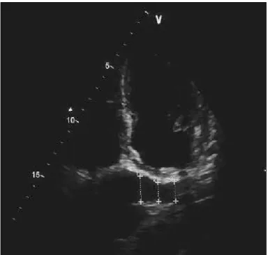
(LV) ejection fraction was estimated from the apical four- and two-chamber views using the Simpson’s biplane method. The LV diameters were measured from the parasternal long-axis view. Left atrium diameter was measured from the apical 4-chamber view at the end of ventricular systole from the free wall of the left atrium to interatrial septum. Pulsed-wave Doppler of the mitral or tricuspid inflow and pulsed-wave tissue Doppler of the mitral or tricuspid annulus were obtained from the apical 4-chamber view with a 5-mm sample volume. Mitral or tricuspid inflow E-waves and early myocardial relaxation velocities (Ea) were then used to calculate the E/Ea ratio, which was used as an indicator of left and right ventricle filling pressure. The sys-tolic pulmonary artery pressure (PAP) was derived from the tri-cuspid regurgitant jet velocity with the modified Bernoulli equa-tion. The RV area was determined by tracing the RV endocardi-um at diastole from the annulus to the apex along the free wall and then back to the annulus along the interventricular septum. The right atrial (RA) area was traced at the end of ventricular systole from the lateral side of the tricuspid annulus to the septal side, discounting the area between the leaflets and annulus, fol-lowing the RA endocardium, excluding the inferior and superior vena cava and RA appendage. The RV myocardial performance index (MPI) was obtained using tissue Doppler echocardiogra-phy (TDE). TDE was performed in the apical four-chamber view using a 5- to 10-mm sample volume placed on the tricuspid annulus lateral wall. The isovolumetric relaxation time (IRT) was measured from the end of the systolic velocity to the start of the early diastolic velocity (Ea). The isovolumetric contraction time (ICT) was measured from the end of the late diastolic velocity to the start of the systolic velocity. Ejection time (ET) was lated as the duration of the systolic velocity. RV MPI was calcu-lated using the equation (ICT+IRT)/ET. The intraobserver and interobserver variability was 3.6% and 4.0%, respectively. The reported upper reference limit of the RV MPI is 0.55 by TDE in healthy subjects (6). HF patients with an RV MPI >0.55 were defined as having impaired RV myocardial function. The CS diameter was measured from the posterior atrioventricular groove in an apical four-chamber echocardiographic view during ventricular systole, because the maximum diameter of the CS occurs during ventricular systole. CS diameter measurements were obtained from the following three areas: at the termination of the CS orifice (prox), 1 cm from the left side (mid), and between the orifice and origin on the left side (dist). The mean CS diameter was calculated as follows: mean CS=(prox CS+mid CS+dist CS)/3 (Fig. 1) (2). The intraobserver and interobserver variability was 3.2% and 4.1%, respectively.
Statistical analysis
Statistical analysis was performed with SPSS 21.0 for Windows (SPSS Inc.). Continuous data were presented as mean±SD, and categorical data were expressed as numbers and percentage. The Kolmogorov-Smirnov test was used to evaluate whether the distribution of continuous variables was normal. One-way analysis of variance (ANOVA) with posthoc Tukey test
or Kruskal-Wallis with posthoc Sidak test was used to compare the three groups. Categorical variables were summarized as percentages and compared with the chi-square test. Correlation analysis was performed using Spearman or Pearson test. Multivariate linear regression analysis was performed in order to determine the predictive factors for CS from variables show-ing significance values <0.1 in the correlation analysis. In con-sideration of the wide range and of the non-skewed distribution, left ventricular EF was log-transformed to reduce the effects of extreme values and to obtain a normal distribution for statistical tests. The accuracy of CS value to predict HF patients with RV MPI >0.55 mm was assessed by receiver operating characteris-tics curve (ROC) analysis. A p value <0.05 was considered statis-tically significant.
Results
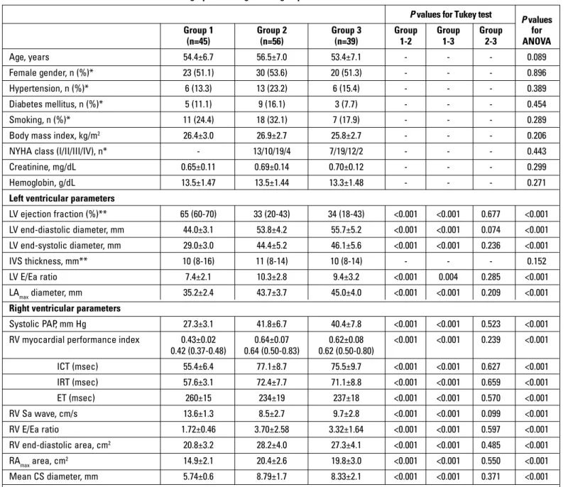
The clinical and echocardiographic characteristics of the three groups are presented in Table 1. There was no significant difference between the groups (Groups 1, 2, and 3) in terms of age, gender, diabetes mellitus, hypertension, body mass index, and smoking (p>0.05). As shown in Table 1, all of the patients were stable in terms of their chronic HF status, with 13 (23.2%) ischemic HF patients in NYHA class I, 10 (17.8%) in class II, 19 (33.9%) in class III, and 4 (7.1%) in class IV and with 7 (17.9%) idiopathic DCM patients in NYHA class I, 19 (48.7%) in class II, 12 (30.7%) in class III, and 2 (5.1%) in class IV. Furthermore, the echocardiographic characteristics were similar between patients with ischemic DCM (Group 2) and with idiopathic DCM (Group 3). The diameter of CS was significantly greater in both Group 2 and Group 3 than in Group 1 (8.79±1.7 mm and 8.33±2.1 mm vs. 5.74±0.6 mm; p <0.001 for both). The MPI measured in the
RV using TDE is presented in Table 1. The RV MPI was signifi-cantly higher in both Group 2 and Group 3 than in the control group (0.64±0.07 and 0.62±0.08 vs. 0.43±0.02; p<0.001 for both). Therefore, the RA area was significantly greater in both Group 2 and Group 3 than in the control group (20.4±2.6 cm2 and 19.8±3.0
cm2 vs. 14.9±2.1 cm2; p<0.001 for both). The echocardiographic
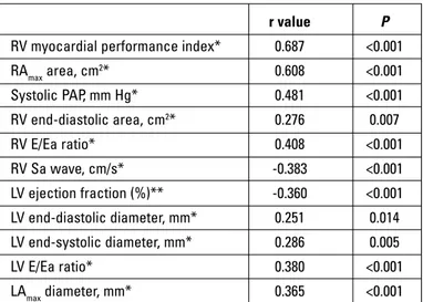
variables that were significantly correlated with the diameter of the CS are presented in Table 2. A strong correlation was found between the mean CS diameters and RV MPI, RA area, and systolic PAP in patients with HF (r=0.687, r=608, and r=0.481; p for each variable <0.001). Those variables showing significant cor-relations according to Table 2 were included in a multivariate linear regression analysis. According to our results, RV MPI (β=0.550, 95% CI: 6.623-15.506, p<0.001), RA area (β=0.232, 95% CI: 0.016-0.194, p=0.021), RV E/Ea ratio (β=0.130, 95% CI: 0.013-0.255, p=0.030), and LV E/Ea ratio (β=0.138, 95% CI: 0.018-0.174, p=0.017) were independent predictors of the CS diameter in patients with HF (Table 3). Patients with HF (n=95) were catego-rized into two groups with respect to the top and bottom 0.55 of their RV MPI. The group with RV MPI >0.55 mm (n=72) was defined to be HF patients with impaired RV myocardial func-tions. For the prediction of HF patients with RV myocardial dys-function, the cut-off value for the diameter of the CS was 7.35 mm, with a sensitivity of 83% and a specificity of 79% (AUC=0.839, 95% CI=0.731-0.946, p<0.001) (Fig. 2).
Discussion
In the current study, we found that the CS diameter was significantly increased in patients with both ischemic and idio-pathic DCM. Additionally, a dilated CS was strongly associated with RV echocardiographic characteristics, including the RV MPI, RA area, systolic PAP, and RV E/Ea ratio. There was a mod-erate correlation between CS diameter and echocardiographic characteristics of the left ventricle, including the end-systolic and-diastolic diameters, left atrial diameters, and EF value. Furthermore, RV MPI, RA area, RV E/Ea ratio, and LV E/Ea ratio were identified as independent predictors of CS diameter in patients with HF.
The CS is a tubular structure that transmits venous blood to the right atrium. The CS contracts simultaneously with the atria, because its wall contains atrial myocardium (8). Thus, the diam-eter of the CS changes at different phases of the cardiac cycle, peaking at the end of ventricular systole and becoming smallest during atrial contraction (9). For this reason, in our study, the diameter of the CS was measured at the end of ventricular sys-tole. A dilated CS can result from increased blood flow due to abnormal venous drainage in the left superior vena cava, total anomalous intra-cardiac pulmonary venous drainage, severe tricuspid regurgitation, CS diverticulum, or a coronary artery to CS fistula. The absence of primary abnormalities is generally a manifestation of high RA pressure due to functional tricuspid regurgitation (10-12). Koberstein et al. (13) showed that CS dila-tation with RA blood reflux to the CS was due to increased RA
Figure 1. Echocardiographic measurements of coronary sinus are shown
pressure. They also found that 15% of the blood sampled from the CS originated from the RA in dogs with elevated RA pressure caused by partial obstruction of the pulmonary artery. In com-parison, Mahmud et al. (14) reported that the size of the CS was significantly correlated with the size of the right atrium (r=0.60, p<0.001) and RA pressure (r=0.59, p<0.001) but not with the size of the right ventricle in patients who underwent right heart cath-eterization for the evaluation of pulmonary hypertension. In addition, a dilated right atrium is a sign of increased RA pres-sure. Machraouni et al. (15) reported a strong correlation
between RA pressure and the RA area index (r=0.64). However, this is a qualitative measure that does not allow the interpreter to assess RA pressure (6). Also, in our study, CS diameter was positively correlated with the RA area, and therefore, the RA area was found as an independent predictor of CS dilatation.
RV systolic and diastolic dysfunction is common in patients with HF and is associated with a poor long-term prognosis (16-19). The MPI, which is also known as the Tei index, has been described as a non-invasive measurement of ventricular func-tion. The TDE-derived MPI reflects both the systolic and
dia-P values for Tukey test P values Group 1 Group 2 Group 3 Group Group Group for
(n=45) (n=56) (n=39) 1-2 1-3 2-3 ANOVA Age, years 54.4±6.7 56.5±7.0 53.4±7.1 - - - 0.089 Female gender, n (%)* 23 (51.1) 30 (53.6) 20 (51.3) - - - 0.896 Hypertension, n (%)* 6 (13.3) 13 (23.2) 6 (15.4) - - - 0.389 Diabetes mellitus, n (%)* 5 (11.1) 9 (16.1) 3 (7.7) - - - 0.454 Smoking, n (%)* 11 (24.4) 18 (32.1) 7 (17.9) - - - 0.289
Body mass index, kg/m2 26.4±3.0 26.9±2.7 25.8±2.7 - - - 0.206
NYHA class (I/II/III/IV), n* - 13/10/19/4 7/19/12/2 - - - 0.443
Creatinine, mg/dL 0.65±0.11 0.69±0.14 0.70±0.12 - - - 0.299
Hemoglobin, g/dL 13.5±1.47 13.5±1.44 13.3±1.48 - - - 0.271
Left ventricular parameters
LV ejection fraction (%)** 65 (60-70) 33 (20-43) 34 (18-43) <0.001 <0.001 0.677 <0.001 LV end-diastolic diameter, mm 44.0±3.1 53.8±4.2 55.7±5.2 <0.001 <0.001 0.074 <0.001 LV end-systolic diameter, mm 29.0±3.0 44.4±5.2 46.1±5.6 <0.001 <0.001 0.236 <0.001 IVS thickness, mm** 10 (8-16) 11 (8-14) 10 (8-14) - - - 0.152 LV E/Ea ratio 7.4±2.1 10.3±2.8 9.4±3.2 <0.001 0.004 0.285 <0.001 LAmax diameter, mm 35.2±2.4 43.7±3.7 45.0±4.0 <0.001 <0.001 0.209 <0.001 Right ventricular parameters
Systolic PAP, mm Hg 27.3±3.1 41.8±6.7 40.4±7.8 <0.001 <0.001 0.523 <0.001 RV myocardial performance index 0.43±0.02 0.64±0.07 0.62±0.08 <0.001 <0.001 0.239 <0.001
0.42 (0.37-0.48) 0.64 (0.50-0.83) 0.62 (0.50-0.80) ICT (msec) 55.4±6.4 77.1±8.7 75.5±9.7 <0.001 <0.001 0.627 <0.001 IRT (msec) 57.6±3.1 72.4±7.7 71.1±8.8 <0.001 <0.001 0.659 <0.001 ET (msec) 260±15 234±19 237±18 <0.001 <0.001 0.570 <0.001 RV Sa wave, cm/s 13.6±1.3 8.5±2.7 9.7±2.8 <0.001 <0.001 0.099 <0.001 RV E/Ea ratio 1.72±0.46 3.70±2.58 3.32±1.64 <0.001 <0.001 0.597 <0.001 RV end-diastolic area, cm2 20.8±3.2 28.2±4.0 27.3±4.1 <0.001 <0.001 0.485 <0.001 RAmax area, cm2 14.9±2.1 20.4±2.6 19.8±3.0 <0.001 <0.001 0.550 <0.001 Mean CS diameter, mm 5.74±0.6 8.79±1.7 8.33±2.1 <0.001 <0.001 0.371 <0.001
Continuous variables were expressed as mean±standard deviation or median (min-max), and categorical variables were expressed as number of cases and percentage. One-way analysis of variance (ANOVA) with posthoc Tukey test was used for comparison
*The chi-square test was used to analyze categorical variables. **The Kruskal-Wallis test with posthoc Sidak test was used for the analysis of nonparametric variables Group 1- Control, Group 2-Patients with ischemic cardiomyopathy, Group 3- Patients with idiopathic dilated cardiomyopathy.
CS - coronary sinus; ET - ejection time; ICT - isovolumetric contraction time; IRT - isovolumetric relaxation time; IVS - interventricular septum; LA - left atrium; LV - left ventricle; LV E/ Ea ratio - mitral Doppler inflow E wave velocity-to-annular tissue Doppler Ea wave velocity ratio; NYHA - New York Heart Association; PAP - pulmonary artery pressure; RA - right atrium; RV - right ventricle; RV E/Ea ratio - tricuspid Doppler inflow velocity-to-annular tissue Doppler Ea wave velocity ratio; Sa - annular tissue Doppler systolic velocity
stolic function of the ventricles, defined as the ratio of the iso-volumic time divided by the ET, or [(IRT + ICT)/ET] (20). The RV MPI represents subclinical RV dysfunction and volume overload in patients with congenital heart disease (21) and is increased in patients with primary pulmonary hypertension, RV infarction, and hypertrophic cardiomyopathy (22-24). In accordance with these findings, in our study, we found that the RV MPI was sig-nificantly increased in patients with idiopathic and ischemic DCM compared with normal controls. Furthermore, there was a positive and significant correlation between RV MPI and CS diameter in these patients. Similarly, Vatankulu et al. (25) report-ed that the size of the CS was significantly associatreport-ed with increased RV MPI in patients with mitral stenosis.
Cardiac remodeling is the result of hemodynamic changes in patients with LV systolic dysfunction. The left atrial pressure and diameter increase due to pressure and volume overload in the left ventricle in these patients. An elevated left atrial pressure leads to passive elevation in the pulmonary arterial pressure, which leads to impaired functioning of the right side of the heart through overloading of the right ventricular and atrial pressures (5, 26, 27). Additionally, RV function impairs secondary to myo-cardial infarction or ischemia due to a (proximal) right and/or left anterior descending coronary artery lesion in HF patients (6). In our study, CS diameter was found to be negatively correlated with LV ejection fraction and positively correlated with LV E/Ea ratio, LA diameter, PAP, RV MPI, RV E/Ea ratio, and RA area. However, there were relatively more powerful correlations between the CS diameter and the parameters of RV rather than those of LV. Furthermore, the RV MPI, RA area, and RV E/Ea ratio with LV E/Ea ratio were found as independent predictors of CS
dilatation. In light of these data, we conclude that the diameter of the CS may increase owing to increased RA pressure due to impaired RV function, depending on the mechanisms mentioned above in patients with ischemic and idiopathic DCM.
Study limitations
The first limitation is the relatively small number of patients; subjects were selected from patients with chronic compensated LVSD. Second, invasive measurements of RV parameters were not performed together with RV MPI measurements; however, most of our patients had higher systolic PAP. Third, contrast injection was not used for measurement of the coronary sinus. Fourth, the cross-sectional design limited our ability to deter-mine the significance of changes in the diameter of the CS for the long-term prognosis and mortality of patients with LVSD.
Conclusion
Our results confirm our hypothesis that the diameter of the CS increases, owing to increased RA pressure due to impaired RV functions, and it can be used as a novel echocardiographic
r value P RV myocardial performance index* 0.687 <0.001 RAmax area, cm2* 0.608 <0.001 Systolic PAP, mm Hg* 0.481 <0.001 RV end-diastolic area, cm2* 0.276 0.007 RV E/Ea ratio* 0.408 <0.001 RV Sa wave, cm/s* -0.383 <0.001 LV ejection fraction (%)** -0.360 <0.001 LV end-diastolic diameter, mm* 0.251 0.014 LV end-systolic diameter, mm* 0.286 0.005 LV E/Ea ratio* 0.380 <0.001 LAmax diameter, mm* 0.365 <0.001
*Pearson’s test, **Spearman test. LA - left atrium; LV - left ventricle; LV E/Ea ratio - mitral Doppler inflow E wave velocity-to-annular tissue Doppler Ea wave velocity ratio; PAP - pulmonary artery pressure; RA - right atrium; RV - right ventricle; RV E/Ea ratio - tricuspid Doppler inflow velocity-to-annular tissue Doppler Ea wave velocity ratio
Table 2. The association of coronary sinus diameter with echocardiographic parameters in patients with heart failure
β t 95% confidence interval P RV MPI 0.550 4.929 6.623-15.506 <0.001 RA area 0.232 2.340 0.016-0.194 0.021 RV E/Ea ratio 0.130 2.190 0.013-0.255 0.030 LV E/Ea ratio 0.138 2.423 0.018-0.174 0.017
*Multivariate linear regression analysis.
LV E/Ea ratio - mitral Doppler inflow E wave velocity-to-annular tissue Doppler Ea wave velocity ratio, MPI - right ventricular myocardial performance index; RA - right atrium; RV E/Ea ratio - tricuspid Doppler inflow velocity-to-annular tissue Doppler Ea wave velocity ratio
Table 3. Independent predictors of coronary sinus diameter Figure 2. The cut-off value of the coronary sinus diameter for prediction
of heart failure patients with impaired right ventricular myocardial function was 7.35 mm, with a sensitivity of 83% and a specificity of 79% in the ROC curve analysis AUC=0.839, 95% CI=0.731-0.946, p<0.001).
AUC - area under curve; ROC - receiver operating characteristics curve analysis
AUC: 0.839 CI: 0.731-0.946 p<0.001 1.0 0.8 0.6 0.4 0.2 0.0 Sensitivity 1-Specificity 0.0 0.2 0.4 0.6 0.8 1.0
marker to provide information about impaired RV function in patients with ischemic and idiopathic DCM. However, further study will be necessary to confirm these preliminary findings.
Conflict of interest: None declared. Peer-review: Externally peer-reviewed.
Authorship contributions: Concept - M.Ç., A.D., M.O.; Design - M.Ç.; Supervision - E.A., A.S.; Resource - A.S., M.P., F.Ü.; Data collection and/ or processing - M.Ç., A.S., E.A., M.P.; Analysis and/or Interpretation - M.Ç., E.A., F.Ü., M.Çetin.; Literature search - S.A., A.D., M.O.; Writing - M.Ç.; Critical review - M.Ç., E.A.
References
1. Potkin BN, Roberts WC. Size of coronary sinus at necropsy in sub-jects without cardiac disease and in patients with various cardiac conditions. Am J Cardiol 1987; 60: 1418-20. [CrossRef]
2. Isaacs D, Hazany S, Gamst A, Stark P, Mahmud E. Evaluation of the coronary sinus on chest computed tomography in patients with and without pulmonary artery hypertension. J Comput Assist Tomogr 2009; 33: 513-6. [CrossRef]
3. Kızılkan N, Davutoğlu V, Erbağcı H, Karagöz A, Akçay M, Sarı I, et al. Coronary sinus dilatation. A simple additional echocardiograph-ic indechocardiograph-icator of severe rheumatechocardiograph-ic mitral and trechocardiograph-icuspid valve disease. Saudi Med J 2010; 31: 153-7.
4. Yüce M, Davutoğlu V, Yavuz S, Sarı I, Kızılkan N, Ercan S, et al. Coronary sinus dilatation is associated with left ventricular systolic dysfunction and poor functional status in subjects with chronic heart failure. Int J Cardiovasc Imaging 2010; 26: 541-5. [CrossRef] 5. Drazner MH, Velez-Martinez M, Ayers CR, Reimold SC, Thibodeau
JT, Mishkin JD, et al. Relationship of right- to left-sided ventricular filling pressures in advanced heart failure: insights from the ESCAPE trial. Circ Heart Fail 2013; 6: 264-70. [CrossRef]
6. Rudski LG, Lai WW, Afilalo J, Hua L, Handschumacher MD, Chandrasekaran K, et al. Guidelines for the echocardiographic assessment of the right heart in adults: a report from the American Society of Echocardiography endorsed by the European Association of Echocardiography, a registered branch of the European Society of Cardiology, and the Canadian Society of Echocardiography. J Am Soc Echocardiogr 2010; 23: 685-713. [CrossRef]
7. Pearlman AS, Gardin JM, Martin RP, Parisi AF, Popp RL, Quinones MA, et al. Guidelines for optimal physician training in echocardiog-raphy recommendations of the American Society of Echocardiography Committee for physician training in Echocardiography. Am J Cardiol 1987; 60: 158-63. [CrossRef] 8. Coakley JB, King TS. Cardiac muscle relations of the coronary
sinus, the oblique vein of the left atrium and the left precaval vein in mammals. J Anat 1959; 93: 30-5.
9. D’Cruz IA, Johns C, Shala M. Dynamic cyclic changes in coronary sinus caliber in patients with and without congestive heart failure. Am J Cardiol 1999; 83: 275-7. [CrossRef]
10. Paul W, Beisel B, Krug A, Vagt A, Timpe A, Brase R, et al. Dilated coronary sinus. A preoperative transesophageal echocardiograph-ic diagnosis. Anaesthesist 2000; 49: 25-8. [CrossRef]
11. Zamorano J, Almería C, Alfonso F, Angeles Perez M, Grauper C, et al. Transesophageal Doppler analysis of coronary sinus flow a new
method to assess the severity of tricuspid regurgitation. Echocardiography 1997; 14: 579-88. [CrossRef]
12. Andrade JL, Somerville J, Carvalho AC, Campos O Jr, Mitre N, Martinez EE Jr, et al. Echocardiographic routine analysis of the coronary sinus by an apical view: Normal and abnormal features. Texas Heart Institute J 1986; 13: 197-202.
13. Koberstein RC, Pittman DE, Klocke FJ. Right atrial admixture in coronary venous blood. Am J Physiol 1969; 216: 531.
14. Mahmud E, Raisinghani A, Keramati S, Auger W, Blanchard DG, DeMaria AN. Dilation of the coronary sinus on echocardiogram: prevalence and significance in patients with chronic pulmonary hypertension. J Am Soc Echocardiogr 2001; 14: 44-9. [CrossRef] 15. Machraoui A, von Dryander S, Hinrichsen M, Jäger D, Lemke B,
Ulmer WT, et al. Two-dimensional echocardiographic assessment of right cardiac pressure overload in patients with chronic obstruc-tive airway disease. Respiration 1993; 60: 65-73. [CrossRef] 16. Ghio S, Gavazzi A, Campana C, Inserra C, Klersy C, Sebastiani R, et
al. Independent and additive prognostic value of right ventricular systolic function and pulmonary artery pressure in patients with chronic heart failure. J Am Coll Cardiol 2001; 37: 183-8. [CrossRef] 17. Alp H, Karaaslan S, Baysal T, Oran B, Örs R. Comparison of left and
right ventricular pulsed and tissue Doppler myocardial perfor-mance index values using Z-score in newborns with hypoxic-ischemic encephalopathy. Anadolu Kardiyol Derg 2011; 11: 719-25. 18. Demirkol S, Ünlü M, Arslan Z, Baysan O, Balta S, Kurt IH.
Assessment of right ventricular systolic function with dP/dt in healthy subjects: an observational study. Anadolu Kardiyol Derg 2013; 13: 103-7.
19. Şimşek E, Şimşek Z, Taş MH, Kuçur C, Günay E, Üçüncü H. Evaluation of right ventricular functions in patients with nasal polyposis: an observational study. Anadolu Kardiyol Derg 2013; 13: 251-6.
20. Özkan G, Adar A, Ulusoy S, Bektaş H, Kırış A, Fidan M, et al. Presence of fragmented QRS and its correlation with myocardial performance index in patients with nephrotic syndrome. Anadolu Kardiyol Derg 2014 in press.
21. Eidem BW, O’Leary PW, Tei C, Seward JB. Usefulness of the myo-cardial performance index for assessing right ventricular function in congenital heart disease. Am J Cardiol 2000; 86: 654-8. [CrossRef] 22. Møller JE, Søndergaard E, Poulsen SH, Appleton CP, Egstrup K.
Serial Doppler echocardiographic assessment of left and right ventricular performance after a first myocardial infarction. J Am Soc Echocardiogr 2001; 14: 249-55. [CrossRef]
23. Mörner S, Lindqvist P, Waldenström A, Kazzam E. Right ventricular dys-function in hypertrophic cardiomyopathy as evidenced by the myocar-dial performance index. Int J Cardiol 2008; 124: 57-63. [CrossRef] 24. Işılak Z, Yalçın M, İncedayı M, Çay S. Persistent left superior vena
cava associated with giant coronary sinus. Anadolu Kardiyol Derg 2012; 12: E39.
25. Vatankulu MA, Koç F, Gül EE, Bacaksız A, Sönmez O, Demir K, et al. The relationship between coronary sinus and impaired right ven-tricular myocardial performance index in mitral stenosis. Echocardiography 2013; 30: 936-9. [CrossRef]
26. Davutoğlu V, Yıldırım C, Küçükaslan H, Yüce M, Sarı I, Tarakcıoğlu M, et al. Prognostic value of pleural effusion, CA-125 and NT-proBNP in patients with acute decompensated heart failure. Kardiol Pol 2010; 68: 771-8.
27. Kavarana MN, Pessin-Minsley MS, Urtecho J, Catanese KA, Flannery M, Öz MC, et al. Right ventricular dysfunction and organ failure in left ventricular assist device recipients: a continuing problem. Ann Thorac Surg 2002; 73: 745-50. [CrossRef]