Igor D. Alexandrov
1,2*
, Margarita V.
Alexandrova
1,2, Svetlana V.
Korablinova
1and Larisa N. Korovina
1 1Joint Institute for Nuclear Research, Dubna 141980,Moscow Region-Russia
2Vavilov Institute of General Genetics, Russian Academy of Sciences, 119991, Moscow-Russia
Abstract
At present, given a bulk of models for higher-order chromatin structure in interphase nuclei of animal somatic cells, little is known about the spatial chromosome organization in animal male germ cells mainly due to the lack suitable methods for detailed observation and analysis without disruption of the existing organization. We pioneered in the study of this issue via analysis of radiation-induced inversion patterns in Drosophila male germ cell genome taking into account the fact that the formation of inversion requires the spatial proximity and contact of its ends. Analysis of 72 γγ-ray- or neutron-induced vg inversions in which the first break is invariantly associated with the vestigial (vg) gene in the middle of 2R autosome shows the second inversion breakpoints that highly non-randomly distributed over the entire second chromosome clustering at the three autosome 2 “hot” chromosome areas. These findings show that the polar Rabl-configuration of interphase chromosomes in various somatic cells is not typical for the haploid genome of Drosophila
mature sperms. The specific megarosette-loopstructure of haploid chromosome in the mature animal sperms is proposed and justified. Key words: Radiation, inversions, chromosome
folding arrangement, sperm genome, Drosophila.
Introduction
Shortly after the second meiotic division of the secondary spermatocyte during spermatogenesis in animal kingdom, the telophase chromosomes decondense and diffuse to form the haploid “interphase” nucleus of early spermatid where important morphogenetic and synthetic processes of spermiogenesis get under way. Among these processes, organization of the cytoplasmic organelles into ordered complexes of normal architecture accompanied by realignment of spermatid genome from the loose chromatin fibers to closely packed paracrystal chromatin structures (Demerec, 1950; Meyer, 1969) and transition from a typical somatic or lysine-rich (Tokuyasu et al., 1972) chromosomal histone to a highly arginine-rich type late in sperm maturation (Das et al., 1964) appear to be a key events in animal spermiogenesis (Kimmins and Sassone-Corsi, 2005) resulting in the specific sperm nucleus evolutionarily adapted for insulation of the genetic information during the period of its transit from one generation to the next.
The complexity of these processes rise many questions about the large-scale organization of “interphase” chromatin in developing spermatids and a spatial arrangement of haploid chromosomes in the motile mature sperm as well.
The experimental data that could provide the answers to these questions are entirely absent so far. Conversely, a bulk of models for spatial organization of chromosomes in interphase nuclei of plant and animal somatic cells have been considered in related literature, beginning with the most early “polar” (telomere and centromere are located at opposite
Spatial arrangement of the animal male germ cell genome:
I. Non-random pattern of radiation-induced inversions
involving the vestigial region in autosome 2 of Drosophila
melanogaster
*Correspondence author:
Joint Institute for Nuclear Research, Dubna 141980, Moscow Region-Russia
E-mail: [email protected]
nuclear poles) model classically defined as “Rabl’s configuration” (Rabl, 1885) and, later really described in a variety of cell nuclei (Comings, 1980; Saumweber, 1987; Hiraoka et al., 1990), and ending with diverse current chromatin loop models which variously involve irregularly folded fibers or moderate / “giant” loop domains (Manuelidis, 1990; Sachs et al., 1995; Cremer and Cremer, 2001).
It is not clear however, what kind of these models may be best matched to genome architecture in developing spermatids and mature male germ cells, particularly, taking into account the specific function brought about by the latter. Furthermore, the current models have given an insight into the conformation of isolated chromosomal domains rather than of an entire chromosome. Therefore, to get a notion on the issue of interest, it is essential, as a first step, to clarify whether so-called Rabl orientation of chromosomes still persists in sperm genome after the second meiotic division and spermatid differentiation.
In order for this elucidate, it is necessary to use the experimental approaches other than a classical cytology or current high-resolution in situ hybridization techniques due to impossibility to examine the sperm chromatin under the microscope.
In this connection, the investigation of the pattern of radiation-induced intrachromosomal exchanges (inversions) may be considerable promise as a means for clarification of the global arrangement of one or another chromosome in sperm nucleus. The following three known fundamental facts form the basis for such promissory approach:
i. The direct, local, and stochastic action of ionizing radiation on the genetic matter resulting in the double-strand breaks of DNA, chromosome breaks and rearrangements subsequently (Ozalpan, 2001).
ii. Chromosome breaks induced by radiation in mature sperm stay open until fertilization (Maddern and Leigh, 1974) and the formation of chromosome exchanges is confined by the short time before the first chromosome replication in male pro-nucleus after fertilization and the occurrence of zygote (Würgler, 1971).
iii. The formation of any one of chromosome exchanges (translocation, inversion, transposition etc.) requires that the two interacting chromosome regions were spatially close to each other enough.
Taking into account these facts, pattern (the size, position, and frequency) of radiation-induced inversions at the large metacentric autosome 2 of
Drosophila sperm genome was studied. Hereat, the
genetic experiments were designed so that to isolate not all of possible randomly arising intrachromosomal exchanges but only those that have had one of inversion breaks (so-called the “first break”) invariably associated with one or another selected genetic loci of the autosome. Then, location of the second inversion breaks should indicate which chromosome regions, and as often in different nuclei, are spatially close to the genetic loci selected showing thereby the loops of appropriate sizes. Therefore, enough large sets of locus-specific inversions can give an insight into the global loop arrangement and topographic parameters of autosome under study in haploid sperm nucleus.
Using these new feasible, and, in our opinion, considerable promise approaches, a series of inversions associated with the three genetic loci of different location on the autosome 2, namely, black body (b), cinnabar eyes (cn), and vestigial wings (vg) loci, were obtained after irradiation of Drosophila adult male sperms by gamma-rays or fission neutrons. The present paper focuses on the pattern of the vg inversions in respect of which the most extensive data were collected in spite of a fairly low frequency of the vg inversion induction (2.0x10⁻⁸per locus per rad and
7.5x10⁻⁸ per locus per rad for γ-rays and neutrons,
accordingly) (Alexandrov et al., 2001). The results for the 72 vg inversions analyzed show that
i. The vg locus lying at the middle of 2R arm of this autosome (subsection 49E) interacts the most frequently (20/72 or the same arm of autosome 2 (section 41) forming the loop of 9 sections or about ten million pairs of DNA bases (10 Mb); ii. In other 35 cases, the vg locus alternatively
interacts equally frequently (24-25%) either with neighbouring proximal/distal chromosome regions (sections 48/50-51, respectively) forming the small loops of 0.3-3 Mb DNA or with the highly separated distal ends of 2L as well as 2R arms of autosome (sections 21-25 and 56-60, respectively) giving rise to the “giant” loops of about 30 and 12 Mb DNA, accordingly;
iii. The rest of inversions (17 cases) show that the vg locus can rarely interact with some internal areas of the 2L or 2R chromosome arms resulting in the large loops of various sizes in the separate sperm nuclei.
An often contacts and interactions of the middle 2R arm (vg region) with its centromeric and telomeric ends suggest that the global arrangement of chromosome in sperm nucleus is not in line with the Rabl configuration but rather matches to the specific giant rosette-loop structure the qualitative features of which are discussed. 2D and 3D models of this structure will be dealt in the next paper.
Material and methods
Random samples of 43 γ-rays- and 29 neutron-induced vg inversions were obtained over large-scale
experiments as a part of the extensive project on the RBE- neutron energy relationship under the locus-specific mutation induction in Drosophila. The biological as well as physical details of experiments such as Drosophila stocks and germ cells irradiated, the sources of radiation (γ-rays of 60Co; fission
neutrons), doses, and regimes of irradiation were described earlier (Alexandrov, 1984). The main results of the fine genetical and cytogenetical analysis of all γ-rays and neutron-induced vg inversions being essential for the answer to the issue of interest were published elsewhere too (Alexandrov and Alexandrova, 1987; Alexandrov et al., 2004). Here, it is
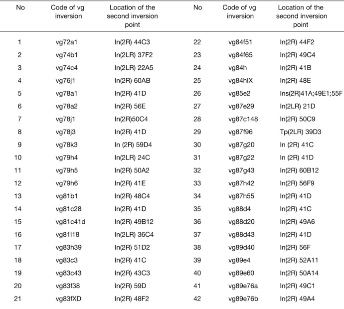
No Code of vg Location of the No Code of vg Location of the
inversion second inversion inversion second inversion
point point
1 vg72a1 In(2R) 44C3 22 vg84f51 In(2R) 44F2
2 vg74b1 In(2LR) 37F2 23 vg84f65 In(2R) 49C4
3 vg74c4 In(2LR) 22A5 24 vg84h In(2R) 41B
4 vg76j1 In(2R) 60AB 25 vg84hIX In(2R) 48E
5 vg78a1 In(2R) 41D 26 vg85e2 Ins(2R)41A;49E1;55F
6 vg78a2 In(2R) 56E 27 vg87e29 In(2LR) 21D
7 vg78j1 In(2R)50C4 28 vg87c148 In(2R) 50C9
8 vg78j3 In(2R) 41D 29 vg87f96 Tp(2LR) 39D3
9 vg78k3 In (2R) 59D4 30 vg87g20 In (2R) 41C
10 vg79h4 In(2LR) 24C 31 vg87g22 In (2R) 41D
11 vg79h5 In(2R) 50A2 32 vg87g43 In(2R) 60B12
12 vg79h6 In(2R) 41E 33 vg87h42 In(2R) 56F9
13 vg81b1 In(2R) 48C4 34 vg87h55 In(2R) 41D
14 vg81c28 In(2R) 41D 35 vg88d4 In(2R) 41C
15 vg81c41d In(2R) 49B12 36 vg88d20 In(2R) 49A6
16 vg81l18 In(2LR) 36C4 37 vg88d43 In(2R) 41D
17 vg83h39 In(2R) 51D2 38 vg89d40 In(2R) 56F
18 vg83c3 In(2R) 41C 39 vg89e4 In(2R) 52A11
19 vg83c43 In(2R) 43C3 40 vg89e60 In(2R) 50A14
20 vg83f38 In(2R) 59D 41 vg89e76a In(2R) 49C1
21 vg83fXD In(2R) 48F2 42 vg89e76b In(2R) 49A4
Table 1. The location of the second inversion breakpoints on the polytene autosome 2 of D. melanogaster for γ-ray-induced vg
worth noting that the molecular position of the “first” inversion breakpoints inside or outside (but in the immediate vicinity) of the vg gene (subsections 49D4-E1 on the polytene 2R arm of autosome 2) was determined by the in situ hybridization technique using
3H-labeled proximal (OR8) and distal (OR2) genomic
fragments overlapping the gene under study (Alexandrova et al., 1997). The precise chromosome location of the second inversion breakpoints was performed by the standard cytological technique for
Drosophila polytene chromosomes in the mutant
chromosome/wild-type chromosome heterozygotes and estimated accurately within subsection of the polytene autosome 2 guided by its classical map under the light microscopy (Lefevre, 1976).
Results and discussion
As the first step of analysis of the pattern of vg inversions, all of them were classified as γ-ray- and neutron-induced ones (Table 1 and 2, respectively) with indication of chromosome location of their second inversion breaks whereas the “first” breakpoints are always intimately associated with the vg gene (subsection 49D4-E1 of 2R arm of autosome 2) resulting in the appropriate vg phenotypes in mutant homozygotes.
A comparative consideration of these data shows that both γ-rays and neutrons induce basically the same “simple” double breaks (on the chromosome level) inversions with the closely similar seats of the second breaks on 2L or 2R arm of the chromosome. However, thereat it should be pointed out that, in the same irradiated nucleus, more complex chromosome exchanges of two inversions with one break in common (at the vg region) can occur after action of both radiations but neutrons appear to be more effective than γ-rays in induction of such rearrangements (vg inversions No 15, 16, 22, and 23 for neutrons, Table 2 and No 26, Table 1 for γ-rays, respectively). As a result, the overall numbers of exchanges isolated and analyzed add up to 43 inversions for γ-rays and 29 ones for neutrons. Which is their second inversion breakpoints distributed along the entire autosome?
As indicated in the tables and as mentioned above already, the seats of γ-ray-induced second inversion breakpoints coincide very closely with that of neutron-induced ones. This enables us to integrate the findings for both radiations within a single set of data to detect the “hot” chromosome areas with which the vg region is most often brought into spatial proximity in sperm
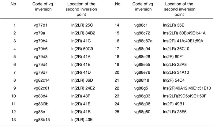
No Code of vg Location of the No Code of vg Location of the
inversion second inversion inversion second inversion
point point
1 vg77d1 In(2LR) 25C 14 vg88c1 In(2LR) 36E
2 vg79a In(2LR) 34B2 15 vg88c72 Ins(2LR) 30B;49E1;41A
3 vg79b4 In(2R) 41C 16 vg88c87a Ins(2R) 41A;49E1;59A
4 vg79b6 In(2R) 50C9 17 vg88c94 In(2LR) 36C10
5 vg79d3 In(2R) 41A 18 vg88e28 In(2R) 60F1
6 vg79d4 In(2R) 41E 19 vg88e55 In(2LR) 22A8
7 vg79d7 In(2R) 41D 20 vg88e76 In(2LR) 34A10
8 vg82c14 In(2LR) 36D 21 vg88f18 In(2R) 54C4
9 vg82c61 In(2LR) 24E2 22 vg88g5 Ins(2R)49A12;49E1;51E10
10 vg83d4 In(2R) 48F 23 vg88g33 Ins(2LR)39D5;49E1;59F
11 vg83l3b In(2R) 41E 24 vg88g38 In(2R) 49B1
12 vg85c In(2R) 41B 25 vg88g80 In(2LR) 25E6
13 vg88b15 In(2LR) 40E
Table 2. The location of the second inversion breakpoints on the polytene autosome 2 of D. melanogaster for neutron-induced vg
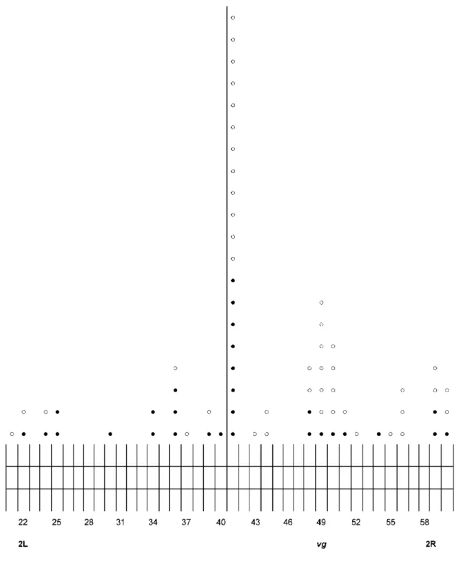
Figure 1. The distribution of the second inversion breakpoints over the polytene autosome 2 for 43 Á-ray- (O) and 29 neutron-induced (•) vg inversions arising in mature sperms of D. melanogaster wild type adult males.
nuclei resulting in the radiation-induced recombination and cytologically visible inversion with the vg phenotype.
Analysis of the pooled data (Figure 1) shows that a middle of 2R arm of autosome 2 marked by the vg gene (section 49) and a block of centromeric heterochromatin of the same arm (section 41) are the most frequently (20 cases out of 72 or 27.8 %) interacted to give rise to inversion and, therefore, neared to each other in space of sperm nucleus forming a sizable loop of the nine sections or almost ten million pair of DNA bases (10 Mb). Further, as it can seen from Figure 1 in other sperm nuclei, the telomeric areas of 2L (sections 21-25) as well as 2R (sections 56-60) arms systematically interact (in toto, 17 cases out of 72 or 23.6 %), with the vg region resulting in the “giant” loops comprising of 24-27 sections (29-32 Mb), in the first case, or 7-11 sections (8-13 Mb),in the second case, if it is taken into account that the physical length of the 2R euchromatin DNA is 21.4 Mb, and that of 2L arm is 23 Mb, and each section contains on average about 1.2 Mb (Adams et al., 2000). Therefore, it is thought that these telomeric areas are spatially relatively close (but are farther than heterochromatin) to the vg region. At the same time, in the genome of other sperms, smaller inversions (0.3-3 Mb) with the second breakpoints in sections 48 or 50-51 occur regularly (18/72 or 25.0 %), testifying to a possibility of induced contact and exchange of the vg region with these adjacent chromosome areas. The rest of inversions (17/72 or 23.6 %) have the second breakpoints which are rarely and randomly distributed within the internal areas of 2L or 2R. arms of autosome forming the large loops of various sizes in the separate sperm nuclei
Thus, the results obtained show that there are at least three “hot” areas in Drosophila autosome 2 (telomeric, centromeric, and adjacent to vg region areas) with which a middle of the 2R arm marked by the vg gene can the most often and highly non-randomly (X2= 32.4x10⁶; df = 4; p < 2.6x10⁻³ for the
Poisson distribution) interacts. Hence it follows that all of these chromosome areas as the bases of potential loops are preferentially grouped within defined nuclear region "sensitive microvolume". When taken into account that in sperm nucleus, as in interphase nucleus of somatic cells (Zhimulev, 1993), centromeric heterochromatin is located on the nuclear envelope as a kind of “anchor” or "bearing" point for the entire chromosome it is felt that the vg region (a middle of 2R arm) is flexible within the microvolume and interacts
with chromosome areas in question with probability determined by their spatial proximity (radius of interaction). Thereat, the type of the “alternative” partner (heterochromatin or section of chromosome area) should point out to the nearest proximity and realized probability of interaction of the vg region with this partner in this sperm nucleus.
The pattern of the vg inversions described unequivocally testifies against the polar Rabl-configuration of major autosome 2 in Drosophila sperm genome and provides first indication of its specific dimensional arrangement in the form of one tightly packed megarosette-loop structure which obviously best matches to extremely small sperm nucleus and a specific function of sperm genome. This structure contains all kinds of loop which have been identified in interphase nuclei of somatic animal and plant cells such as genetic (up to 0.3 Mb) and chromomeric or banding (0.3 ≥ 3 Mb) loop subdomains (Manuelidis, 1990) as well as a large-scale “giant” chromatin loop domains (Sachs et al., 1995; Cramer and Cramer, 2001). Therefore, it is safe to assume that somatic and germ cell chromosomes are built up from the same structural units but their global spatial arrangement (macroarchitecture) is quite different. To reconstruct and visualize the postulated here megarosette-loop structure of “interphase” chromosome for Drosophila male germ cell nucleus, the computer simulation of its two- and three-dimensional model on the base of inversion pattern described above was performed and the results obtained will be presented at the next paper.
Acknowledgement
We are grateful to Mikle V. Repin for software development and A.L.Karpovsky for technical assistance
References
Adams MD, Celniker SE, Holt RA et al., The genome sequence of Drosophila melanogaster. Science. 287: 2185-2195, 2000.
Alexandrov ID. Quality and frequency patterns of γ-and neutron-induced visible mutations in
Drosophila spermatozoa. Mutat Res. 127: 123-127,
1984.
Alexandrov ID and Alexandrova MV. Genetics and cytogenetics of the vestigial induced by gamma-rays, 252Cf and fission neutrons. DIS on Drosophila
Alexandrova MV, Lapidus IL, Zinkevich and Alexandrov ID. Radiation Induced Breaks Are Clustered in Gene Introns. Doklady Akademii Nauk (Russ). 354:256-258, 1997.
Alexandrov ID, Alexandrova MV, Lapidus IL and Korablinova SV. RGE of fission neutrons under the recessive mutation induction in Drosophila
melanogaster. Radiation Biol Radioecology (Russ).
41: 245-258, 2001.
Alexandrov ID, Zakharov IA and Alexandrova MV. The Moscow Regional Drosophila melanogaster Stock Center. Drosophila on Inform Serv. 87: 1-22, 2004. Comings DE. Arrangement of chromatin in the
nucleus. Hum Genet. 53: 131-143, 1980.
Cremer T and Cremer C. Chromosome territories, nuclear architecture and gene regulation in mammalian cells. Nat Rev Genet. 2: 292-301, 2001.
Das CC, Kaufmann BP and Gay H. Histone-protein transition in Drosophila melanogaster. Expr Cell
Res. 35: 507-514, 1964.
Demerec M. Biology of Drosophila. John Wiley and Sons, Inc. N.Y. 1-632, 38-44, 1950.
Hiraoka Y, Agard D and Sedat JW. Temporal and spatial coordination of chromosome movement, spindle formation and nuclear envelope breakdown during prometaphase in Drosophila
melanogaster embryos. J Cell Biol. 111:
2815-2828, 1990.
Kimmins S and Sassone-Corsi P. Chromatin remodelling and epigenetic features of germ cells.
Nature. 434:583-589, 2005.
Lefevre G Jr. A photographic representation and interpretation of the polytene chromosomes of
Drosophila melanogaster salivary glands. In: The
Genetic and Biology of Drosophila. M. Ashburner and E. Novitski (Eds.). Acad Press. London, V 1a: 31-66, 1976.
Maddern RH and Leigh B. The timing of the restitution of chromosome breaks induced by X-rays in mature sperm of Drosophila melanogaster. Mutation Res. 41: 255-268, 1976.
Manuelidis L. A View of Interphase chromosomes.
Science. 250: 1533-1540, 1990.
Meyer GF. Experimental studies on spermiogenesis in
Drosophila. Genetics Suppl. 61: 79-92, 1969
Ozalpan A. Basic Radiobiology. Published in Golden Horn University, Istanbul-Turkey, 353, 2001. Rabl C. Über Zelltheilung. Morphologisches Jahrbuch.
10: 214-330, 1885.
Sachs RK, Engh G. Van den, Trask B, Yokota H and Hearst JE. A random-walk/giant-loop model for interphase chromosomes. Proc Natl Acad Sci. USA. 92: 2710-2714, 1995.
Saumweber H. Arrangement of chromosomes in interphase cell nuclei In: Results and problems in cell differentiation. Hennig W. (Ed.). Springer, Berlin Heidelberg New York, Vol XIV. 223-234, 1987. Tokuyasu K, Peacock WJ and Hardy RW. The
dynamics of spermiogenesis in Drosophila
melanogaster. I. Individualization process. Zeit Zellforsch. 124: 479-506, 1972.
Würgler FE. Radiation-induced translocations in inseminated eggs of Drosophila melanogaster.
Mutation Res. 13: 353-359, 1971.
Zhimulev IF. Heterochromatin and position effect of the gene. Nauka, Novosibirsk, Russ. 491,1993.http://www.nsu.ru/education/biology/ge netics/.