Corresponding author: Dervis Mansuri YILMAZ E-mail: [email protected]
Original Investigation
Published Online: 30.03.2017Dervis Mansuri YILMAZ
1, Ersin HACIYAKUPOGLU
2, Serkan DIRIL
1, Leman SENCAR
3, Erol AKGUL
4, Sait POLAT
3,
Sebahattin HACIYAKUPOGLU
5, Ersin NAZLICAN
61Cukurova University, School of Medicine, Department of Neurosurgery, Adana, Turkey 2Umraniye Education and Research Hospital, Neurosurgery Clinic, Istanbul, Turkey
3Cukurova University, School of Medicine, Department of Histology and Embryology, Adana, Turkey 4Cukurova University, School of Medicine, Department of Radiology, Adana, Turkey
5Acibadem University, School of Medicine, Department of Neurosurgery, Adana, Turkey 6Cukurova University, School of Medicine, Department of Public Health, Adana, Turkey
Effects of Arginine Vasopressin and V1 Receptor Antagonist on
Cerebral Vasospasm Secondary to Subarachnoid Hemorrhage:
An Experimental Study
ABSTRACT
important organ that synthesizes and secretes mediators effective in vascular homeostasis. Vasoactive substances secreted from the endothelium induce both contraction and dilatation in CVS, but the balance of vasoactive substances deteriorates in favor of contraction. The most important vasoactive proteins secreted from the endothelium are endothelium-derived relaxing factor (EDRF) or nitric oxide
█
INTRODUCTION
C
erebral vasospasm (CVS) secondary to subarachnoid hemorrhage (SAH) is a reversible contraction of the brain vessels as a result of mechanic and physiologic multifactorial stimuli. Vascular endothelium is not only a passive barrier between the tissues and blood, it is also anAIM: To examine morphological, radiological and biochemical effects of arginine vasopressin (AV) and V1 receptor antagonist on cerebral vasospasm (CVS) after subarachnoid hemorrhage (SAH) in rabbits.
MATERIAL and METHODS: Forty male New Zealand white rabbits were randomly divided into four groups comprising 10 rabbits each. The groups were; 1) Control group, 2) SAH group, 3) SAH+AV group, 4) SAH+V1 antagonist group. Diameters of the basilar artery in all groups were measured on angiograms. All animals were sacrificed two days following basilar angiography and tissue samples of basilar artery were obtained under microscope immediate after craniectomy for ultrastructural and biochemical examinations.
RESULTS: The artery diameters were found to be 50% and 50% at the 30th minute in the groups 2 and 3 respectively. In group 3,
CVS was 13% more in comparison with the 2nd group. In group 4, vascular constriction was 34.5% at the 30th minute and about
30.9% at the 300th minute. Despite the increase in regional blood flow, AV did not provide morphological change. Histological
appearance was related to vascular stenosis due to CVS. Histological outcome was the best in group 4 because of less CVS. CONCLUSION: Arginine vasopressin plays an important role in CVS. We detected morphological and radiological recovery in basilar artery, besides moderate improvement due to AV receptor antagonist in CVS.
(NO), endothelin (ET), prostacyclin (PGI2), growth factor (GF), and cytokines (1,4,5,26-28,32).
HbO2 increases in SAH and destructs the nitric oxide synthetize
enzyme (NOS) in the adventitia and provokes superoxide anion synthesis. Also, increased HbO2 and superoxide anions in the
medium neutralize NO and transforms it to nitrate and nitrite in about 10 seconds. Indeed, apart from the endothelium, smooth muscles, perivascular nerves, thrombocytes, glia and macrophages also synthesize NO but this is inadequate in the CVS.
Oxyhemoglobin (oxy Hb), methemoglobin (met Hb), trans-forming growth factor secreted from thrombocytes, PDGF, epidermal growth factor, angiotensin, L Arginine Vasopres-sin (AV) secreted from endothelium control the amount of ET. HbO2 inhibits NO through ET. Two hours after SAH, cells of the
endothelium become circular with a rough surface, binds of endothelium split and separate from the lamina elastic interna, degenerate and fall into the vascular lumen and lead to throm-bus formation. This status occurs earlier in experimental sub-jects (6,7). Therefore, application of AV before desquamation of endothelium cells is expected to repair CVS by increasing the synthesis of NO and PGI2 that have vasodilator effects. It also controls ET1 secretion. ET1 level in vascular wall and cerebrospinal fluid (CSF) increases in two days following SAH and drops at the 7th day. However, in human CVS, it starts to
increase at the 3rd day and reaches the maximal level at the 7th
day following SAH. Therefore, even if ET1 is effective in CVS it will be valid at the first 2 or 3 days. In normal circumstances, it is secreted from supraoptic and paraventricular nuclei as a result of increased plasma concentration of AV and decrease in blood pressure. It is transported to the neurohypophysis and participates in the systemic circulation. It has antidiuretic and vasopressor effects. Its effect on V1 receptor results in intracellular increase of Ca+2 and vascular smooth muscle
contraction due to activation of phospholipase C synthesis of inositol triphosphate. There is a large amount of receptors in the brain. AV stimulates vasoactive proteins, cytokines secreted from endothelium and growth factor. NO is synthesized by the oxidation of guanidino nitrogen atom at the terminal of L-Arginine. Therefore, intraarterial application of AV constitutes a substrate for NO synthesis. Furthermore, L-Arginine increases regional blood flow, thus, may improve decreased circulation in CVS (6,7,9,10,15-17,23,25).
Therefore, we injected autologous arterial blood into the cisterna magna of rabbits in order to generate SAH and observed basilar artery spasm for a period of 5 hours, by angiography. Changes in vascular wall were detected by electron microscopic examination of the basilar artery. In order to determine antioxidant substances effective in CVS, we measured superoxide dismutase (SOD) and malondialdehyde (MDA) levels in the basilar artery wall.
█
MATERIAL and METHODS
Forty male New Zealand white rabbits weighing 3-3.5 kg were used in this study. The animals were anesthetized and intubated. After canalising the central ear artery with a 22 gauge
teflon catheter, mean blood pressure, pulse rate, temperature and ventilation were monitored (Hewlett-Packard, Ca, USA). Following regional cleaning, the right femoral artery was canalized at the inguinal level and the cannula was introduced to the right vertebral artery and angiography was performed with 5 cc Ultravist 300 (lopromide, Bayer, Leverkusen, Germany) at 0, 30, 60, 120, 180 and 300th minutes. During
this process, the ventilator was arranged to be at pCO2 35-40
mmHg.
All the subjects were sacrificed two days following basilar angiography and the basilar artery was dissected under the microscope after immediate craniectomy (1,18). Dissected basilar artery was put in clean up washed with 0.2 biphosphate buffered saline and divided into 2 equal segments from distal to proximal. For electron microscopic examination, arterial tissues were immediately placed in 5% glutaraldehyde buffered at pH 7.4 with millonig phosphate buffer for 3 hours. Afterwards the arterial tissues were subsequently fixed in 1% osmium tetroxide solution for 2 hours. The tissue pieces were then dehydrated in graded ethyl alcohol, embedded in araldite, and processed for transmission electron microscopy using conventional methods and evaluated with the Jeol JEM 1400 transmission electron microscope (Japan)(19). The ½ distal segments were used for assessment of MDA levels and SOD activity (Table I). Lumen width of the basilar artery was measured in pixels (19,21) (Table II).
The subjects were divided into four groups:
1. Group 1 (Control group)(n=10): Ten subjects were kept in
a room with an average temperature of 27 °C and adequate air circulation. The right femoral artery was canalized at the inguinal level and the cannula was introduced to the right vertebral artery. Angiography was performed with 5 cc celtrevist and normal basilar artery image was obtained.
2. Group 2 (n=10): Other than the 1st group, following
basilar artery angiography the rabbits were placed in the 30° Trendelenburg poisition and the inguinal region was washed with povidone-iodine; 2 ml non-heparinized autologous arterial blood was withdrawn from the ear artery and injected percutaneously into the cisterna magna with a 23 gauge needle under sterile conditions. Blood was injected at a rate of 1 cc/min after withdrawing 2 cc CSF. The rabbits were positioned in the ventral recumbent position for 15 minutes to facilitate setting of blood around the basilar artery (18).
3. Group 3 (n=10): As distinct from the 2nd group, 15 minutes
after the generation of SAH we administered 100 ng/min AV [2-Amino-5-guanidinopentanoic acid] (Sigma-Aldrich) for 5 hours through the angiography catheter.
4. Group 4 (n=10): As distinct from the 3rd group, instead of
AV, we administered 100 ng/min V1 receptor antagonist [deamino-Pen1, O-Me-Tyr2, Arg8]-Vasopressin (Sigma-Aldrich) in the same way.
The Mann-Whitney U test was used for all statistical analyses and the p value was considered as significant at 0.05.
█
RESULTS
In group 1 (Control Group), angiograms did not reveal any change in the diameter of the basilar artery (Figure 1). Whereas in group 2 (SAH) (Figure 2), which underwent SAH formation, mean basilar artery diameter was found to be narrowed at a proportion of 50%, with the diameter of 6.33 pixels decreased to 3.16 at the 30th minute, 2.98 at the 60th minute, 3.01 at
the 120th minute, 3.05 at the 180th minute and 3.13 at the
300th minute. In group 3 (SAH + AV), the artery diameter was
found to be narrowed about 50% at the 30th minute and CVS
was 13% higher in comparison to the 2nd group (Figure 3).
In group 4 (SAH+V1 receptor antagonist) (Figure 4), vascular constriction was 34.5% at the 30th minute and about 30.9%
at the 300th minute.
Electron Microscopic Findings
Group 1 (Control): Electron microscopic examination of the
arterial wall revealed normal ultrastructure. All of the endothelial cells of tunica intima were regularly arranged with intact junctional complexes. The subendothelial layer and internal elastic membrane were normal in structure. Also, the smooth muscle cells of tunica media disclosed normal ultrastructure in nuclear chromatin and cytoplasmic organelles (Figure 5).
Group 2 (SAH): Electron microscopic examination of the arterial
wall revealed separation and disruption of the endothelial cells and protrusion to the arterial lumen. Furthermore, endothelial cells of the tunica intima showed loss of cell junctions. Although a few of endothelial cells exhibited slight cellular alterations, most of the endothelial cells disclosed swelling of
the clumping of nuclear chromatin in nuclei and cytoplasmic organelle alterations of the smooth muscle cell in the tunica media were also noted (Figure 6).
Group 3 (SAH+AV)
Ultrastructural observations of the arterial wall revealed separation and protrusion toward the arterial lumen of endothelial cell and loss of junctional complexes between the cells. Furthermore we saw slight to moderate cellular changes
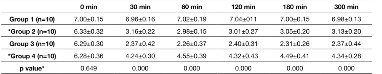
Table II: The Mean Value of Basilar Artery Diameters among Groups
0 min 30 min 60 min 120 min 180 min 300 min
Group 1 (n=10) 7.00±0.15 6.96±0.16 7.02±0.19 7.04±011 7.00±0.15 6.98±0.13 *Group 2 (n=10) 6.33±0.32 3.16±0.22 2.98±0.15 3.01±0.27 3.05±0.20 3.13±0.20 Group 3 (n=10) 6.29±0.30 2.37±0.42 2.26±0.37 2.40±0.31 2.31±0.26 2.37±0.44 *Group 4 (n=10) 6.28±0.36 4.24±0.30 4.55±0.39 4.32±0.43 4.49±0.41 4.34±0.28 p value* 0.649 0.000 0.000 0.000 0.000 0.000 *: p<0.001
⃰ In group 4, we found statistically higher mean basilar artery diameters in comparison with group 2 (Except 0 min).
Table I: Superoxide Dismutase Activity (SDO), Malondialdehyde (MDA) Levels in 40 Rabbits*
Groups SOD (U/g protein) MDA (mmol/m2)
1 1064±33.8 119±21
2 3210±822 468±36
3 8365±421.2 527±52
4 1144±42.1 226±28
p value 0.000 0.000
*Values are expressed as the mean ± SD
Figure 1: Vertebrobasilar angiography in the control group showed normal vertebrobasilar artery.
Figure 2: Vertebrobasilar angiography after SAH group showed spasmed vertebrobasilar artery. Note, the distal end could not be observed.
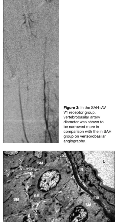
Figure 3: In the SAH+AV V1 receptor group, vertebrobasilar artery diameter was shown to be narrowed more in comparison with the in SAH group on vertebrobasilar angiography.
Figure 4: Vertebrobasilar angiography in
SAH+antagonist of AV V1 receptor group showed spasmed artery eliminated.
Figure 5: Group 1. The endothelial cells (EN) of tunica intima showed normal ultrastructure. The subendothelial layer (SE) and internal elastic membrane (IEM) were regularly arranged. The smooth muscle (SM) of the tunica media also revealed normal ultrastructures. The nucleus (N), collagen fibers (COL) and lumen (L) of the vessel were indicated. Bar= 1 µm.
mitochondria with degeneration of their cristae and increase in heterochromatin in nuclei with enlargement of nuclear membrane. Moreover, many of the endothelial cells showed various sized large vacuoles in the basal cytoplasm. In most of the areas, the thickening of the subendothelial layer and the internal elastic membrane were also seen. Additionally,
substances. Prostanoids, nitric oxide, prostacyclin, endothelin, angiotensin, oxygen free radicals and thromboxane A2 are endothelium-derived vasoactive agents that cause smooth muscle cell hyperpolarization. Vascular tonus depends upon a balance between the endothelial vasodilators and vasoconstrictors, and in SAH, this balance deteriorates in favor of constriction (4,8,10,11,13).
Endothelial structural and functional changes occur in CVS due to SAH. HbO2, TGF, PDGF, EGF, angiotensin and ET
secreted from the clot and thrombocyte in subarachnoid space decreases the amount of NO. This decrease occurs by means of arginine vasopressin that stimulates endothelium. including cytoplasmic vacuolation and increase in nuclear
heterochromatin similar to those in the SAH group. Moreover, the arterial wall showed the thickening of the subendothelial layer and the internal elastic membrane. The smooth muscle in the tunica media revealed degeneration of some organelles and various sized cytoplasmic vacuoles (Figure 7).
Group 4 (SAH+AV V1 Antagonist)
In this group, some of the endothelial cells exhibited swelling of the mitochondria, disruption of their cristae and variously sized cytoplasmic vacuoles, but many of the cells in the tunica media were normal. The cell junctions were frequently marked between endothelial cells. The subendothelial layer was regularly arranged and internal elastic membrane was normal in general. Although a few of the smooth muscle cells in tunica media revealed increase in nuclear chromatin and swelling of mitochondria, and cytoplasmic vacuolation, many cells showed normal ultrastructure (Figure 8).
█
DISCUSSION
CSV following SAH is known to be due to multifactorial stimuli. Mechanic, metabolic and inflammatory factors providing smooth muscle contraction and inhibiting vasodilatation play an important role in CVS. These factors arising from blood elements in the subarachnoid space stimulate vasoconstrictor substances secreted from vascular endothelium and inhibit vasodilator substances. The cerebral vessel endothelium constitutes selective blood and brain barrier besides providing homeostasis. Mediators secreted from the endothelium regulate coagulation, fibrinolysis, vascular tonus, blood flow and blood pressure besides playing a role in immune and inflammatory reactions (1,5,13,14,24,32).
The endothelium secretes vasoactive proteins, growth factor, cytokines, procoagulants, antithrombotic and anticoagulant
Figure 8: Group 4. Most of the endothelial cells (EN) reveal normal ultrastructure. Junctional complexes between endothelial cells were intact (*). Regular arrangement of the subendothelial layer (SE) and internal elastic membrane (IEM) were also seen. The internal elastic membrane was normal. Smooth muscle cells (SM), nucleus (N), mitochondria (M), and lumen (L) of the vessel were indicated. Bar= 0.5 µm.
Figure 7: Group 3. Most of the endothelial cells (EN) showed separation and protrusion toward the arterial lumen. Thickening and irregular arrangement of the subendothelial layer (SE) and internal elastic membrane (IEM) were also seen. Smooth muscle cells (SM), nucleus (N), collagen fibers (COL) and erythrocyte (E) in the lumen (L) of the vessel were indicated. Bar= 2 µm.
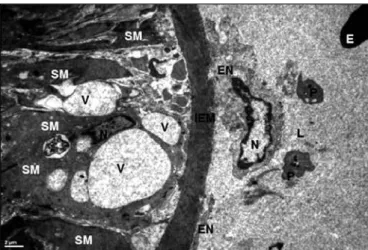
Figure 6: Group 2. Most of the endothelial cells (EN) showed separation from the basal lamina of the tunica intima. Thickening of the internal elastic membrane (IEM) were also seen. Most of the smooth muscle cells (SM) of the tunica media reveals cytoplasmic vacuolation (V). Nucleus (N), collagen fibers (COL), erythrocyte (E) and blood platelets (P) in the lumen (L) of the vessel were indicated. Bar= 2 µm.
Cobb et al. demonstrated that systemic vascular resistance decreases in septic shock due to endotoxin, IL 1 and TNF and application of vasopressor agents does not provide benefit (2).
AV receptor antagonists, ET receptor antagonists, and converting enzyme inhibitors may reduce the severity of CVS. Therefore, following SAH, we applied AV receptor antagonist in group 4 rabbits. V1 receptor antagonists have been used in cardiac failure, Raynaud disease, myocardial infarction, CVS, cerebral thrombus, dysmenorrhea, early abortion and ocular hypertension (17,29). Vakili et al. applied V1 antagonist in arteria cerebri media (ACM) occlusion and detected decrease in early ischemia. This report indicates that V1 receptor antagonist improves brain damage, edema and reperfusion besides morphological recovery in the basilar artery (30). In our study, we detected morphological recovery in the basilar artery besides moderate improvement due to AV V1 receptor antagonist in CVS. These findings indicate that AV and ET play an important role in CVS.
Prostaglandins, eicosanoids, leukotrienes, free oxygen radicals occur due to increase in unsaturated lipid peroxidation in the arterial wall following trauma and anoxia due to SAH. These are among the causes of CVS and they also induce degeneration with harmful effect on tissue. Hemolysis of clot in subarachnoid area results with arise of oxy-Hb, met-Hb, bilirubin, especially Fe+2 and plays an important role in
generation of CVS. Also arise of cytokines, angiotensin, and histamine due to inflammation. These phenomena result with morphological changes in vascular wall (22,24,27).
Electron microscopic examinations of basilar artery samples were found to be normal in group 1. We observed significant cellular changes in the basilar artery due to SAH in group 2. Electron microscopic examination of the arterial wall revealed separation and disruption of the endothelial cells and protrusion to the arterial lumen. Furthermore, endothelial cells of the tunica intima showed loss of cell junctions. Although a few endothelial cells exhibited slight cellular alterations, most endothelial cells disclosed swelling of mitochondria with degeneration of their cristae and increase in heterochromatin in nuclei with enlargement of the nuclear membrane. Moreover, many of the endothelial cells showed various sized large vacuoles in the basal cytoplasm. In most of the areas, thickening of the subendothelial layer and the internal elastic membrane were also seen. Additionally, the clumping of nuclear chromatin in nuclei and cytoplasmic organelle alterations of the smooth muscle cell in the tunica media were also noted.
In group 3, ultrastructural observations of the arterial wall revealed separation and protrusion toward the arterial lumen of endothelial cell and loss of junctional complexes between the cells. Furthermore, there were slight to moderate cellular changes including cytoplasmic vacuolation and increase in nuclear heterochromatin similar to those in the SAH group. Moreover, the arterial wall showed the thickening of the subendothelial layer and the internal elastic membrane. The smooth muscle in the tunica media revealed degeneration of some organelles and various sized cytoplasmic vacuoles. Actually, AV is a potent vasopressor, but we estimated that
intravenous (IV) application of AV before destruction of endo-thelium results in stimulation of endoendo-thelium and secretion of vasodilator agents like NO and prostaglandin I2. We applied
intraarterial AV 5 minutes after SAH before destruction of va-sopressor potent endothelium. NO is derived from guanidino nitrogen atom oxidation in L-Arginine terminal. We considered that IV application of AV following SAH can be a good option for the replacement of decreased substrate.
AV is an important neurotransmitter secreted from supraoptic paraventricular nucleus. Its vascular effect is to increase inositol triphosphate synthesis and intracellular Ca+2 concentration
mediated by V1 receptor. V1 receptor is dominant in central nervous system (CNS) and CSF. It plays an important role in ion homeostasis and regulation of cerebral microvascular resistance. Therefore, we aimed to investigate the effects of both AV and AV V1 receptor specific antagonist on CVS and morphological changes in vessels (3,6,8,9,17,23).
In group 1 (Control Group), angiograms did not reveal change in diameter of basilar artery. Whereas, in group 2 (SAH) which underwent SAH formation, mean basilar artery diameter was found to be narrowed at a rate of 50%. In group 3 (SAH+AV), artery diameter was found to be narrowed about 50% at the 30th minute and CVS was 13% more in comparison to the
2nd group. In group 4 (SAH+V1 receptor antagonist), vascular
constriction was 34.5% at the 30th minute and about 30.9%
at the 300th minute. This means that AV causes more severe
CVS than SAH, and selective antagonist of AV V1 receptor eliminates this effect widely. Half life of AV is 20 minutes. It affects by means of V1 receptors in smooth muscles and by controlling ET secretion. Following SAH, ET is increased at the second day and decreased at the 7th day. It is not in relation
with CVS in humans, because in humans CVS starts at the 3rd day and reaches maximum level at the 7th day following
SAH. HbO2 inhibits NO and this probably occurs through ET
(16,29,31-33).
Recovery due to endothelial stimulation occurs in the long term because of increase in CVS due to AV. However, Pluta et al. demonstrated that L arginine does not reverse CVS but improvement in regional blood flow results with remission (25). Following injection of AV into the 3rd ventricle of rabbits
Gondim et al. detected increase in intraocular pressure, contraction of pupillary diameter (9). These findings were found to be prevented by V1 receptor antagonist application. Transition of AV through the blood-brain barrier is limited. Therefore, we planned to stimulate endothelium by applying AV directly to intraarterial basilar artery. It is reported that intraventricular and intracisternal application of AV is effective in preventing of CVS in the late period.
Nitroxidergic neural endings in adventitia and eNOS ruins and consequently, NO synthesis decreases, vasodilator effect disappears in SAH. AV application into the cisterna results with vasodilatation in a long time period by regulation of NOS, INOS and increased NO and this plays an important role in potential therapy (2,20,25).
3. Cowley AW, Hinojosa-Laborde C, Barber BJ, Harder DR, Lombard JH, Greene AS: Short-term autoregulation of systemic blood flow and cardiac output. News Physiol Sci 4: 219-225, 1989
4. Emre M: General structure and properties of endothelial cells. Singing M (ed). Endothelial and Our System. Istanbul: Printas Printing Inc, 2005:1-29
5. Findlay MS: Cerebral vasospasm. In: Winn RH (ed). Youmans Neurological Surgery. Philadelphia, Pa: WB Saunders, 2004: 1839-1867
6. Gardiner SM, Campton AM, Bennet T, Palmer RMJ, Moncode S: Regional hemodynamic changes during oral ingestion of NG-monomethyl-l-arginine or NG-nitro-l-arginine methyl ester
in conscious Brattleboro rats. Br J Pharmacol 101(1):10-12, 1990
7. Gibo H, Carver CC, Rhoton A, Lenkey C, Mitchell RJ: Microsurgical anatomy of the middle cerebral artery. J Neurosurg 54(2):151-169, 1981
8. Goksel MH, Ozum U, Oztoprak I: The therapeutic effect of continuous intracisternal L-arginine infusion on experimental cerebral vasospasm. Acta Neurochir 143(3): 277-285, 2001 9. Gondim EL, Liu JHK, Costa VP, Weinreb RN: Exogenous
vasopressin influences intraocular pressure via the V1 receptors. Curr Eye Res 22(4): 295-303, 2001
10. Gottlieb Al, Langille BL, Wong MK, Kim DW: Structure and function of the endothelial cytoskeleton. Lab Invest 65: 123-137, 1991
11. Hacein-Bey L, Harder RD, Meier TH, Varelas NP, Miyata N, Lauer KK, Cusick FS, Roman JR: Reversal of delayed vasospasm by TS-011 in the dual hemorrhage dog model of subarachnoid hemorrhage. Am J Neuroradiol 27: 1350-1354, 2006
12. Hacıyakupoğlu S, Yılmaz DM, Özsoy KM, Erman T, Hacıyakupoğlu E, Özgür H, Polat S: Effects of amiloride and Na Selenite on the ATPase levels, malondialdehyde levels, and on superoxide dismutase activity and the histopathologic findings after subarachnoid hemorrhage in the pigs. Neurosurg Q 17(4): 263-267, 2007
13. Haefliger IO, Meyer P, Flammer J, Lüscher TF: The vascular endothelium as a regulator of the ocular circulation: A new concept in ophtalmology. Survey Ophthalmol 39:123-132, 1994
14. Harder DR: Pressure-inducted myogenic activation of cat cerebral arteries is dependent on intact endothelium. Circ Res 60:102-107, 1987
15. Jackson EK: Vasopressin and other agents affecting the renal conservation of water. In: Hardman JG, Limbird LE, Molinoff PB, Ruddon RW, Gilman AG (eds). Goodman and Gilman’s The Pharmacological Basis of Therapeutics. 9thed New York:
Mc Graw Hill, 1996:295-303
16. Khurana GV, Benarroch EE, Katusic SZ, Meyer FB: Cerebral blood flow and metabolism. In: Winn RH (ed). Youmans Neurological Surgery. Chapter 86, Part II, Philadelphia: Elsevier, 2004:1467-1494
17. Khurana GV, Smith AL, Baker AT, Eguchi D, O’Brien T Katusic SZ: Protective vasomotor effects on in vivo recombination endothelial nitric oxide synthase gene expression in a canine model of cerebral vasospasm. Stroke 33: 182-789, 2002
In this degeneration, endothelin secreted from endothelium of AV contributes to the mitogenic, spastic and proliferative effects of vessels (4,10,12,17,30).
In group 4, some of the endothelial cells exhibited swelling of the mitochondria, disruption of their cristae and variously sized cytoplasmic vacuoles, but many of the cells in the tunica media were normal. The cell junctions were frequently marked between endothelial cells. The subendothelial layer was regularly arranged and internal elastic membrane was normal in general. Although a few of the smooth muscle cells in the tunica media revealed an increase in nuclear chromatin and swelling of mitochondria, and cytoplasmic vacuolation, many cells showed normal ultrastructure.
The ultrastructural appearance was similar in groups 2 and 3. Despite the increase in regional blood flow, AV did not provide a morphological change. Histological appearance was related to vascular stenosis due to CVS. Histological features were better in group 4 in comparison to groups 2 and 3 and accordingly occurrence of CVS was less frequent.
The SOD enzyme is an intermediate product found in every cell that metabolizes oxygen in order to dismutase free oxygen radicals. An increase in superoxide radicals is parallel to an increase in SOD enzyme. Free oxygen radicals react with unsaturated lipid acids in the target cell wall following neutralization by antioxidants such as SOD, catalase, glutathione peroxidase, vitamin E, C, B, A carotene flavonoids, and co-enzyme Q. In our samples, the SOD level was high in tissues with SAH, and higher in AV-administered subjects. Despite similar cellular degeneration in both groups, we detected increased SOD level in group 3. This can be explained with inadequate SOD synthesis to fight against free oxygen radicals. Although the same degeneration was observed in groups 2 and 3, the high SOD level can be based on the increase in rCBF due to AV and acceleration in SOD synthesis. In group 4, the SOD levels were decreased compared to those of groups 2 and 3, and cellular degeneration was less. We can state that V1 receptor antagonists decrease cellular degeneration and production of free oxygen radicals accordingly (2,12,13,16,19,25,30).
█
CONCLUSION
We detected morphological and radiological recovery in the basilar artery besides moderate improvement in CVS due to AV V1 receptor antagonist application. These findings indicate that AV and ET play an important role in CVS.
█
REFERENCES
1. Alexander JS, Hechtman HB, Shepro D: Phalloidin enhances endothelial barrier function and reduces inflammatory permeability in vitro. Microvas Res 35: 308-315,1998
2. Cobb PJ, Natanson C, Hoffman WD, Lodato FR, Banks S, Koev CA, Solomon AM, Elin JR, Hosseini MJ, Danner LR: N omega-amino-L-arginine, an inhibitor of nitric oxide synthase, raises vascular resistance but increases mortality rates in awake canines challenged with endotoxin. J Exp Med 176(4): 1175-1182, 1992
26. Schini VB, Vanhoutte PM: Endothelium-derived vasoactive factors. In: Loscalzo J, Schafer AI (eds). Thrombosis and Hemorrhage. Oxford: Blackwell Scientific Publications, 1994: 349-367
27. Stemerman MB, Colton C, Morell E: Perturbations of the endothelium. In: Spaet TH (ed). Progress in Hemostasis and Thrombosis. Orlando: Grune & Stratton, Inc, 1984: 289-324 28. Takenake K, Yamada H, Sakai N, Ando T, Nakashima T,
Nishimura Y, Okano Y, Nozawa Y: Cytosolic calcium changes in cultured rat aortic smooth-muscle cells induced by oxyhemoglobin. J Neurosurg 74: 620-624, 1991
29. Vajkoczy P, Meyer B, Weidauer S, Raabe A, Thome C, Ringel FA, Breu V, Schmiedek P: 803 Cerebral vasospasm after aneurysmal subarachnoid hemorrhage is inhibited by clazosentan an endothelin receptore antagonist. Neurosurgery 57: 396, 2005
30. Vakili A, Hiroharu K, Plesnila N: Role of arginine vasopressin V1 and V2 receptors for brain damage after transient focal cerebral ischemia. J Cereb Blood Flow Metab 25: 1012-1019, 2005
31. Vanhoutte PM: The endothelium: Modulator of vascular smooth muscle tone. N Engl J Med 319: 512-513, 1988 32. Weir B: Intracranial aneurysms and subarachnoid hemorrhage.
In: Vascular diseases of the nervous system, Part VII, New York: Raven Press, 1996: 2191-2213
33. Zubkow AY, Ogigara K, Bernanke DH, Parent AD, Zhang J: Apoptosis of endothelial cells in vessels affected by cerebral vasospasm. Surg Neurol 53: 260-266, 2000
18. Kırış T, Karasu A, Yavuz C, Erdem T, Unal F, Hepgül K, Baloğlu H: Reversal of cerebral vasospasm by the nitric oxide donor SNAP in an experimental model of subarachnoid hemorrhage. Acta Neurochir 141: 1323-1329, 1999
19. Mc Cord JM, Fridovich I: Superoxide dismutase an enzymatic function for erythrocuprein (hemocuprein) J Biol Chem 244: 6049-6055, 1969
20. Millonig G: Advantages of a phosphate buffer for Os SO4 solutions and fixation. J Appl Physics 32: 1637, 1961
21. Ohkawa H, Ohishi N, Yagi K: Assay for lipid peroxides in animal tissues by thiobarbituric acid reaction. Anal Biochem 95: 351-358, 1979
22. Osuka K, Suzuki Y, Watanabe Y, Takayasu M, Yoshida J: Inducible cyclooxygenase expression in canine basilar artery after experimental subarachnoid hemorrhage. Stroke 29: 1219-1222, 1998
23. Palmer RMJ, Ferrige AG, Moncada S: Nitric oxide release account for the biological activity of endothelium derived relaxing factor. Nature 327: 524-526, 1987
24. Peterson JW, Kuwan BO, Keramura A: Immunological reaction against of the aging human subarachnoid erythrocyte. J Neurosurg 71: 718-726, 1989
25. Pluta MR, Afshar BKJ, Thompson GB, Boock JR, White HJ, Oldfield HE: Increased cerebral blood flow but no reversal or prevention of vasospasm in response to l-arginine infusion
after subarachnoid hemorrhage. J Neurosurg 92:(1) 121-126, 2000