See discussions, stats, and author profiles for this publication at: https://www.researchgate.net/publication/6215917
Persistent left superior vena cava: The anatomical and surgical importance
Article in The West Indian medical journal · February 2007DOI: 10.1590/S0043-31442007000100013 · Source: PubMed
CITATIONS 26
READS 117 6 authors, including:
Some of the authors of this publication are also working on these related projects:
Physiotherapy and cardiac surgery View project
endovascular intervention View project Mustafa Bilge Erdoğan
Bahçeşehir University 23PUBLICATIONS 77CITATIONS SEE PROFILE Pınar Karakaş Cukurova University 14PUBLICATIONS 332CITATIONS SEE PROFILE Birol Yamak
Natomed Hospital ANKARA TURKEY
103PUBLICATIONS 459CITATIONS SEE PROFILE
All content following this page was uploaded by Birol Yamak on 06 February 2015.
THE
SURGICAL IMPORTANCE
OF
A
PERSISTENT LEFT SUPERIOR VENA CAVA*
BY
ELLIOTT S. HURWITTt
From the Surgical Division, the Montefiore Hospital, New York City
A left superiorvena cava may be present simul-taneously with a normal right superior vena cava, or, even more infrequently, as the only superior
vena cava. The surgical importance ofapersistent
left superior vena cava varies depending on its
termination into the rightorleft atrium, as wellas
thenature and severity of any associated
cardio-vascular anomalies.
Left Superior Vena Cava Entering Right Atrium. This is not a particularly uncommon anomaly, the
veinterminating either directly into the right atrium,
or indirectly by way of the coronary sinus. The
embryology and frequency of this configuration have been reviewed in a previous communication
(Hur-witt, Escher and Citrin, 1955). Since anomalies of the heart andgreatvessels tendtooccurin
associa-tion, one may encounter a left superior vena cava
draining into the right atrium in theprocessof other
operations, such as in the correction of a patent ductus arteriosus. Under such circumstances the anatomical course ofthe left superior vena cavais
such that it does not interfere with the stated operation, and the vein usually has no surgical
importance.
With the advent ofopen heartsurgery, however,
employing either cardiopulmonary by-pass or
hypothermia, aleft superiorvena cava assumes real
surgical significance, regardless of whether it drains into the rightorleft atrium. Failuretoidentify and mobilize this vessel, with temporary occlusion for hypothermia and cannulation or occlusion during
cardiopulmonary by-pass,mayseriously compromise
the procedure. The necessity for this manoeuvre
has been described inanumber ofarticles,including one from this department (Robinson, Glotzer,
Gilbert, Escher and Hurwitt, in press).
Left Superior Vena Cava Entering Left Atrium. Asacontributingcauseofcyanosis, the importance
ofdrainage ofanysystemic vein into the left auricle has usually been overshadowed by the severity of associated cardiac defects. Subsequent to the
summaryof thereportedcases(Hurwittetal.,1955),
two additional such patients have come to surgery atthishospital.
Case Histories Uncorrected Lesions
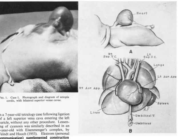
Case1. A 4 lb. prematureinfant, oneof maletwins, was brought to the hospitalonJanuary 21, 1957, with
complete ectopia cordis (Fig. 1)andseverecyanosisand dyspnoea; the twin was normal. A covering of skin wasfashionedoverthe heart, and the baby diedon the-evening of the operation. A left superior vena cava entering the left atriumwasonlyoneof theeight major anomalies of the heart andgreatvesselsfoundatautopsy.
Anormalright superiorvenacavawasalso present. A
motion picture and complete description of this
case-have beenprepared (Hurwitt and Lebendiger, inpress).
Case 2. Severecyanosis andnearlyfatalepisodesof
syncopewerethepresenting problemsin this
18-month-old girl. Investigation in another hospital had
estab-lished thepresenceof botharight and left superiorvena cavaandalevocardia inthepresenceofasitus inversus
abdominalis. On June 17, 1958, anattempt wasmade
toconstructaBlalockshunt between the left subclavian
artery and the left pulmonary artery, but irreversible
cardiac arrest developed during mobilization of the
pulmonary artery. Post-mortem examination disclosed
anessentiallybilocularheart,with the leftsuperiorvena cava draining into the left side ofa chamber that was practicallyacommonauricle. Splenic agenesis,described
inseveral suchcases, wasnotpresent.
Corrected Lesions
Even when severe associated anomalies of the heart
arepresent,however,interruptionof the flowof systemic-venousblood into theleft auriclemaybebeneficial,with
or without attempted correction of the other lesions. Diaz and Anido(1949) reported significant improvement
* PresentedattheFifth AnnualMeeting,theBritish Association of PaediatricSurgeons, London, July 23, 1958.
t Aidedbyagrant from the New York Heart Association.
ARCHIVES
OFDISEASE
INCHILDHOOD
t.
A
Lt.
R,t Aur.App.
FIG.1. Case1. Photographanddiagramofectopia
cordis,withbilateral superiorvenae cavae.
in a7-year-old tetralogycasefollowingligation
ofa left superiorvena cava enteringtheleft auricle,without any otherprocedure.
Lessen-ing ofcyanosis wassimilarlydescribed inan
8-year-old with Eisenmenger's complex, by Feindtand Hauch(1953). Ekstrom(personal
communication) supplemented construction
of a Blalock shunt in a 12-year-old with
tetralogy of Fallot, situs inversus totalis and double superior venae cavae, by ligating the anomalous vena
cavaasitenteredthearterial auricle.
Obviouslytheoutlookismuchmorefavourable when the left superior vena cava entering the left auricle constitutes the major cause ofcyanosis, or when any
concomitant intracardiac defects mayalso beamenable
tocorrection. Examplesof each of thesesituations have nowcome tooperationatthishospital.
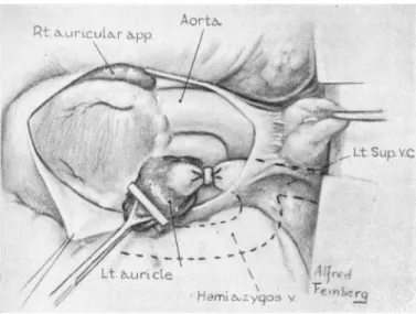
Case 3. This has previously beenreported in detail
(Hurwittetal., 1955). Surgical relief ofcyanosis in an
8-year-old girl, due to entrance ofaleft superior vena cavainto theleftauricle,wasaccomplished by intraperi-cardialligationof the leftsuperiorvenacavaon Decem-ber 29, 1954 (Fig. 2). Thevenous return tothe heart after theprocedureisdepictedinFig.3. Post-operatively
there wasmarkedimprovementinclubbing of the fingers, cyanosis. dyspnoea, appetite, andexerciseperformance. Slightperipheralarterialoxygenunsaturation persisted, presumably related to anatypical moderate hypoplasia
oftheposteriorlyplacedmainpulmonaryartery.
Case 4. A 10-year-old negro girl with acyanotic congenitalheartdiseasewasfoundoncardiac
catheteriza-Liver
tion to have an atrial septal defect with a significant
flow from left to right and a left superior vena cava.
Operation,underhypothermia,wasperformedon July 1, 1957,through an anterior thoracicincision, enteringthe
right pleural cavityinthefourth interspaceand the left
pleural cavity in the third interspace, with transverse
division of the
sternum.
Ontheright there was asmall
superiorvenacava with anormalazygos tributary. The left superior vena cava was considerably larger, was
joinedby a large hemiazygos vein, and entered the left atrium anteriorly to theentranceof the left pulmonary
veins. Digital explorationof theright auricleconfirmed
the presence of a large atrialseptaldefect, andoxygen
determinations from both auricles showed complete
admixtureoftheblood atthis levelunder the operative conditions(Fig. 4a).
Atabody temperature of
880
F.the inferiorand the twosuperiorvenaecavae wereoccluded for six and a half minutes while the right auricle was opened and anatrialseptal defect measuring4cm.inlength and
21
cm. inwidth wasrepairedby acontinuoussilk
suture. While the right auricle now contained blood with a venous oxygensaturation,blood from theleftauricle at the siteof insertionof theleftsuperiorvena cava wasalsohighly
2
group.bmj.com on February 13, 2013 - Published by
adc.bmj.com Downloaded from
PERSISTENT
SUPERIOR
VENA CAVAFIG. 2. Case 3. Intrapericardialligationof leftsuperiorvenacava. Note large hemiazygos entering left superior vena cava
extraperi-cardially. (From Hurwitt etal., 1955.)
unsaturated (Fig. 4b). Apparently the return from the left superior vena cava had been streaming practically directly through the atrial defect, and did not produce
significant oxygen desaturation in the left auricle until thedefect was closed.
Intrapericardial ligation of the leftsuperior vena cava resulted in eliminating this source of systemic venous blood from the left atrium, with a restorationtonormal of cardiac physiology (Fig. 4c). At no time
post-operativelywasthere anyevidence of oedema, cyanosis, or increased venous pressure, confirming the presence of adequate communicating channels between the two
superior vena caval systems. Thechild was re-examined
Rt.SupVC f L oygol
t.iru'Vi,
'A
!-Com*. ...i'. ...:!
IlfiacC.
~~~~~~~~~~~~~~~ilac
i
V.'X
FiG.3. Case 3. Diagrammaticreconstruction of vascular anomalies
andligationofleftsuperiorvenacava. (FromHurwittelal., 1955.)
Both figures are reproduced by courtesy of C. V. Mosby Co., St. Louis, Mo. 101.5% I Lt.SuP.V.C. \ .J Y I OXYGEN SATURATION
FiG. 4a, b, c.-Case 4. Statusatoperation: (a), initially; (b), after closure of the atrial septal defect; and (c), after ligation of the left superior
venacava.
3
V. OXYGEN SATURATION 1/0# 5 %, Rtt Sup.V.C. 66LS Vj 11 LtSup.VC. OXYGEN SATURATION 81.501, I I I I group.bmj.com on February 13, 2013 - Published by adc.bmj.com Downloaded from4
ARCHIVES OF
DISEASE
INCHILDHOOD
one year after operation and found to be in excellenthealth.
Comment
Drainage of any systemic vein into the left atrium contributes to the cyanotic state, and as such merits consideration for surgical correction; the only categorical contra-indication would be atresia of the tricuspid valve. The anomalous vein will usually be a superior vena cava, although the inferior vena
cava (Gardner and Cole, 1955), coronary sinus
(Mankin and Burchell, 1953), andlevo-atriocardinal
veins (Gould, 1953) have also been reported
terminating in the left auricle.
The degree of improvement following ligation of aleft superior vena cava will be determined bythe severity of any uncorrected associated lesions. It may be substantial, however, even in the presence of complex defects of the tetralogy type (Gould, 1953; Gardner and Cole, 1955; Hurwitt etal., 1955). When the associated anomalies are also corrected,
asin Case 4, or when no major concomitant abnor-malities are found, the result may be excellent. Cases havebeen described with no associatedseptal
defects (Potter, 1948; Gardner and Cole, 1955; Peel, Semple, Kelly and Blum, 1956; Tuchman, Brown, Huston, Weinstein, Rowe and Crumpton, 1956), and none was found in our Case 3. When the
flow from the left superior vena cava is streaming
almost directly through the atrial septal defect,
significant arterial oxygen unsaturation may not be
detected until the defect is closed, as in Case 4. Occlusion of the left superior vena cava then restoresthe situation to normal.
A disadvantage of approaching repair of an
atrial septal defect through an incision limited to
opening the right pleural cavity is the possibility of
overlookingthe presence ofaleft superiorvena cava.
Even in the hands of those who employ 'closed' techniques for these operations, the opportunityfor
correctinga left superior vena cavaentering the left auricle would be missed. Most surgeons currently
close atrial septal defects under direct vision with
'open' techniques. If venous inflow occlusion with
hypothermia is practised, failure to take a left superior venacavainto account may result both in excessive blood loss and poor visualization of the
defect, regardless of which atrium receives the
anomalous vein; the same considerations apply equally to cardiopulmonary by-pass. For these reasonseither a verticalsternum-splitting incision or
a bilateral anterior thoracotomy with division of the
sternum are preferable to right thoracotomy alone.
Interruption of aleft superiorvena cavaentering
the left auriclemustbeperformedintrapericardially
inorderalso to divertthe flowfromthehemiazygos
vein to the right side (Figs. 2, 3). The interruption may be accomplished simply by ligation, as in the reported cases, or by division. If a solitary left superior or inferior vena cava were found entering the left atrium, transplantation to the right side wouldbe necessary, either by direct suture or by a graft. One unsuccessful attempt to re-route blood in a solitary left superior vena cava from the left to the right auricle has been reported by Tuchman et al. (1956). When bilateral superior venae cavae are present, apparently the communicating veins across. the mediastinum or through the azygos system are usually adequate, so that closure of the left superior vena cavaresults in shunting the blood to the right side, rather than the syndrome of superior vena caval compression. When such communications are absent, however, as suggested in the report by Peel et al. (1956), interruption of the left superior vena cavashould be supplemented by an anastomosis.
totheright superiorcaval system.
Summary
The surgical importance of a left superior vena
cavahas been analysed.
Regardless of whether the left superior vena cava drains into therightorleftatrium, itmustbe either occluded or cannulated during open heart surgery
witheitherhypothermiaorcardiopulmonaryby-pass.
Anysystemic vein entering the left auricle may bea
majororcontributingcauseofcyanosis,andshould be
corrected,except in the presence oftricuspidatresia.
Dataare presented on four cases of leftsuperior vena cava entering the left atrium, with surgical
correction intwo instances, one of which has been
reportedpreviously.
Ligation ofa leftsuperiorvenacavaenteringthe left auricle and closure ofa large atrialseptaldefect
wereaccomplishedin a 10-year-old girl.
This represents the fifthreportedcase ofrelief of
cyanosis following interruption of a left superior
vena cavaterminating in the left auricle.
REFERENCES
Ekstrom, G.(1954). Personalcommunication.
Feindt, H. R. andHauch,H.J.(1953). Z.Kreisl.-Forsch.,42, 53.
Gardner,D. L.andCole,L.(1955). Brit. HeartJ.,17, 93.
Gould,S. E.(1953). PathologyoftheHeart,pp.491, 496. CharlesC.
Thomas,Springfield, Illinois.
Hurwitt, E.S., Escher, D.J. W. andCitrin, L.I.(1955). Surgery,
38, 903.
and Lebendiger, A. A.M.A. Arch. Surg. In the press. PresentedattheSixth ScientificMeeting,the NorthAmerican Chapter of the International Cardiovascular Society, San Francisco,June21, 1958.
Mankin,H.T. andBurchell,H. B.(1953). Proc.MayoClin., 28,463.
Peel,A. A. F.,Semple, T.,Kelly,J.C. andBlum,K.(1956). Scot.
med.J.,1, 83.
Potter,E. L. (1948). Arch.Path.(Chicago), 46,87.
Robinson, G.,Glotzer, P.,Gilbert, M., Escher,D.J. W. andHurwitt,
E.S. Amer. J.Cardiol. Inthepress. Presented intheNew
YorkSociety for ThoracicSurgery, May 9,1958.
Rodiguez Diaz,A.andAnido, H.(1949). Dis.Chest, 15, 684. Tuchman,H., Brown,J. F., Huston,J. H.,Weinstein,A.B., Rowe,
G.G. andCrumpton, C.W.(1956). Amer. J.Med., 21, 481 group.bmj.com
on February 13, 2013 - Published by adc.bmj.com
doi: 10.1136/adc.34.173.1
1959 34: 1-4
Arch Dis Child
Elliott S. Hurwitt
Cava
Persistent Left Superior Vena
The Surgical Importance of a
http://adc.bmj.com/content/34/173/1.citation
Updated information and services can be found at:
These include:
References
http://adc.bmj.com/content/34/173/1.citation#related-urls
Article cited in:
service Email alerting
the online article.
article. Sign up in the box at the top right corner of Receive free email alerts when new articles cite this
Notes
http://group.bmj.com/group/rights-licensing/permissions
To request permissions go to:
http://journals.bmj.com/cgi/reprintform
To order reprints go to:
http://group.bmj.com/subscribe/
To subscribe to BMJ go to:
View publication stats View publication stats