Original Article / Özgün Araştırma
Assessment of Myocardial Function by Myocardial Performance
Index in Patients with Acute Ischemic Stroke
Unal Ozturk1, Onder Ozturk2, Yusuf Tamam3
1 Univ of Health Sciences, Diyarbakır Gazi Yasargil Education and Research Hospital, Dept of Neurology, Diyarbakir Turkey ORCID: 0000-0002-8341-5070 2 Univ of Health Sciences, Diyarbakır Gazi Yasargil Education and Research Hospital, Dept of Cardiology, Diyarbakir Turkey ORCID: 0000-0003-4204-0599 3 Dicle University Medical Faculty, Department of Neurology, Diyarbakir Turkey ORCID: 0000-0002-9886-2017
Received: 14.05.2019; Revised: 19.08.2019; Accepted: 19.09.2019
Abstract
Objective: Acute stroke is an important cause of morbidity and mortality. Myocardial injury is an important complication of acute cerebrovascular events. In this study, we aimed to investigate the relationship between myocardial function assessed by myocardial performance index (MPI) and National Institutes of Health Stroke Scale (NIHSS) score in patients with acute ischemic stroke.
Method: The study comprised 97 patients (males, 42; females, 55; 65 ± 16 years) diagnosed with acute ischemic stroke. 17 patients were excluded. Patients were divided into two groups based on the calculated NIHSS score (Group 1, NIHSS score < 16; Group 2, NIHSS score ≥ 16). Demographic, clinical, and laboratory data for all patients were collected. Cardiac function was evaluated by transthorasic echocardiography within 48 hours of admission to the neurology care unit.
Results: There were no significant differences among demographic parameters of patients. MPI was significantly higher in Group 2 patients than in Group 1 patients (0.59±0.25 vs 0.48±0.20, p=0.004).
Conclusion: Our results suggest that MPI is associated with stroke severity on admission in patients with acute ischemic stroke. MPI is an indicator of global myocardial dysfunction with a different from LVEF. MPI, which includes both systolic and diastolic time intervals is an option to assess the global cardiac dysfunction.
Keywords: Ventricular dysfunction, left ventricular, echocardiography, stroke.
DOI: 10.5798/dicletip.661265
Yazışma Adresi / Correspondence: Onder Ozturk, University of Health Sciences, Diyarbakir Gazi Yasargil Education and Research Hospital Department of Cardiology, Uckuyular, Kayapinar, Diyarbakir, Turkey e-mail: [email protected]
716
Akut İskemik İnmeli Hastalarda Miyokard Fonksiyonunun Miyokardiyal Performans İndeksi ile Değerlendirilmesi
Öz
Amaç: Akut inme morbidite ve mortalitenin önemli nedenlerinden biridir. Miyokardiyal hasarlanma, akut serebrovasküler olayların önemli bir komplikasyonudur. Yapmış olduğumuz bu çalışmanın amacı, akut iskemik inmeli hastalarda Ulusal Sağlık İnme Enstitüleri Ölçeği (NIHSS) ve miyokard performans indeksi (MPİ) ile değerlendirilen miyokard fonksiyonları arasındaki ilişkiyi araştırmaktır.
Yöntemler: Çalışmaya akut iskemik inme tanılı 97 hasta (erkek,42; kadın,55; yaş, 65 ± 16) alındı. 17 hasta çalışmadan çıkarıldı. NIHSS skoruna göre hastalar iki gruba ayrıldı (Grup 1, NIHSS skor <16; Grup 2, NIHSS skoru ≥ 16). Bütün hastalardan demografik, klinik ve laboratuvar verileri toplandı. Hastaların kardiyak fonksiyonları, nöroloji yoğun bakım ünitesine yatırıldıktan sonraki 48 saat içerisinde transtorasik ekokardiyografi değerlendirildi.
Bulgular: Hastaların demografik özellikleri açısından gruplar arasında anlamlı farklılık saptanmadı. MPİ, Grup 2 hastalarında, Grup 1 hastalarına göre anlamlı derecede daha yüksek bulundu (0.59±0.25 ile 0.48±0.20, p=0.004). Sonuç: Yapmış olduğumuz bu çalışmada, akut iskemik inme ile yatan hastalarda, inme şiddeti ile MPİ arasında bir ilişki olduğu sonucuna varılabilir. MPİ, LV EF’dan farklı olarak, global miyokardiyal disfonksiyonunun bir göstergesidir. MPİ, hem sistolik, hem de diyastolik zaman intervallerini içermekte olup, global kardiyak disfonksiyonu değerlendirir.
Anahtar kelimeler: Ventrikül disfonksiyonu, sol ventrikül, ekokardiyografi, inme.
INTRODUCTION
Acute stroke is an important cause of morbidity and mortality1, and cardiovascular
complications are common after an acute stroke2. Myocardial injury is an important
complication of acute cerebrovascular events. Autonomic nervous system dysregulation after acute cerebrovascular events possibly give rise to sympathetic system activation resulting in hypercontraction of cardiac myocytes and subsequent myocardial injury. Neurogenic stress cardiomyopathy (NSC) is a acute left ventricular myocardial systolic dysfunction that can be observed after acute cerebrovascular events, such as acute ischemic or hemorrhagic stroke, cranial trauma, subarachnoid hemorrhage, and seizures3.
Often, NSC can be very similar to myocardial infarction, presenting with ischemic ECG changes, including ST segment and T wave deviations, reduced ejection fraction, elevated troponin levels, and ventricular wall motion abnormalities. However, there is no significant obstruction of the coronary arteries in patients with NSC. Acute systolic heart failure due to
NSC is an important cause of lethal ventricular arrhythmias and mortality4.
The Doppler echocardiographic assessment of MPI, combining time intervals of left ventricular (LV) contraction and relaxation, is an important predictor of death and congestive systolic heart failure after myocardial infarction (MI). Noninvasive measurements of time intervals have historically been used for evaluation of both systolic and diastolic myocardial function, but because of practically difficulties in calculating the intervals, these measurements have not often been applied clinically. The MPI is obtained by measuring the Doppler time intervals from the mitral inflow, and LV outflow velocity time intervals. MPI index can be calculated easily, is reproducible and independent of the ventricular geometry, and has been shown to have important for clinical application in the assessment of overall cardiac function in many disorders5,6.
In this study, we aimed to investigate the relationship between myocardial performance index and NIHSS score in patients with acute ischemic stroke.
METHOD Study participants and design Patient selection
We prospectively studied consecutive 97 adult patients (males, 42; females, 55; mean age, 65±16 years; range 41–80 years) with acute ischemic stroke (≤24 hours of symptom onset) admitted to the neurology care unit, between July 2017 and December 2018. 17 patients were excluded. Demographic and baseline clinical parameters, including neurological deficit severity assesment with NIHSS on admission to the neurology care unit were recorded. Patient clinical data, history of cardiovascular risk factors, and stroke onset were determined, and neurologic examination was conducted at the time of admission. The diagnosis of acute ischemic stroke was made on the basis of neurologic examination and cranial imaging within 24 hours of symptom onset. Patients with a well-defined time of ischemic stroke symptom onset were included in the study and those with any previous history of cerebrovascular disease or transient ischemic attack, cerebral hemorrhage, documented atrial fibrillation, ischemic heart disease, congestive heart failure, severe valvular heart disease, congenital heart disease, chronic obstructive pulmonary disease, chronic renal failure were excluded. In addition, patients in whom a proper position could not be obtained during echocardiography and those with poor echocardiographic image quality, were excluded. Seventeen patients were excluded because of previous history of cerebrovascular disease (n=2), documented atrial fibrillation (n=4), congestive heart failure (n=3), coronary heart disease (n=5), serious valvular heart disease (n= 2), a poor echocardiographic image quality (n=1). Baseline stroke severity was assessed using the NIHSS score7.
All patients underwent immediate computed tomography after being admitted to emergency care unit. Troponin values were measured and
electrocardiogram (ECG) was recorded after admission to the neurology care unit. Echocardiographic examination was performed within the first 48 hours of admission to the neurology care unit. The NIHSS evaluation and echocardiographic examination were conducted by blinded investigators (U.O and O.O, respectively). The study was approved by the Diyarbakir Gazi Yasargil Education and Research Hospital Ethics Committee. Ethics Committee number of study is 57. The study was conducted in accordance with the principles of the Declaration of Helsinki.
ECHOCARDIOGRAPHIC EXAMINATION Two-dimensional echocardiography
All patients underwent comprehensive transthoracic echocardiography examinations, which were conducted according to the guidelines of the American Society of Echocardiography. An echocardiography system (Philips EPIQ 7C, Philips Healthcare, Andover, MA, USA) equipped with a multifrequency transducer (3–8 MHz) and tissue harmonic imaging capability was used. Single-lead electrocardiogram was continuously recorded. Patients were kept in the left lateral decubitus position. Images were obtained from the parasternal long- and short-axes, apical two- and four-chamber, and long-axis views. All echocardiograms included at least three consecutive cardiac cycles and were digitally stored for offline analysis. In addition to LVEF, end-systolic and end-diastolic volumes were measured from the apical two- and four-chamber views, using the standard biplane Simpson’s technique. Interventricular septum, posterior wall thickness, LV end-diastolic diameter, and left atrial antero-posterior diameter were measured from a parasternal long-axis view8. Echocardiographic parameters
were calculated by qualified physician (O.O.), and echocardiographic examination was conducted by an investigator who was blinded to the patients’ clinical information.
718
Assessment of LV Function
MPI calculation Doppler time intervals were calculated from mitral inflow and left ventricular outflow Doppler tracings, as described by Tei and co-workers9. Mitral inflow
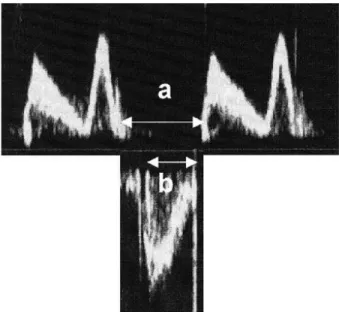
parameter was recorded with the transducer in the apical 4-chamber view. The echocardiaographic pulsed wave Doppler beam was aligned as perpendicular as possible to the plane of the mitral annulus. The pulsed Doppler sample volume was placed between the tips of mitral leaflets during diastole. The LV outflow tract velocity curve was recorded from the apical long-axis view with the Doppler sample volume positioned just below the aortic valve. The interval 'a' calculated from cessation to onset of mitral inflow is equal to the sum of isovolumic contraction time (ICT), ejection time (ET), and isovolumic relaxation time (IRT). ET 'b' is calculated from the duration of the left ventricular outflow Doppler velocity profile. The sum of ICT and IRT was obtained by subtracting b from a. The MPI was measured as shown in Fig. 1: (a-b)/b.
Figure 1. Pulsed wave Doppler recordings of left ventricular
inflow (Top) and outflow (Bottom). (a) Cessation to onset of tricuspid inflow. (b) Pulmonary ejection time. The myocardial performance index (MPI) was calculated as (a–b)/b, where a–b is equal to the sum of isovolumic relaxation time and isovolumic contraction time
Definition of Stroke and Assesment of Stroke Severity
According to the updated definition of acute ischemic stroke in the American Heart Association/American Stroke Association guidelines, ischemic stroke is diagnosed based on the combination of symptoms and/or signs of typical neurological impairment and cranial imaging evidence of central nervous system infarction. Therefore, acute ischemic stroke is defined as a neurological dysfunction episode caused by focal cerebral, spinal, or retinal infarction on cranial imaging10.
NIHSS is a simple, valid, and realiable systematic assessment tool that measures acute stroke-related neurologic deficit7. The NIHSS
score is very important scale for clinical assessment as it enables determination of appropriate treatment, prediction of lesion size, measurement of stroke severity, and prediction of patient outcome in patients with acute ischemic stroke. The NIHSS comprises11
different elements evaluating specific ability. Each ability is scored between 0 and 4, where 0 corresponds to normal functioning and 4 corresponds to complete impairment. A patient’s NIHSS score is calculated by adding the score for each parameter of the scale; 42 is the highest score possible. A higher NIHSS score corresponds to greater impairment of cerebral function in a stroke patient. According to NIHSS score, there are five stroke severity groups: NIHSS =0 (no stroke), NIHSS=1-4 (minor stroke), NIHSS=5-15 (moderate stroke), NIHSS=16-20 (moderate to severe stroke), NIHSS=21-42 (severe stroke). A baseline NIHSS score greater than 16 indicates a strong probability of patient disability and death7.
Stroke severity at admission to the neurology care unit was assessed by the NIHSS score by a neurologist (U.O). Patients were categorized into two groups; Group 1 comprised of patients with non-severe stroke (NIHSS<16; n=58),
whereas Group 2 comprised of patients with severe stroke (NIHSS≥16; n=22).
Statistical Analysis
Statistical analysis was conducted with the SPSS statistical package (Version 12.0; SPSS Inc., Chicago, IL,USA). All baseline parameters were analyzed. Continuous variables are expressed as mean±SD, and categorical variables are expressed as percentages. Intra-observer variability was calculated as the absolute difference between the two measurements as a percentage of their mean. Mann–Whitney U test and Chi-square test were used for comparison of data as appropriate. p values <0.05 were considered statistically significant. Pearson’s correlation was used to determine the relationship between LV MPI and other echocardiographic parameters. The Pearson’s or Spearman’s correlation was used for assessing correlations between variables.
RESULTS Baseline Characteristics
Baseline characteristics of patients are summarized in Table I. Clinical characteristics of patient were similar with respect to age, gender, hypertension, diabetes, smoking (p>0.05). Systolic blood pressure (BP), diastolic BP, heart rate, dyslipidemia, and troponin levels in Group 2 patients were significantly higher than Group 1 patients (p<0.05). Serum hemoglobin HbA1c and low density lipoprotein cholesterol levels were significantly higher in Group 2 patients than Group 1 patients (p<0.05).
Echocardiographic Findings
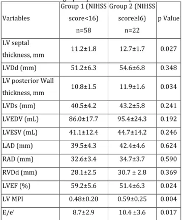
Echocardiographic parameters are summarized in Table II. LV wall thickness and E/e’ values were significantly higher in Group 2 patients than in Group 1 patients (p < 0.05). LV MPI were significantly lower in Group 1 patients having lower NIHSS scores than in Group 2 patients having higher NIHSS. LVEF were significantly higher in Group 1 patients having
lower NIHSS scores than in Group 2 patients having higher NIHSS. Intra-observer and inter-observer variability were calculated. Ten patients (12%) were randomly assigned to assess variability. Interobserver variability was 5% and intraobserver variability was 4%. Table I: Clinical characteristics of patients
Variables Group 1 (NIHSS score<16) n=58 Group 2 (NIHSS score≥l6) n=22 p Value Age (years) 65.4±12.7 69.8±17.7 0.619 Gender (F/M), n 32 / 26 13 / 9 0.718 Hypertension, n % 28 (48%) 13 (59%) 0.062 SBP (mmHg) 143.5±15.2 159.8±18.5 0.043 DBP (mmHg) 76.4±8.6 84.3±12.7 0.048 Heart Rate (bpm) 92.3±14.7 115±16.5 0.027 Diabetes Mellitus, n % 14 (24%) 6 (29%) 0.091 Smoking, n % 7 (12%) 4 (16%) 0.076 Dyslipidemia, n % 9 (15%) 8 (36%) 0.043 Troponin (ng/L) 6.143 14.705 0.037 HbA1c (%) 6.24±1.43 9.56±1.53 0.009 Glucose (mg/dl) 143.5±37.4 192.7±48.5 0.026 Creatinine (mg/dL) 1.3±0.4 1.8±0.6 0.023 LDL cholesterol (mg/dL) 103.3±27.1 128.4±39.5 0.008 HDL cholesterol (mg/dL) 42.1±12.3 39.4±10 0.384
*F:Female, † M:Male, ‡ SBP: Systolic Blood Pressure, § DBP: Diastolic Blood Pressure, || LDL: Low Density Lipoprotein, ¶ HDL: High Density Lipoprotein.
Correlation analysis performed to investigate the relationship between NIHSS score and echocardiographic parameters, showed a negative correlation between the NIHSS score and LVEF. In addition, there was a positive correlation between absolute value of the NIHSS score and LV MPI, age, heart rate and E/e’. (Table III).
720 Table II: Echocardiographic parameters of patients
Variables Group 1 (NIHSS score<16) n=58 Group 2 (NIHSS score≥l6) n=22 p Value LV septal thickness, mm 11.2±1.8 12.7±1.7 0.027 LVDd (mm) 51.2±6.3 54.6±6.8 0.348 LV posterior Wall thickness, mm 10.8±1.5 11.9±1.6 0.034 LVDs (mm) 40.5±4.2 43.2±5.8 0.241 LVEDV (mL) 86.0±17.7 95.4±24.3 0.192 LVESV (mL) 41.1±12.4 44.7±14.2 0.246 LAD (mm) 39.5±4.3 42.4±4.6 0.624 RAD (mm) 32.6±3.4 34.7±3.7 0.590 RVDd (mm) 28.1±2.5 30.7 ± 2.8 0.369 LVEF (%) 59.2±5.6 51.4±6.3 0.024 LV MPI 0.48±0.20 0.59±0.25 0.004 E/e’ 8.7±2.9 10.4 ±3.6 0.017
* NIHSS: National Institutes of Health Stroke Scale, † LV: Left Ventricle, ‡ LVDd: Left ventricular diastolic diameter, § LVDs: Left ventricular systolic diameter, || LVEDV: Left ventricular end-diastolic volume, ¶ LVESV: Left ventricular end-systolic volume, ** LAD: Left atrial diameter, †† RAD: Right atrial diameter, ‡‡ RVDd: Right ventricular diastolic diameter, *** LVEF: Left ventricular ejection fraction, ††† LV GLS: Left ventricular global longitudinal strain.
Table III: Correlation between NIHSS score and clinical
parameters in patients with acute ischemic stroke
Parameters Pearson’s correlation
coefficient (r value) p Value
LV MPI 0.432 0.019
LVEF -0.314 0.038
E/e’ 0.217 0.026
Age 0.320 0.042
Heart rate 0.419 0.023
* NIHSS: National Institutes of Health Stroke Scale, † LV GLS: Left ventricular global longitudinal strain, ‡ LVEF: Left ventricular ejection fraction.
DISCUSSION
Ischemic stroke and congestive heart failure are very important causes of morbidity and mortality. It has been previously reported that heart failure is associated with an increased risk of acute ischemic stroke. Heart failure is also reportedly associated with a poor prognosis in patients with stroke10,11. In
addition, LV dysfunction occurs frequently after cerebrovascular events12-14. LV systolic and
diastolic dysfunctions result from complex and interrelated alterations in myocardial function and structure, peripheral vasculature, and neurohumoral regulation. Neurogenic stunned myocardium (NSM) is defined as left myocardial injury and dysfunction of the autonomic nervous system, that occurs after cerebrovascular events. Previous studies have suggested that severe neurologic injury, female gender, and elevated plasma troponin and brain natriuretic peptide levels are independent predictors of NSM15,16. LVEF is a
important marker of systolic function; however, it was not detected in diastolic dysfunction. The effect of ischemic stroke severity on LV function is not very well known, and there are only a few studies investigating this relationship17,18. Sung PH et al. found that
the severe acute ischemic stroke patients had lower LVEF19. In our study we found that LV EF
value was significantly lower in severe stroke patients. MPI is an indicator of global myocardial dysfunction with a different from LVEF. Myocardial Performance Index, which includes both systolic and diastolic time intervals can be considered as a parameter to assess the global cardiac dysfunction.
LV MPI is a useful marker for assesment of global LV function. Very less is known about the relationship between acute ischemic stroke severity and LV MPI. Importantly, this is the first study assessing LV function by LV MPI in the early period of acute ischemic stroke. The novel and most important finding of our study is that both LV systolic dysfunction and LV
diastolic function was detected with LV MPI after acute ischemic stroke. We found that MPI was significantly higher in patients with higher NIHSS scores than in those with lower NIHSS scores.
Hypertension, hyperlipidemia, smoking, and Diabetes Mellitus are important risk factors for atherosclerotic cerebrovascular disease. We found that, blood pressure at admission is significantly higher in severe ischemic stroke patients. However, Bonardo P et al found that, among young patients with acute ischemic stroke, large infarct volume was not associated with high blood pressure at admission20. In our
study, we found that LDL cholesterol and HbA1c were signficantly higher in patients with higher NIHSS scores than in those with lower NIHSS scores. Hendrix P et al. found that diabetes mellitus history is an important predictor of stroke severity21.
Acute ischemic stroke is characterized by severe autonomic dysregulation, including alterations in the autonomic reflex tract, central autonomic areas, and hormonal factors. Stroke-related sympathetic activation is high in patients with higher NIHSS score. Regardless of prior cardiovascular status, an acute period of stroke importantly influences systemic BP, heart rate, LV function, and biochemical parameters (Glucose, troponin, creatinin)14. In
this study, we found that troponin levels were significantly higher in severe acute ischemic stroke patients. Chang et al observed that cardiac biomarkers, especially serum troponin levels, are associated with acute large vessel occlusion in patients with acute ischemic stroke22. Lindsberg et al observed elevated
blood glucose level is common in the early phase of ischemic stroke. In our study blood glucose levels were significantly higher in severe stroke patients on admission. Although up to one-third of severe acute ischemic stroke patients have diagnosed diabetes, likely a major proportion of patients have stress related hyperglycemia mediated partly by the release
of cortisol and norepinephrine23. In our study
we found that E/é value was significantly higher in severe stroke patients. Ryu WS et al. suggested that E/e' ratios were associated with cranial arterial occlusion in AF-related stroke and may play a role in identifying patients at high risk of acute severe stroke24. In this study
we found that creatinine levels were significantly higher in severe stroke patients. Mostofsky E et al suggesting that, shared risk factors underlying vascular diseases including age, diabetes mellitus, hypertension, left ventricular hypertrophy may represent an important vascular pathogenesis resulting from reduced renal clearence. Renal function predicts survival in patients with acute ischemic stroke25.
Study Limitations
Our study has several limitations. Numerical differences between groups 1 and 2 are limitation of this study. Effects of stroke localisation especially stroke involving the insular cortex may be associated with adverse cardiac outcomes. In our study we didn’t investigate stroke localisation on the cardiac function. Although we excluded patients with major cardiac history, it is possible that chronic systolic heart failure was missed. However, we excluded patients with echocardiographic parameters that are compatible with chronic heart failure, such as segmental dyskinesis, dilated or hypertrophic cardiomyopathy, or severe valvular disease. Moreover, elevation of cardiac troponin levels over time is in accordance with the current definition of stress induced cardiomyopathy. Therefore, we suggest that stress cardiomyopathy features should be studied in patients with acute ischemic stroke and without chronic asymptomatic heart failure.
722
CONCLUSIONS
The results of our study suggest that LV MPI is associated with stroke severity on admission in patients with acute ischemic stroke. Myocardial Performance Index, which includes both systolic and diastolic time intervals to assess the global cardiac dysfunction. LV MPI can help to evaluate LV systolic and diastolic function in patients with acute neurologic diseases.
Therefore, this study suggests that LV MPI can asess LV myocardial dysfunction in patients with acute ischemic stroke.
Ethical approval: The study was approved by
the Ethics Committee of our hospital. The study was conducted in accordance with the principles of the Declaration of Helsinki.
Conception and design: Unal Ozturk and
Onder Ozturk
Acquisition, analysis, and interpretation of data: Unal Ozturk and Onder Ozturk
Conflicts of interest: The authors have no
conflict of interests to declare.
Financial Disclosure: The authors declared
that this study has received no financial support.
REFERENCES
1. Rojek A, Gąsecki D, Fijałkowski M, et al. Left ventricular ejection fraction and aortic stiffness are independent predictors of neurological outcome in acute ischemic stroke. J Hypertens. 2016; 34: 2441-8. 2. Malik AN, Gross BA, Rosalind Lai PM, et al. Neurogenic
Stress Cardiomyopathy After Aneurysmal Subarachnoid Hemorrhage. World Neurosurg. 2015; 83: 880-5.
3. Kerro A, Woods T, Chang JJ. Neurogenic stunned myocardium in subarachnoid hemorrhage. J Crit Care. 2017; 38: 27-34.
4. Ermis E, Demirelli S, Ceylan M, et al. The evaluation of myocardial function of patients in the early stage of acute ischemic stroke by two-dimensional speckle tracking echocardiography. J Clin Ultrasound. 2016; 44: 305-11.
5. Ozturk O, Ulgen MS, Tekes S, Ozturk U, Toprak N. Influence of the Angiotensin Converting Enzyme I/D Gene Polymorphisms on Right Ventricular Myocardial Performance Index in Patients With a First Acute Anterior Myocardial Infarction. Circ J. 2005; 69: 211-5. 6. Öztürk Ö, Toprak N. Akut Anteriyor Miyokard
İnfarktüslü Hastalarda Geliş Miyokardiyal Performans İndeksi ile Lökositoz Arasındaki İlişki. Dicle Med J. 2004; 2: 36-41.
7. Lyden P. Using the National Institutes of Health Stroke Scale: A Cautionary Tale. Stroke. 2017; 48: 513-19. 8. Lang RM, Badano LP, Mor-Avi V, et al.
Recommendations for cardiac chamber quantification by echocardiography in adults: an update from the American Society of Echocardiography and the European Association of Cardiovascular Imaging. J Am Soc Echocardiogr. 2015; 28: 1-39.
9. Tei C, Nishimura RA, Seward JB, Tajik AJ. Noninvasive Doppler derived myocardial performance index: Correlation with simultaneous measurement of cardiac catheterization measurements. J Am Soc Echocardiogr. 1997; 10: 169–78.
10. Zawadka-Kunikowska M, Slominski K, Slomko J, et al. Comprehensive non-nvasive cardiac and autonomic assessment in acute ischaemic strokepatients: pilot study. Minerva Cardioangiol. 2018; 66: 376-85.
11. Vemmos K, Ntaios G, Savvari P, et al: Stroke etiology and predictors of outcome in patients with heart failure and acute stroke: a 10-year follow-up study. Eur J Heart Fail. 2012; 14: 211-8.
12. Holmström A, Fu ML, Hjalmarsson C, Bokemark L, Andersson B. Heart dysfunction in patients with acute ischemic stroke or TIA does not predict all-cause mortality at long-term follow-up. BMC Neurol. 2013; 23: 122.
13. Ripoll JG, Blackshear JL, Díaz-Gómez JL. Acute Cardiac Complications in Critical Brain Disease. Neurosurg Clin N Am. 2018; 29: 281-97.
14. Constantinescu V, Matei D, Cuciureanu D, et al. Cortical modulation of cardiac autonomic activity in ischemic stroke patients. Acta Neurol Belg. 2016; 116: 473-80.
15. Biso S, Wongrakpanich S, Agrawal A, et al. A Review of Neurogenic Stunned Myocardium. Cardiovasc Psychiatry Neurol. 2017; 2017: 5842182.
16. Mierzewska-Schmidt M, Gawecka A. Neurogenic stunned myocardium - do we consider this diagnosis in patients with acute central nervous system injury and
acute heart failure? Anaesthesiol Intensive Ther. 2015; 47: 175-80.
17. Kim WJ, Nah HW, Kim DH, et al. Association between Left Ventricular Dysfunction and Functional Outcomes at Three Months in Acute Ischemic Stroke. J Stroke Cerebrovasc Dis. 2016; 25: 2247-52.
18. Milionis H, Faouzi M, Cordier M, et al. Characteristics and early and long-term outcome in patients with acute ischemic stroke and low ejection fraction. Int J Cardiol. 2013; 168: 1082-7.
19. Sung PH, Chen KH, Lin HS, et al. The Correlation between Severity of Neurological Impairment and Left Ventricular Function in Patients after Acute Ischemic Stroke. J Clin Med. 2019; 8. pii: E190. doi: 10.3390/jcm8020190.
20. Bonardo P, Pantiú F, Ferraro M, et al. Impact of Infarct Size on Blood Pressure in Young Patients with Acute Stroke. J Vasc Interv Neurol. 2018; 10: 14-6.
21. Hendrix P, Sofoluke N, Adams MD, et al. Risk Factors for Acute Ischemic Stroke Caused by Anterior Large Vessel Occlusion. Stroke. 2019; 50: 1074-80.
22. Chang A, Ricci B, Grory BM, et al. Cardiac Biomarkers Predict Large Vessel Occlusion in Patients with Ischemic Stroke. J Stroke Cerebrovasc Dis. 2019 Mar 19.
pii: S1052-3057(19)30057-6. doi:
10.1016/j.jstrokecerebrovasdis 2019.02.013.
23. Lindsberg JP, Roine RO. Hyperglycaemia in acute stroke. Stroke. 2004; 35: 363-4.
24. Ryu WS, Bae EK, Park SH, et al. Increased Left Ventricular Filling Pressure and Arterial Occlusion in Stroke Related to Atrial Fibrillation. J Stroke Cerebrovasc Dis. 2018; 27: 1275-82.
25. Mostofsky E, Wellenius GA, Noheria A, et al. Cerebrovasc Dis. 2009; 28: 88-94.