INSTITUTE OF NATURAL AND APPLIED SCIENCES
ISOLATION AND INVESTIGATION OF BIOREMEDIATION
POTENTIALS OF PETROLEUM- DEGRADING BACTERIA FROM
KURDISTAN REGION IN IRAQ
SHELAN SHUKRI KARAM
MASTER THESIS DEPARTMENT OF BIOLOGY
DIYARBAKIR January - 2016
FEN BİLİMLERİ ENSTİTÜSÜ
KURDISTAN IRAK BÖLGESİ’NDEN PETROLÜ PARÇALAYAN
BAKTERİLERİN İZOLASYONU VE BİOREMEDİASYON
POTANSEYİLLERİNİN ARAŞTIRILMASI
SHELAN SHUKRI KARAM
YÜKSEK LİSANS TEZİ BİYOLOJİ ANABİLİM DALI
DİYARBAKIR Ocak– 2016
ACKNOWLEDGEMENTS
First and foremost, I would like to thank my Merciful Allah for giving me strength and health to do this work. This thesis was prepared to fulfill the Master of Science degree in the Faculty of Sciences of Dicle University in Diyarbakir. I would like to express my sincere gratitude and thankfulness to my supervisor Prof. Dr. Kemal GÜVEN, for his priceless guidance, help, patience, encouragement, excellent guidance from the initial to the final level and enabled me to develop an understanding of this work. I would like to express my deepest gratitude to Dr. Ömer ACER, for excellent guidance from the initial to the final level, enabled me to develop an understanding of this work and for helping me at laboratory work. I would also like to thank Prof. Dr. A. Selçuk ERTEKİN for helping me during Master thesis at Biology Department. I would like to thank Ress. Assist. Dr. Fatma MATPAN BEKLER, Assoc. Prof. Dr. Reyhan GÜL GÜVEN, Prof. Dr. Erhan ÜNLÜ. My special thanks are dedicated to Hawre Ismail ROZHBAYANI and Delsher Mohammed to support and help me to collect samples. Finally, I would like to thank my parents and my husband for their love, support, encouragements, and care. Additionally, this master thesis is dedicated to my son (Seepan) for his unconditional love.
Page
ACKNOWLEDGEMENTS... I CONTENTS... II ABSTRACT... V ÖZET... VI LIST OF TABLES... VII LIST OF FIGURES... IX ABREVIATIONS AND SYMBOLS... XI
1. INTRODUCTION... 1
2. REVIEW... 5
2.1. History of Bacterial Systematic……... 5
2.2. Bacterial Identification... 8
2.2.1. Phenotypic Methods... 8
2.2.1.1. The Morphologic Identification of Bacteria... 9
2.2.1.2. Physiological Properties... 10 2.2.1.3. Biochemical Properties... 11 2.2.2. Genotypic Methods... 11 2.2.2.1. GC Ratios... 12 2.2.2.2. 16S rRNA Analysis... 12 2.2.2.3. DNA-DNA Hybridization... 13 2.2.3. Chemotaxonomical Methods... 14
2.2.3.1. Fatty Acid and Lipid Analysis... 15
2.3. Classification of Bacteria... 15
2.3.1. Phylogenetic Overview of Bacteria………...…… 16
2.4. Proteobacteria... 17
2.4.1. Cronobacter sp……… 19
2.4.2. Serratia sp………..….……… 21
2.6. Microbial Treatment of Crude Oil………..….. 34
2.6.1. Microbial Improvement of Crude Oil………. 34
2.6.2. Microbial Degradation of Crude Oil….………..…… 35
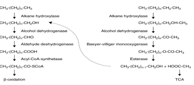
2.6.2.1. Metabolic Machinery………...………… 40
2.6.2.2. Pathways for Degradation of n-Alkanes………..……… 40
2.7. Summary of Relevant Literature by Subject Coverage... 42
3. MATERIALS AND METHODS... 51
3.1.1. Collection of Samples... 51
3.1.2. Media Compositions Used ……… 52
3.2. Method... 53
3.2.1. Isolation of Microorganisms………... 53
3.2.2. Determining the Optimum Growth Conditions... 53
3.3. Morphological, Physiological and Biochemical Tests... 54
3.3.1. Gram Staining... 54
3.3.2. Spore Painting... 54
3.3.3. Mobility and Oxygen Requirement... 55
3.3.4. Starch Hydrolysis Test... 55
3.3.5. Catalase Test... 55
3.3.6. Gelatin Hydrolysis Test... 56
3.3.7. Oxidase Activity... 56
3.3.8. Degradation of Tyrosine... 56
3.3.9. Urease Test... 57
3.3.10. Citrate Test... 57
3.3.11. Sodium Azide Sensitivity... 57
3.3.12. NaCl Tolerance... 58
3.3.13. Lysozyme Sensitivity ……….……… 58
3.3.14. Antibiotic Sensitivity Test... 58
3.4. 16S rRNA Gene Analysis... 59
4.2. Morphological Analysis... 61
4.3. Determination of the Optimal Growth Conditions... 64
4.3.1. Determination of the Optimal Growth Temperature…….……….…….……… 64
4.3.2. Determination of the Optimal Growth pH……….……….………… 65
4.3.3. Determination of the Optimum Growth Time……… 66
4.4. Biochemical and Physiological Tests... 67
4.4.1. Mobility and Oxygen Requirement... 67
4.4.2. Starch Hydrolysis Test... 68
4.4.3. Catalase Test... 69
4.4.4. Gelatin Hydrolysis Test... 70
4.4.5. Oxidase Activity... 70
4.4.6. L-tyrosine Test... 71
4.4.7. Urease Test... 72
4.4.8. Sodium Citrate Test... 73
4.4.9. Sodium Azide Sensitivity... 73
4.4.10. NaCl Tolerance... 75
4.4.11.Lysosome Hydrolysis Test……… 75
4.4.12. Antibiogram (Disc Diffusion Method) Test... 76
4.5.16s rRNASquence Analysis……….………..………… 87
4.6. Degradation Test and GC Analyses of Petroleum Products………….………… 91
5. RESULTS AND DISCUSSION……… 99
6. CONCLUSIONS AND FUTURE PROSPECTS……….…………...…. 119
7. REFERENCE... 121 C.V………...
KURDISTAN REGION IN IRAQ MASTER THESIS
SHELAN SHUKRI KARAM DEPARTMENT OF BIOLOGY
INSTITUTE OF NATURAL AND APPLIED SCIENCES UNIVERSITY OF DICLE
2016
In our daily life and in industry, petroleum hydrocarbons are important as primary energy resources. Crude oil is used as raw matter in numerous industries, including the refinery-petrochemical industry, where crude oil is refined through different technological processes into usable products. Most of the petroleum goes in the ecosystem via leaks and accidental spills of coastal oil refineries. As a result, it directly or indirectly affects human safety and ecological systems. Toxic organic materials in crude oil can be carcinogenic, mutagenic and potent immunotoxicants and discharged into the soil and groundwater. One of these toxic materials is aliphatic hydrocarbons, particularly alkanes of shorter chain length (C10-C20) are more toxic.
The aims of this study are to isolate and identify the bacterial strains from petroleum contaminated sites as well as determine the ability for the biodegradation of crude oil.
For screening of hydrocarbon degrading microorganisms, different bacterial strains were isolated from different petroleum areas of Iraqi Kurdistan. The final isolates selected as the superiors hydrocarbon degraders were identified by morphological, physiological, biochemical screening tests and 16SrRNA sequence analysis. According to 16S rRNA gene sequencing analysis, five strains designated as 2B, 3B, 4A, 6C and 8F were found to be closely related to Cronobacter malonaticus (96.31% similarity), Cronobacter dublinesis subsp. dublinesis (95.48% similarity), Serratia marcescens subsp. marcescens (92.94% similarity), Pseudomonas aeruginosa (96.23% similarity) and Acinetobacter calcoaceticus (99.08% similarity) respectively.
GC-MS analysis of hydrocarbons with chain length (C9- C34) showed that 2B, 4A, 6C and 8F were able to degrade 84.76%, 73.25%, 91.62%, and 41% n-alkanes, respectively, in the crude oil incubated for 10 days. Along with the selected individual strains, a mixed bacterial consortium prepared using the above strains were also used for degradation studies. The mixed bacterial consortium did not lead to enough growth and degradation. The mixed bacterial consortium degraded 69.50% percentage of n-alkanes ranging from C11 to C34.
Key Words: n-Alkanes, crude oil, bacterial identification, biodegradation, 16S rRNA squence analysis, GC-MS analysis.
ARAŞTIRILMASI YÜKSEK LİSANS TEZİ SHELAN SHUKRI KARAM
DİCLE ÜNİVERSİTESİ FEN BİLİMLERİ ENSTİTÜSÜ
BİYOLOJİ ANABİLİM DALI
2016
Günlük yaşamda ve sanayide, petrol hidrokarbonları birincil enerji kaynağı olarak önemli bir yere sahiptir. Hampetrol rafine edildiği yerlerde, kullanılabilir ürünler haline farklı teknolojik süreçler yoluyla Rafineri-Petrokimya endüstrisi dahil, birçok endüstride hammadde olarak kullanılmaktadır. Petrol büyük oranda sızıntı ve kıyı petrol rafinerilerinin kazayla dökülmesi yoluyla ekosistem içerisine girer. Sonuç olarak, doğrudan veya dolaylı olarak insane ve güvenliğini ve ekolojik sistemleri etkiler. Hampetrolün içerisindeki toksik organic maddeler, kanserojen, mutajenik ve güçlü immünotoksik olabilmekte aynı zamanda toprak ve yeraltısularına deşarj olabilmektedirler. Bu toksik maddelerden biri olan alifatik hidrokarbonlar, özellikle alkanların daha kısa zincirli (C10-C20) olanları çok toksiktir.
Bu çalışmanın amacı, petrolle kirlenmiş bölgelerden bakteri izolasyonu ve tanımlanmasına ilaveten petrol parçalama yeteneklerini belirlemektir.
Hidrokarbonları parçalayan bakterilerin taranması için, Irak’ın Kürdistan bölgesindeki farklı petrol alanlarından farklı bakteri türleri izole edilmiştir. Petrolü parçaladığı belirlenen son izolatlar, morfolojik, fizyolojik, biyokimyasal testler ve 16S rRNA dizi analizi yapılarak tanımlanmıştır. 16S rRNA dizi analizine gore, 2B, 3B, 4A, 6C ve 8F şeklinde adlandırılan bakteri suşlarının sırasıyla Cronobacter malonaticus (% 96.31 benzerlik), Cronobacter dublinesis subsp. Dublinesis (%95.48 benzerlik), Serratia marcescens subsp. marcescens (%92.94 benzerlik), Pseudomonas aeruginosa (%96.23 benzerlik) ve Acinetobacter calcoaceticus (%99.08 benzerlik) türlerine yakın oldukları bulunmuştur.
%1 Ham petrolde bulunan C9-C34 aralığındaki hidrokarbonların GC-MS analizi, 2B, 4A, 6C ve 8F’in 10 günlük inkübasyon ile %1’lik ham petrolde bulunan n-alkanları sırasıyla %84.76, %73.25, %91.62 ve %41 oranlarında parçaladıkları tespit edilmiştir. Seçilen saf türlerin yanında yukarıda belirtilen izolatlardan oluşan karışık bakteriyel konsorsiyumu da degradasyon çalışmaları için hazırlanmıştır. Karışık bakteri konsorsiyumu, yeterli üreme ve degradasyona yol açmamıştır. Karışık bakteriyel konsorsiyumu, C11 den C34’e kadar olan n-alkanları %69,50 oranında parçalamıştır.
AnahtarKelimeler: n-alkanlar, ham petrol, bakteriyel identifikasyon, biyodegradasyon, 16S rRNA dizi analizi, GC-MS.
Table 2.1. Classification methods: history of bacteria and Archaea... 8
Table 2.2. Some of the characteristics and methods are used in classification methods of Bacteria and Archaea... 9
Table 2.3. Some genotypic methods utilized in bacterial taxonomy... 19
Table 2.4. Applications for Acinetobacter sp. and their products ... 29
Table 2.5. Enzyme classes oxidizing alkanes ... 38
Table 3.1. Oven temperature program of gas chromatography ... 60
Table 4.1. Sodium azide sensitivity (OD600)... 74
Table 4.2. Tolerance of ısolated bacteria on different NaCl concentration (OD600)... 75
Table 4.3. Tolerance of isolated bacteria on % 0.001 Lysosome concentration (OD600)……….. 75
Table 4.4. Effects of antibiotics on the isolated bacteria... 86
Table 4.5. 16S rRNA sequence analysis about 976 base pairs in length of Cronobacter sp. strain 2B... 87
Table 4.6. 16S rRNA sequence analysis about 805 base pairs in length of Cronobacter sp. strain 3B ... 88
Table 4.7. 16S rRNA sequence analysis about 856 base pairs in length of Serratia sp. strain 4A... 88
Table 4.8. 16S rRNA sequence analysis about 796 base pairs in length of Pseudomonas sp. Strain6C... 88
Table 4.9. 16S rRNA sequence analysis about 797 base pairs in length of Acinetobacter sp. strain 8F... 89
Table4.10. The Utilization range and degradation percentage of n- alkanes in crude Oil by isolated strains... 97
Table 5.1. Morphological, physiological and biochemical tests on selected Coronobacters sp……….. 103
Table 5.2. Morphological, physiological and biochemical tests on isolated Serratia sp ... 107
Table 5.4. Morphological, physiological and biochemical tests on isolated
Figure 2.1. The three branches of life all possess a DNA-based genome... 16
Figure 2.2. Classification of bacteria by 16S rRNA sequence analysis... 17
Figure 2.3. Hierarchical arrangement in taxonomy modified... 18
Figure2.4 16S rRNA sequence analysis of Cronobacter sp... 21
Figure 2.5 16S rRNA sequence analysis of Serratia sp. by relation between types 23 Figure 2.6. Phylogenetic tree of some Pseudomonas species drawn by using 16S rRNA sequence analysis……… 26
Figure 2.7 Phylogenetic tree of some Acinetobacter species drawn by using 16S rRNA sequence analysis……….. 30
Figure 2.8. Zakho region of Kurdistan is polluted through crude oil, as well as hazard from h2s gas……… 32
Figure.2.9. Schematic of alkane degradation in gram-negative bacteria………….. 39
Figure 2. 10 Most frequent pathways for the degradation of n-alkanes by terminal and sub-terminal oxidation………. 42
Figure 3.1. Samples were taken from oil wells (a and b) in Zakho region……….. 51
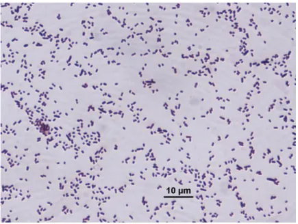
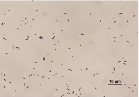
Figure 4.1. Gram staining of 2B……….. 61
Figure 4.2. Gram staining of 3B……….. 62
Figure 4.3. Gram staining of 4A……….. 62
Figure 4.4. Gram staining of 6C……….. 63
Figure 4.5. Gram staining of 8F……… 63
Figure4.6. Effect of temperature on the growth of isolated bacterial strains………. 64
Figure 4.7. Effect of pH on the visolated bacterial strains…..………. 65
Figure 4.8. Effect of time-course on bacterial growth………. 66
Figure 4.9. Motility and oxygen requirement test on the isolates………. 67
Figure 4.10. Isolated bacteria showing the zone of clearance with iodine solution on starch plates………. 68
Figure4.11. Catalase test on isolated bacteria……… 69
Figure4.15. Urease test after 24 h incubation at 30°C……….. 72
Figure 4.16. Ability of isolated bacteria utilizing citrate……… 73
Figure 4.17. Antibiotics Disc diffusion test of 2B………. 77
Figure 4.18. Antibiotics Disc diffusion test of 3B……….. 79
Figure 4.19. Disc diffusion susceptibility test on 4A………. 81
Figure 4.20. Disc diffusion susceptibility test on 6C………. 83
Figure 4.21. Antibiotics disc diffusion test on 8F ………. 85
Figure. 4.22. Partial 16S rRNA sequence-based phylogenetic neighbour-joining….... 90
Figure. 4.23. GC profiles of crude oil remaining in the BSM after incubation without (a) and with (b) strain 2B………... 92
Figure. 4.24. GC profiles of crude oil remaining in the BSM after incubation without (a) and with (b) strain 4A………... 93
Figure. 4.25. GC profiles of crude oil remaining in the BSM after incubation without (a) and with (b) strain 6C………... 94
Figure. 4.26. GC profiles of crude oil remaining in the BSM after incubation without (a) and with (b) strain 8F………... 95
Figure. 4.27. GC profiles of crude oil remaining in the BSM after incubation without (a) and with (b) mixed strain………... 96
BSM L : Liter mL : Milliliter µL : Microliter g : Gram g/L : Grams / liter mg : Kilogram mg/mL : Milligram / milliliter min : Minute
mL/min. : Milliliter / minute
v/v : Volume / Volume (volume / volume)
nm : Nanometer
µm : Micrometer
N : Normal
mm : Millimeter
µg : Microgram
K2HPO4 : Dipotassium phosphate
KH2PO4 :Potassium dihydrogen phosphate
Na2HPO4.7H2O : Disodium hydrogen phosphate heptahydrate
NH4Cl : Ammonium chloride
MgSO4 : Magnesium sulfate
CaCl2 : Calcium chloride
FeCl3 : Iron (III) chloride
MnSO4 : Manganese (II) sulfate
ZnSO4. 7H2O : Zinc sulfate heptahydrate
(NH4) 6 MO7O24.4H2O : Ammonium heptamolybdatetetrahydrate
NMR : Nuclear Magnetic Resonance
GC-MS : Gas Chromatography / Mass Spectrometry
LC-MS : Liquid Chromatography / Mass Spectrometry
I2 : Iodine
NaCI : Sodium-chloride
H2O : Water
H2SO4 : Sulfuric acid
HCI : Hydrogen chloride
NaOH : Hydroxide sodium
DNA : Deoxyribonucleic acid
rRNA : Ribosomal ribonucleic acid
OD : Optical Density
min : Minute
PCR :Polymarase chain reaction
PPM : Parts per million
PH : Potential of hydrogen
RFLP : Restriction fragment length polymorphism
% : Percentage
1. INTRODUCTION
The use of petroleum over the world is very common and sometimes the petroleum accidents take place. Nearly every year five million tons of crude oil and refined oil to enter the environment as a result of anthropogenic sources such as oil spills (Malatova 2005, Mahjoubi et al. 2013). Most of the oil comes from tankers, barges and other vessels as well as from land pipeline spills. Extensive changes in marine and terrestrial ecosystems resulting from the grounding (Malatova 2005).
Most hydrocarbons be falling in crude oil have toxic effects as result of their chemical structure (Al-Wasify and Hamed 2014). Both aliphatic and aromatic compounds are included in these toxic hydrocarbon compositions, such as polycyclic aromatic hydrocarbons (PAHs), whose toxicity expanded proportionally to the number of carbon atoms in the compound and individually in the case of PAHs with higher than four-member rings in its structure.
Toxic organic materials in crude oil can cause sub-lethal chronic toxicity and acute lethal toxicity, or both, depending on the exposure, dosage, and the organism exposed (Gardner et al. 1991, Orisakwe et al. 2004). One of these toxic materials is Polycyclic aromatic hydrocarbons (PAHs), that is the major problem as environmental contaminants (Al-Wasify and Hamed 2014). These polycyclic aromatic hydrocarbons (PAHs) are a group insolubile organic composites in water, composed of two or more fused aromatic rings in their chemical structure. PAHs are of environmental and humanitarian concern, are dangerous pollutants and health jeopardy, because of their toxic, mutagenic and carcinogenic characteristics (Hesham et al. 2014). Another hydrocarbons are toxic aliphatic hydrocarbons. Alkanes may constitute 50% to 95% of crude oil, related on the oil source. Alkanes compound of larger chain length (C20- 40C) are hydrophobic solids difficult to degrade. Also, they have less toxic effect than hydrocarbons of shorter chain length (C10- C20), while those of Longer chain n-alkanes and PAHs in crude oil were commonly considered to be only slightly biodegradable due to their higher hydrophobicity (Xia et al. 2014).
Various groups of microorganisms have been found in crude oil contaminated site, despite the toxicity of the chemical synthesis occurring in crude oil (Wolicka and Borkowski 2012). The variety and the number of microorganisms at a given location may help to characterize that site with regard to the toxicity of these hydrocarbons to the microbiota, age of the spill and condensation of the pollutant. For that matter, in a crude oil-polluted soil, the biodiversity and microbial spread of certain microbe(s) may indicate how well the soil is suffering the growth of that microbe(s). Fresh spills and/or high levels of pollutants often kill or inhibit large sectors of the soil microbial population, while soils with lower levels or old pollution show greater numbers and variety of microorganisms (Saadoun et al. 2008). In addition, microbial isolates from the soils that are historically subjected to hydrocarbon pollution exhibit a higher potential of biodegradation than others with no history of such showing.
The improvement of oil removal was studied extensively since the 1970s. One of the methods which are suggested is abiotic method. Physical, mechanical, removal of oil is one types of Abiotic methods, ex: using of the oil sorbents. Those sorbents facilitate in transforming oil into a transportable form for short-run storage. But, most of the used sorbents terminate in the landfills. Also, physiochemical methods often chemical organic solvents with or without surfactants used, especially shoreline cleaners (Mills et al. 2003, Malatova 2005). The shoreline cleaners with surfactants emulsify the adsorbed oil, which transported deeper into the shoreline soil or is entrained beside water. The physicochemical techniques such as washing, surfactant and incineration guidance to the initiation of more toxic compounds in the environment (Oboh et al. 2006). Chemical agents are used in most of the physicochemical methods. For this reason, they cause toxicity killing off aquatic organisms. They produce another source of alloy and also augment the oil recovery cost. Another method is chemical methods. The oil-solvent mixtures usually used in skimming methods.Abiotic losses due to dispersion, evaporation of low molecular hydrocarbons and photooxidation (involve only aromatic compounds), which play a major role in the decontamination of the oil spill environments (Mills et al. 2003).
Also, another method is bioremediation, which is a natural method of removing ptroleum and oil-derived products contamination from the habitats. This is a biotic method or biological procedure that uses living organisms to remove completely or decrease pollutants (organic and inorganic compounds) from polluted zones. It is a natural process
without the addition of chemical materials to the habitats and is cheaper, compared to physiochemical methods. Bioremediation offers a practical alternative to oil spill response. Many soil microorganisms, bacteria and fungi, in aerobic condition, convert crude oil hydrocarbons into composites that are non-toxic or deportment their complete mineralization to compounds that are inorganic. This technology is fount to be effective for the treatment of oil pollution. One reason is that most of the molecules in crude oil and refined products are biodegradable (Malatova 2005, Teramoto et al. 2009, Wolicka and Borkowski 2012). So, bioremediation is an effective method for removing hydrocarbon pollution from different environments.
Stimulation of microorganisms in contaminated areas may solve the polluted problem with a low cost. Methods of conventional remediation commonly move the pollution problem to another location or generate preferable costs. In mutuality, bioremediation is a non-perilous and natural process to the environments. Bioremediation can be performed at the contaminated site without changeable the existing land management plans (Vidali 2001). Use any method, bioremediation has its drawbacks, not all compounds are sensitive to relatively fast and perfect degradation. In some of cases, products of degradation of oil paronymous products have toxic effects on microorganisms. Furthermore, bioremediation requires the usage of complex techniques in the presence of mixtures of compounds that are randomly dispersed in the medium. Bioremediation lasts longer than chemical methods; moreover, its correct duration cannot be precisely determined. In-situ bioremediation ways are not always easy to monitor, whilst ex-situ bioremediation methods are rather expensive due to transportation costs and soil storage (Wolicka and Borkowski 2012). Laboratory and field studies let us the designation of the favorable parameters for the bioremediation of soil contaminated by oil hydrocarbons: biomass content over 105 cells/g dry mass, temperature 20- 30 °C, pH:6.5- 7.5, relative humidity 20- 30%, oxygen content at least, 0.2 mg/l hydrocarbons, Carbon: Nitrogen: Phosporous (C: N: P) = 100:10:1 or 70:7:1 (Wolicka and Borkowski 2012).
Hydrocarbon-degrading microorganisms are generally distributed in aquatic, freshwater, and soil ecosystems. Most of these organisms, the superiority of which are bacteria have been characterized and classified utilizing cultural, biochemical and molecular techniques. Bacteria have developed millions of years ago regulatory systems that guarantee the synthesis of enzymes so that the original attack on these compounds is
produced only when required (Oboh et al. 2006). Some of microorganisms in soil and ground water may be adapted exhibiting selective enrichment and genetic changes using some composition of crude oil as a source of energy. These compounds via microorganisms are transferred to other levels of the food chain (Gardner et al. 1991, Orisakwe et al. 2004). The microbial bioremediation method is better than physical and chemical methods like volatilization, photooxidation, chemical oxidation to removal and cleaning up PAHs, and also these methods are not safe and cost effective (Al-Wasify and Hamed 2014). The most important subject is that these processes are natural and the ultimate products of the microbiological degradation are carbon dioxide, water and inorganic compounds. The effectiveness of hydrocarbon degradation by bacteria does not lead on the number of carbon atoms in the compound (Chikere et al. 2011, Jain and Bajpai 2012). Bacterial application as microbial bioremediation have long been considered as one of the predominant hydrocarbon degrading agents found in the environment, which are available (Al-Saleh et al. 2009). Despite the trouble and expensive in cost, it seems that oil will remain a major origin of energy in the next several decades because a reliable option has not yet been determined (Oboh et al. 2006).
The primary aim of this study was to isolate and identify the most better hydrocarbon-degrading microorganisms in the contaminated soil and aquatic places in Kurdistan region that is located at north of Iraq, to investigate the biodegradation potential of any strain and a mixed bacterial consortium by detailed chemical analysis using GC. MS test, in order to provide a microbial mixture that would be suitable in bioremediation processes with crude oil spills.
Therefore, in this study, we have aimed to isolate and identify the crude oil degrading bacteria from petroleum-contaminated sites in the oil fields (Zakho, Bardarash, Mreba, Atrush, Koya) in Kurdistan region of Iraq. Moreover, we have studied the ability of these isolated bacterial strains to degrade the aliphatic hydrocarbons by looking the total hydrocarbon rates.
2. REVIEW
2.1. History of Bacterial Systematic
There is the tremendous diversity of microorganisms in nature for scientists to study the bacterial systematics. The ultimate goal of biological classification and characterization of the organisms is making a canonical way to regulate them in the group (Schleifer 2009). Systematic and identification plays a central role in microbiology disciplines (Ludwig 2007). The discovery of new microorganisms made continuously changing in bacterial systematics, because they have different characters and put new evidence in the appropriate place (Arda 2011). Classification of the named species of bacteria did not rise easily or fast (Garrity et al. 2001). Western scientific taxonomy started in Grecian some hundred years BC. The most important works are cited and the development of taxonomy (with the focus on botanical taxonomy) are described up to the rotation of the Swedish botanist Carl Linnaeus (1707–1778), who discovered modern taxonomy (Manktelow 2010) and are here separated into pre-Linnaean, most had been botanical taxonomies, and post-pre-Linnaean, other taxonomies after Linnaeus taxonomy. So, the starting point of the novel or modern taxonomy was started by Carl von Linnaeus, who discovered a system known as Linnaean classification for categorization of organisms and binomial nomenclature for naming organisms. In this system, a microorganism is defined by two scientific names. One of these is (genus) and another name is (species) (Arda 2011). In 1773 and 1774 the Danish naturalist Otto Muller was the first to try a systematic arrangement of the animalcules, but he did not create a clear difference between what we now call protozoa and bacteria. For his last performance, however, which was published in 1786, he did produce two form genera, Monas and Vibrio, which included bacteria and accommodated the punctiform and elongated kinds (Logan 1992). The mycologist Link (1809) reported the first bacterium that we know today, named Polyangium vitellinum that is now placed by the fruiting myxobacteria (Murray and Holt 2001). Bizio (1823) found Serratia marcescens (Garrity et al. 2001). The famous attempt by Ehrenberg (1838) and Cohn (1872, 1875), to gain more than a few names that still survive (e.g. Bacillus, Spirillum and Spirochaeta). Most explanations could hold only on behavior,
habitat and shape since microscopy was the important tool (Garrity et al. 2001). Ferdinand Cohn (1872), by his attempted to classify the known bacteria, six genera of bacteria (Micrococcus, Bacillus, Bacterium, Spirillum, Vibrio, and Spirochaeta) and next (1875) extended the classification to include the cyanobacteria while adding further bacterial genera (Asco Coccus, Sarcina, Leptothrix, Beggiatoa, Crenothrix, Calothrix, Streptococcus [not those recognized today], and Streptothrix) (Schleifer 2009). Buchanan (1925) introduced that Cohn‘s 1875 classification could be the opportunity date for bacterial nomenclature instead of Linnaeus‘ Species Plantarum of 1753 and presented the different ideas for the suitable starting date for bacterial nomenclature, the actual change in starting date proposed in the revised Bacteriological Code (Garrity et al. 2001).
During most of the nineteenth century to allow all system, Nomenclature rules have developed during the 19th and 20th century, and during the last decade standard nomenclature has been challenged by supporters of the Phylocode (Manktelow 2010), With the immersion of study fields as phylogenetics, cladistics, and systematics, the Linnaean system has improvements to a system of novel biological classification based on the evolutionary relationships between organisms, both living and extinct. Migula in 1897 has developed a system, taking into account not only microorganisms morphology, but also the color (colony) and some physiological characteristics (such as nitrogen fixation) and the work was published under the name ―System of the Bacteria‖. In the same year, Lehmann and Neumann published their atlas under the name of ―Diagnostic Bacteriology‖ (1886) (Arda 2011). The latter was apparently the superlative success of the systems and was used in sequential editions till 1930, especially in Europe. F.D. Chester constructed reports in 1897 and 1898 of bacteria of interest in horticulture to succeed in 1901 by his ―Manual of Determinative Bacteriology‖. Chester had recognized that the lack of an organized assembly of descriptions and a system of the classification made the identification of separated as known species and the identification of new species an impossible duty (Garrity et al. 2001). Orla-Jensen (1909) was influential because he resolved and explicited of ―natural relationships‖, reflecting further physiological characters. He bounded genera and species on the basis of characteristics such as metabolic products, temperature ranges for development and fermentation of several sugars in addition to morphology.
Bergey‘ s Manual of Determinative Bacteriology resulted from the interest and efforts of a group of colleagues in the Society of American Bacteriologists (SAB), a committee was formed by scientists such as L.A. Rogers, C. Krumwiede Jr., R.E. Buchanan, and G.H. Smith, which were published in the Journal of Bacteriology in 1917.In this study, bacteria based on phenotypic features, was classified as single-cell plants (Arda 2011). In 1923, a book ―Manual of Determinative Bacteriology‖ was published by a committee from SAB headed by D.H. Bergey. There were two ―starters‖ for the Manual: one of them was R.E. Buchanan and D.H. Bergey. The 7th edition of this book was published in 1957 and the bacteria was still classified as a plant (Schleifer 2009).
Molecular/genetic technology was well established in 1974 when the 8th edition of Bergey‘ s Manual of Determinative Bacteriology was published, however, was not still widely employed to play a role in broad decisions in taxonomy. The briefer Bergey‘ s Manual of Determinative Bacteriology in 1977 was published (Garrity et al. 2001). The first of four books of Bergey's Manual of Systematic Bacteriology between 1984-1986 were published, which became the best existing bacterial classifications, that were based on phenotypic characters (observable appearance of the genotype) with a notation for producing identification schemes (Logan 1992). In 1994 9th edition of Determinative Bacteriology was published.
A major start forward in the study of bacteria occurred in 1977 when Carl Woese recognized that Archaea have a separate line of evolutionary relationship from bacteria. This new phylogenetic taxonomy depended on the sequencing of 16S rRNA, and distributed prokaryotes into two evolutionary domains, as section of the three-domain system. The history of classification of bacteria and archea is shown in Table. 2.1 (modified from Schleifer 2009).
Table 2.1. Classification methods: history of bacteria and Archaea Time Period Basic Features in classification
Late 19th century Morphology, reproduction conditions, pathogenic potential
1900-1960 Morphology, physiology, biochemistry
1960-1980 Chemotaxonomy, digital classification, DNA-DNA Hybridization
1977 16S rRNA
1980-Present Genotypes Analysis, Multilocus series analysis, nucleotide similarity, all Genome Analysis
2.2. Bacterial Identification 2.2.1. Phenotypic Methods
Phenotypical methods are not directly linked to the DNA or RNA tests. It mostly covers classical or traditional phenotypic tests in the identification, which are used in the basic microbiological laboratory. Classic phenotypical features of bacteria are based on morphological, physiological and biochemical characteristics. Many of these features are shown to be unrelated parameters for genetic associations. However, as a whole, they provide descriptive information to identify taxa (Vandamme et al. 1996, Moraes et al. 2013).
Table 2.2. Some of the characteristics and methods are used in classification methods of Bacteria and
Archaea modified from (Black 1996, Madigan et al. 2010)
Category Characteristics
Morphology Colony morphology, Gram reaction, cell size and shape, pattern of flagellation,
the presence of spores, arranged in pairs, clusters or filaments.
Motility Nonmotile, gliding motility, swimming (flagella) motility, swarming, motile by gas vesicles
Metabolism (Nutrition)
Mechanism of energy conservation (phototroph, chemoorganotroph,
chemolithotroph), utilization of individual carbon, nitrogen, or sulfur compounds; fermentation of sugars, nitrogen fixation, growth factor requirements
Physiology Temperature, pH, and salt ranges for growth; response to oxygen (aerobic, facultative, anaerobic), presence of catalase or oxidase, production of extracellular enzymes
Biochemistry Fatty acids, RNA molecules, nature of cellular components such as cell wall (Presence or absence of peptidoglycan; amino acid composition of cross-links), pigments, biochemical test
Genetics Percentage of DNA base, DNA hybridization
Other traits Luminescence, antibiotic sensitivity, serotype; production of unique compounds, for example, antibiotics
2.2.1.1. The Morphological Identification of Bacteria
The identification and classification of organisms have continually been amongst the pre-eminence of the early scientists. Although botanists and zoologists have a superfluity of morphological traits with that to identify plants and animals, the morphological traits of identifying bacteria are few and limiting. This has not produced a peremptory challenge, but an opening for creativity. Gram staining was as a result of the creating penetration of Hans Christian Joachim Gram (1850-1938) to classify bacteria based on the structural characteristic of their cell walls. It meant that bacteria
could be differently classified by Gram staining as Gram negative or Gram positive, a convenient for identification and classification hand tool that continue useful today. Howsoever there are some morphological traits and few changes in those traits, identity based on morphology Syne has considerable taxonomic value. Another method is growing on the media in order to identify their cultural characteristics since various species can exhibit very different colonies (Christopher and Bruno 2003). Any colony has specifications that may be unique to it and this may be helpful in the primitive identification of a bacterial species. The characteristic of the colonies on solid agar media include theirs shape (round, irregular or rhizoid), size (the width of the colony: small, medium, large), surface (smooth, wavy, rough, granular, papillate or glistening), elevation (convex, concave,umbilicate), margin (entire, wave, created, ciliate or curled), structure/-opacity (opaque, translucent / transparent), color (pigmentation: yellow, green between others), degree of growth (scanty, medium or profuse) and nature (separate or confluent, filiform, spreading or rhizoid). Cell shape has also been employed in the characterization and grouping of bacterial species (Cabeen and Jacobs-Wagner 2005). The most basic shapes of bacteria are cocci (spherical in shape), bacilli (rod-shaped) and spirilla (spiral-shaped). Observations of bacterial morphologies are done by light microscopy, which is helped by the application of stains (Tshikhudo et al. 2013). Gram staining and bacterial morphologies usually are employed as the first steps of identification. Light microscopy was applied for identifying colonies of bacteria and morphologies of individual bacteria (Tshikhudo et al. 2013). An amalgamation of morphological identification with Scanning electron microscopy (SEM) and in situ hybridization (ISH) techniques (SEM-ISH) lead to a better understanding, to achieve the phylogenetic and morphological knowledge about bacterial species to be recognized using in situ hybridization with rRNA-targeted oligonucleotide probes (Kenzata and Tani 2012).
2.2.1.2. Physiological Properties
Physiological characteristics of bacteria show variations depending on bacterial types. Thus, reproduction temperature, incubation time, oxygen requirements, the
composition of the medium and other physiological characteristics needs to be investigated and determined (Gül-Güven 2007).
2.2.1.3. Biochemical Charactersistics
Biochemistry, seldom called biological chemistry, is the study of chemical method within and relating to living organisms. (David 2000). The significance of identifying biochemical activity in the assay of microorganisms is so much as various tests are used for this purpose. These include starch, gelatin, catalase, urea, oxidase, nitrate and nitrite reduction, as well as carbohydrate utilization test, selected and used depending on the type of microorganisms (Gül-Güven 2007).
2.2.2. Genotypic Methods
The traditional methods applied to the finding of either the colony characteristics or morphology of single cells remain reliable for bacterial species identification. However, these common techniques create some disadvantages. Firstly, a variation of culture due to changed environmental conditions may lead to vague results. Secondly, they are time-consuming and difficult. Thirdly, a pure culture is needed to undertake identified, making the identification of particular and unculturable bacteria complex and sometimes impossible. To pass these technical problems, newer and automatic methods which quickly and reliably recognize bacteria have been utilized by many laboratories worldwide. At least one of certain methods, particularly analysis of the 16S rRNA gene, join these traditional methods with the automated systems that produce novel techniques to a higher level of trust for bacterial identification (Tshikhudo et al. 2013). These methods are suitable for studies of unculturable bacteria and fastidious an appropriate tool for the metagenomic analysis of environmental samples (Petrosino et al. 2009, de Oliveira et al. 2011). Metagenomics is defined as unculturable bacteria or culture-independent investigation of the collective set of genomes of mixed microbial crowd in consortia that live in an environment, in plants or in animal hosts (Tshikhudo et al. 2013). Comparative analysis of the genome gives several traits for discriminating
between species of bacteria. Genotypic analysis has particular application in microbial taxonomy because of the insights it affords at the DNA level, with DNA–DNA hybridization and DNA profiling is used in microbial taxonomy (Madigan et al. 2010).
2.2.2.1.GC Ratios
Another method that has been employed to compare and describe bacteria is the GC ratio of their DNA. The GC ratio is the rate of guanine (G) and cytosine (C) in an organism‘s genomic DNA (Ludwig 2007, Madigan et al. 2010). GC ratio of total nucleic acids containing guanine and cytosine bases in an organism's DNA is expressed as a percentage. This ratio is a measure of DNA melting temperature, or determined by various methods such as chromatography (Madigan et al. 2010). The GC ratio in microorganisms variation over a broad range, with an amount as low as 17% and as high as nearly 80% between species of Bacteria and Archaea, a range that is moderately broader than in eukaryotes (Schleifer 2009, Madigan et al. 2010). However, GC ratios are utilized less generally in bacterial taxonomy than used in the past (Madigan et al. 2010)
2.2.2.2.16S rRNA Analysis
Ribosomal RNA genes are a significant part of the protein synthesis machinery. They are omnipresent and hence classification based on the analysis of ribosomal RNA genes does not leave out each of the known bacteria. 16S rRNA is a component of the small ribosomal subunit of RNA (30S Ribosomal subunit) and sometimes referred as SSU rRNA. 98% of 16s rRNA of all bacteria similar, there for its same species (Woese 1987, Glazer and Nikaido 2007). Carl Woese in the early 1970s used rRNA for comparison, because rRNA is present and performs an identical function in every cellarorganisms, and more importantly, it‘s sequence has changed extremely slowly during the course of evolution. According to this criterion, the analysis of rRNA genes is a fitting tool for bacterial species identification and systematic categorization. Therewith, ribosomal RNA genes are saved of evolution, but have sufficient variation to
distinguish between taxa (Woese 1987). In prokaryotes, rRNA genes are situated in copies of three or four in a single genome (Glazer and Nikaido 2007, Tshikhudo et al. 2013). The 16S rRNA gene is a suitable strong tool for identifying and classifying bacteria and its length of nearly 1,500 bp is enough for bioinformatic analysis (Janda and Abbott 2007). Analysis and dissociation of the 16S rRNA gene include amplification of the gene by polymerase chain reaction (PCR) and gene sequencing of the resulting PCR products. The gene sequence can be matched with prior collected sequences obtained from several DNA databases of the 16S rRNA gene. It is realized that almost all new sequences trusted have some matches up to some levels, but a 16S rRNA gene copy which does not match that of any known bacterial species to a particular level then it is supposed to be a new species (Tshikhudo et al. 2013). The 16S rRNA gene sequence is applied to identify bacterial species in natural specimens and to find phylogenetic relationships between them (Murat et al. 2010). It is created conceivable by the reality that all bacterial species include the 16S rRNA gene. So, it has a highly conserved area on which to design common primers, as well as hypervariable regions that are useful to identify the species. For bacterial identification, the 16S rRNA gene is regarded as the most broadly accepted gene (Tshikhudo et al. 2013).
2.2.2.3. DNA–DNA Hybridization
DNA-DNA Hybridization useful for comparing species of bacteria that are very closely related, when two organisms contain many identical or extremely similar genes, their DNAs are required to hybridize in approximate relationship to the similarities in their DNA sequences. DNA–DNA hybridization accordingly is helpful for separating between organisms as a complement to small subunit rRNA gene sequence (Glazer and Nikaido 2007, Madigan et al. 2010, de Oliveira et al. 2011). DNA–DNA hybridization is a sensitive technique for revealing delicate variation in the genomes of two organisms and is hence usually useful for differentiating so similar organisms. Although there is no fixed convention as to how much hybridization between two DNAs is important to select two organisms to the same taxonomic rank, hybridization values of 70% or greater are prescribed as evidence that two isolates are the same
species. Rates of at least 25% are expected to show that two organisms are in the same genus (Glazer and Nikaido 2007, Madigan et al. 2010).
Table 2.3. Some genotypic methods utilized in bacterial taxonomy
Method Description/application
DNA–DNA hybridization Genome-wide comparison of sequence similarity. Useful
for distinguishing species within a genius.
DNA profiling Ribotyping , AFLP, rep-PCR . Rapid method to distinguish between species and strains within a species
Multilocus sequence Typing Strain typing using DNA sequences of multiple genes.
High resolution, useful for distinguishing even very closely related strains within a species.
GC ratio Percentage of guanine–cytosine base pairs in the genome. If the GC ratio of two organisms differs by more than about 5%, they cannot be closely related, but organisms with similar or even identical GC ratios may be unrelated. Not much used now in taxonomy because of poor resolution
Multiple-gene or whole genome phylogenetic analysis
Application of cladistic methods to subsets of genes or to whole genomes of the organisms to be compared. Yields better phylogenetic picture than single-gene analysis
2.2.3. Chemotaxonomical Methods
Chemical structure of cellular components in prokaryotes classification is totally useful feature used in the assay. Chemotaxonomic methods are extensively performed particularly where the identification and classification by morphological and physiological analysis is not enough.
2.2.3.1. Fatty Acid and Lipid Analysis
The types and proportions of fatty acids present in cytoplasmic layer lipids and the external membrane lipids of bacteria are important phenotypic traits of interest. The technique for identifying these fatty acids has been nicknamed FAME, fatty acid methyl ester. FAME analyses are generally used in the characterization of new species of bacteria (Black 1996, Schutter and Dick 2000, Madigan et al. 2010).
FAMEs are abundant in the phospholipid bilayer of bacterial membranes (Black 1996, Tshikhudo et al. 2013). The fatty acids presentation of Bacteria differs from species to species in chain length and in the presence or absence of double bonds, circle, branched chains, or hydroxy groups. So, a fatty acid profile can often identify a special bacterial species (Black 1996, Madigan et al. 2010).
Fatty acid defines bacteria based on their physical properties. Profiles of fatty acids are determined by employing gas chromatography (GC), (Nnzeru et al. 2013). The fatty acid analysis for bacterial identification is based on the specific fatty acid structure of the cell wall. Various profile patterns of the extracted fatty acids are compared with pattern identification programs, that exist in microbial databases (Tshikhudo et al. 2013).
2.3. Classification of Bacteria
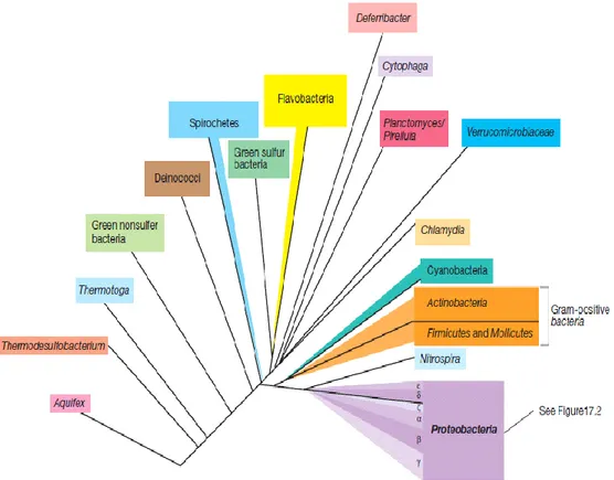
Currently based on genetic relatedness of rRNA, organisms divided into bacteria, archaea and eukaryotes (Figure 2.1). So great that is believed that these three groups may have diverged from an ancient progenitor rather than evolving from one another (Glazer and Nikaido 2007).
Over 7000 species of Bacteria and Archaea are now known, based principally on 16S rRNA gene sequencing, and thousands more, may be as many as 100 000–1000 000 are thought possible to exist. There are ∼1.8 million named species, and most species have yet to be described. Despite decades of effort and thousands of phylogenetic studies on diverse clades, we lack a comprehensive tree of life, or even a summary of our current knowledge. One reason for this shortcoming is lack of data.
GenBank contains DNA sequences for ∼411 000 species, only 22% of estimated named species (Madigan et al. 2010, Smith et al. 2014) . Classification is the organization of organisms into progressively further including groups on the basis of either phenotypic similarity or evolutionary relation. A species is made up of one to various strains, and similar species are arranged into genera (singular, genus). Similar genera are classified into families, families into orders, orders into classes, up to the domain, the highest level taxon. Nomenclature is the actual identifying of organisms and follows the binomial system of nomenclature devised by the Swedish medical doctor and botanist, Carl Linnaeus. The names are Latin or Latinized Greek origin, often representative of some key property of the organism, and are written in italics (Black 1996, Madigan et al. 2010, Staton 2015).
Figure 2.1. That the three branches of life all possess a DNA-based genome (Joseph & Schild 2010).
2.3.1. Phylogenetic Overview of Bacteria
Many major ancestors, called phyla, of Bacteria are known from laboratory cultures study, and several others have been identified from the sequencing and retrieval of ribosomal RNA (rRNA) genes of microbial communities in natural habitats. Figure
2.2 gives a summary of the phylogeny of main phyla of Bacteria for which laboratory cultures have been obtained based on 16S rRNA sequences. The gram-positive bacteria are a large group of essentially chemoorganotrophic Bacteria that can be separated into two subgroups called the Firmicutes plus the Actinobacteria (Manteca et al. 2006, Madigan et al. 2010). The Cyanobacteria are oxygenic phototrophic bacteria, with solstitial roots nearby those of the Gram-positive Bacteria. The remaining phylum of cultured Bacteria, the Proteobacteria (Figure 2.3), is by far the largest and most metabolically diverse of all Bacteria. Proteobacteria constitute the majority of known bacteria of industrial, medical, and agricultural significance. As a group, the Proteobacteria are all gram-negative bacteria (Madigan et al. 2010).
Figure 2.2. Classification of bacteria by 16S rRNA Sequence Analysis
2.4. Proteobacteria
The Proteobacteria is the largest and most metabolically different of all Bacteria. Proteobacteria form the majority and importance of known bacteria for industrial,
medical, and agricultural technology. As a group, the Proteobacteria are all Gram-negative bacteria. They show an exceptionally wide diversity of energy-generating mechanisms, with chemolithotrophic, chemoorganotrophic, and phototrophic species (Figure 2.2). The Proteobacteria are correspondingly diverse in terms of their relationship to oxygen (O2), with anaerobic, microaerophilic, and facultatively aerobic species known. Morphologically, they also show a wide type of cell shapes, including straight cocci, arched rods, filamentous, spirilla, budding, and appendaged forms.
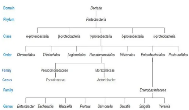
Based on 16S rRNA gene sequences, the phylum Proteobacteria (Figure 2.3) can be classified into six classes, Alphaproteobacteria, Betaproteobacteria, Gammaproteobacteria, Deltaproteobacteria, Epsilonproteobacteria, and Zetaproteobacteria, each containing many genera and species (Madigan 2010). The Proteobacteria are a larger group (phylum) of gram-negative bacteria. They include a wide diversity of pathogens, such as Escherichia, Salmonella, Helicobacter, Vibrio, and Yersinia, and various other important genera (Madigan 2010). Others are free-living (non parasitic), and include multiple of the bacteria responsible for nitrogen fixation (Woese 1987). Alphaproteobacteria grow at extremely low levels of nutrients and have abnormal morphology such as stalks and buds. They include agriculturally significant bacteria capable of inducing nitrogen fixation in symbiosis with plants. They are as well as motile by flagella and are microaerophilic, but several are non-motile. (htp://en.wikipedia.org/wiki/proteobacteria#external links).
Figure 2.3. Hierarchical Arrangement in Taxonomy modified from (modified from Palleroni et al. 1973,
Prescott 2002, Doughari et al. 2011).
2.4.1. Cronobacter sp.
The Cronobacter genus was recognized first in 2007 by Iversen et al (2006). As a clarification of the taxonomic relationship of the biogroups found among strains of Enterobacter sakazakii and revised in 2008. Differentiation among the newly defined Cronobacter sp. It is at first based on genotypic (DNA-based) analysis and is largely supported by biochemical traits.
Kingdom: Bacteria Phylum: Proteobacteria
Class: Gamma Proteobacteria Order: Enterobacteriales Family: Enterobacteriaceae Genus: Cronobacter (Joseph et al. 2011)
Cronobacter genus was identified as the species Enterobacter sakazakii by Farmer et al. in 1980 to honor the Japanese bacteriologist Riichi Sakazaki. They were phenotypically near to E. cloacae, so were put in the Enterobacter genus (Iversen et al 2007, Forsythe 2010).
The recently designated genus, Cronobacter is Gram negative rods that are approximately 3 μm by 1 μm in size, facultative anaerobic. The genus Cronobacter comprises oxidase negative, catalase positive. They are generally motile. Previously, G+C ratios of 57% and 56.7% have been reported for strains belonging to the type species (Iversen et al 2007). They were found to include in sections of the Enterobacteriaceae family and closely connected to Citrobacter and Enterobacter. Cronobacter can amplify over a broad temperature range. The lowest is near refrigeration (~5°C) and the maximum replication temperature (44- 47°C) is strain dependent. At normal room temperature (21°C), Cronobacter had a replicated time of 40-94 minutes. Last researches show the organism was extremely thermotolerant, but, subsequent work clarified that the organism was less thermotolerant. The microorganism has achieved with superiority due to its entirety with hard neonatal infections; necrotizing enterocolitis, meningitis and septicaemia , which can be fatal. Moreover, these are rare in infants, and infections arise in all age groups, although fortunately with less intense clinical sequels (Iversen et al 2004, 2006, Forsythe 2010).
Figure 2.4. 16S rRNA Sequence Analysis of Cronobacter species (Tall et al. 2015).
2.4.2. Serratia sp.
The generic name Serratia used since the 1920s, refers to the physicist Serafino Serrati. It was proved to be responsible for a case of food contamination (Caprette 2009).
Domain: Bacteria Phylum: Proteobacteria Class: Gammaproteobacteria Order: Enterobacteriales Family: Enterobacteriaceae Genus: Serratia (Bizio 1823)
Serratia was of great interest in medicine due to its antibacterial, antifungal, antiprotozoal, immunosuppressive, antimalarial, and anticancer activities (Juo 2001, Caprette 2009). It is motile, facultatively anaerobic, very small straight rods, between 0.5 and 1 µm in diameter and < 2 µm in length, arranged singly. These characteristics caused to rule out all other genera of Gram negative, rods, facultatively anaerobic, not assigned to one of these three families, placing the isolate in family Enterobacteriaceae. Typical colonies were 1-2 mm in size when not round, convex, crowded, glabrous, and slightly umbonate with entire margins, and opaque. New colonies were red in color. Cultures produced an unpleasant and somewhat sweet odor. strongly catalase positive, oxidase negative. Negative for indole production and mixed acid fermentation of glucose (methyl red test). Liquifies gelatin at 22˚C. All observations were consistent with the description of the genus in Bergey's Manual p. 477. 23 Serratia species were listed previously in the first edition book of Bergey‘s Manual. The only Serratia species recognized in the eighth edition of Bergey‘s Manual was S. marcescens (Dworkin and Falkow 2006, Grimont and Grimont 2006).
Serratia marcescens, which belongs to the Enterobacteriaceae family, has been reported to be an important agent of health care-related infections (HCRI), specifically standing out due to its dissemination potential and its high level of intrinsic resistance to the drugs used in Neonatology, and to antiseptic agents. This pathogen persists for long periods in the hospital environment because it colonizes the skin and the gastrointestinal tract of adults and newborns (Grimont and Grimont 2006, Maragakis et al. 2008). S. marcescens is known for its opportunistic characteristic that is most of their strains are resistant to most antibiotics (Caprette 2009). When food is contaminated with S.
marcescens it yields a pink to red discoloration due to the pigment prodigiosin production (Bennett and Bentley 2000).
The presence of R-factors makes most S. marcescens strains effectively resistant to used antibiotics, R-factors are a type of plasmid that holds one or more genes that causes the resistance; examples of the resisted antibiotics are ampicillin, first-generation cephalosporins and macrolides (http://en.wikipedia.org/wiki/Serratia_marcescens 2015.11.17).
2.4.3. Pseudomonas sp.
The genus Pseudomonas includes Genus I of the family Pseudomonadaceae. Five genera (Pseudomonas, Azomonas, Azotobacter, Azorhizophilus and Cellvibrio) have belonged to the family. Known to all chooser genera are some physiological properties, example aerobic, chemoorganotrophic metabolism, lack of fermentation, lack of photosynthesis, and ability to grow in the consumption of a large variety of organic substrates.
A more universal explanation of the genus carried in 1900 (Migula 1900), studying 75 species and registering Pseudomonas aeruginosa, previously identified as ―Bacterial aeruginosa‖, as the type species of the genus (Moore et al. 2006). Palleroni and his colleagues at the University of California at Berkeley were able to recognize different intrageneric groupings among, that time, the species comprising the genus Pseudomonas, by the usage of rRNA similarities it is currently recognized as the class ―Gammaproteobacteria‖ (Palleroni et al. 1973).
Kingdom: Bacteria Phylum: Proteobacteria
Class: Gamma Proteobacteria Order: Pseudomonadales Family: Pseudomonadaceae Genus: Pseudomonas
The original development of the genus Pseudomonas established a taxon based only on characteristics of cell morphology (Moore et al. 2006), with straight or slightly bent rods, 0.5–1.0 micrometer in diameter by 1.5–5.0 micrometer in length. They are Gram negative, motile by one or more than one polar flagella; rarely nonmotile. Endospore formation is seen in several species, but few; e.g., Pseudomonas violacea (Migula 1900, Moore et al. 2006).
Pseudomonas species are rich in the environment, available from different soil and water environments to animal and plant tissues (Moore et al. 2006, Silby et al. 2011). Particularly each habitat has temperatures between about 4 to 42° C, mesophilic,
a pH of 4 - 8, neutral PH condition. Oxidase-positive or negative, catalase-positive, chemoorganotrophic using organic compounds (simple or complex) as a potential habitat (Migula 1900, Moore et al. 2006, Franzetti and Scarpellini 2007, Raaijmakers et al. 2010). They are aerobic, in some cases anaerobic. They were not found in extreme thermophilic and acidophilic habitats (Moore et al. 2006).
Pseudomonas represents a genus able of using a large margin of organic and inorganic compounds and capable of living in different environmental conditions (Palleroni 2005, Moore et al. 2006, Franzetti and Scarpellini 2007, Raaijmakers et al. 2010 ). They may possess various strategies to tolerate periods of ―starvation‖, therefore they are present in water and soil ecosystems and are significant as human, plant and animal pathogens. In nature, Pseudomonas species are present as saprophytes and parasites (Moore et al. 2006, Silby et al. 2011).
The genus Pseudomonas is famous for its metabolic diversity and genetic flexibility. The species of Pseudomonas, in general, grow quickly and are solely regenerateed for their ability to metabolize a vast number of substrates. Pseudomonas is a genus of directly agreeable organisms, which looks to be a result of their simple nutritional requirements and their genetic and metabolic compatibility, the range of carbon compounds their usage, including toxic organic chemicals, like aliphaticand aromatic hydrocarbons. Strains of Pseudomonas species are frequently resisting antibiotics, disinfecting agents, deterging agents, metals, and organic solvents. Many of of the species are not accumulating poly-β-hydroxybutyrate granules, however, accumulating polyhydroxyalkanoates of monomer lengths higher of C4 can happen when growing on alkanes or gluconate (Migula 1900, Moore et al. 2006, Franzetti and Scarpellini 2007, Raaijmakers et al. 2010).
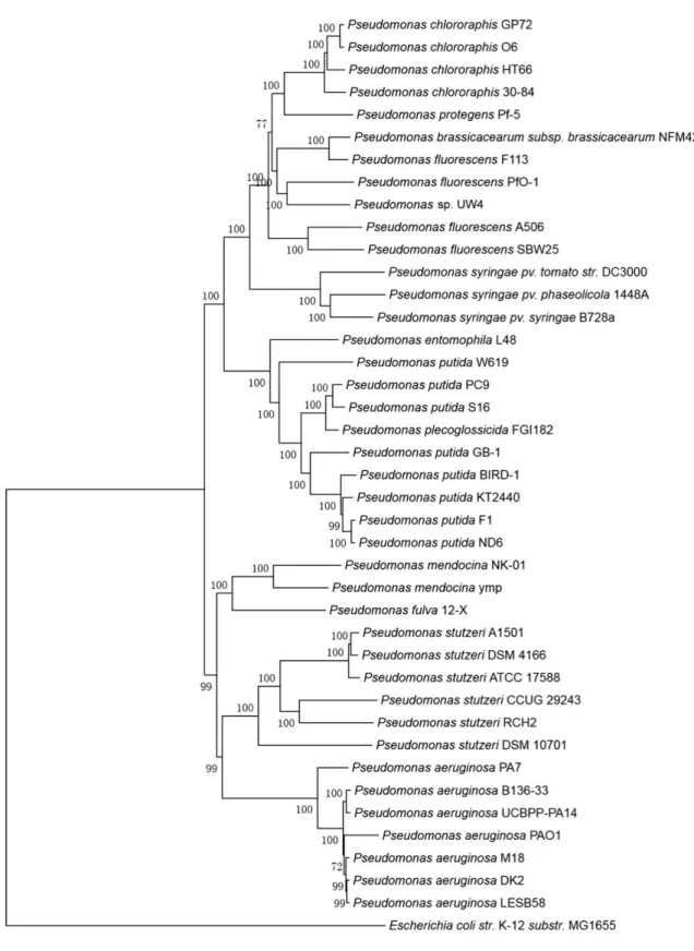
0.05
Figure 2.6. Phylogenetic tree of some Pseudomonas species drawn by using 16S rRNA Sequence
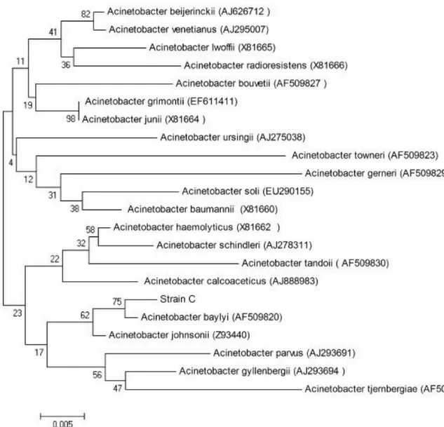
2.4.4. Acinetobacter sp.
The name ―Acinetobacter‖ originates from the Greek word ―akinetos‖ indicate ―unable to move‖, since these bacteria are not motile but they represent a twitching kind of motility (Doughari et al. 2011). The first strain of Acinetobacter sp. was separated from soil and classified as Micrococcus calcoaceticus by Beijerinck in 1911. Acinetobacter groups were incompletely known for long time classified as many different genera (Achromobacter, Alcaligenes, Cytophaga, Diplococcal, Bacterium, Herellea, Mima, Lingolsheim, Micrococcus, Moraxella and Neisseria). In fact, the Bergey‘s Manual of Bacteriology placed these bacteria, A. calcoaceticus, in the family Neisseriaceae with just A. calcoaceticus as a species and the pair subspecies A. anitratum and Acinetobacter lwoffii. The genus Acinetobacter was first discovered in 1954 by Brisou and Prevot to isolate the non motile forms from the motile one of the family ―Achromobactereae‖ (Barbe et al. 2004). Acinetobacter species are comprehensive in nature and the earth, the water surface, sewerage waters also It can be isolated from human skin. For example, A. calcoaceticus, A. pittii, A. johnsonii and A. guillouiae have been isolated from soil and water. Furthermore, Acinetobacter sp. can also be commensals of the human skin (Doughari et al. 2011).
Modern classifications which seem to have gained deep recognition amongst bacterial taxonomists have recognized this group of heterogeneous bacteria as gamma Proteobacteria classified in the order Pseudomonadales and the family Moraxellaceae (Doughari et al. 2011). So the taxonomical classification is given as;
Domain; Bacteria,
Phylum; Proteobacteria, Class; Gammaproteobacteria, Order; Pseudomonadales, Family; Moraxellaceae,
Genus —Acinetobacter (DNA G+C content 39–47%).
New classifications using cell form, absence of flagella, G+C content of DNA and nutritional characteristic, placed these organisms (A. baumannii, A. haemolyticus and A. calcoaceticus also other Acinetobacters) in the genus Moraxella, now known as
Acinetobacter (Barbe et al. 2004), depending on DNA-DNA hybridization studies. Thirty two species of Acinetobacter have now been distinguished, with 22 specific reliable names and the rest assigned numbers and referential to as a ‗genomic group delineated by DNA-DNA hybridization (Prashanth and Badrinath 2005) between the named species.
The genus Acinetobacter is composed of aerobic, nonmotile, catalase-positive, oxidase-negative, indole negative, Gram negative, non fermentative encapsulated coccobacilli rods (Garrity et al. 2005, Doughari et al. 2011). In the exponential stage of growth, they are bacilli 0.9 to 1.6 μm in diameter plus 1.5 to 2.5 micrometers in length, often in pairs or group into longer chains of alternate length. Many strains are weak to reduce nitrates to nitrites (Doughari et al. 2011). Acinetobacter cell wall is generic of that of Gram negative bacteria, but destaining is hard due to an orientation to hold crystal violet and this causes a wrong identification as Gram positive cocci. Acinetobacter generally makes smooth and seldom mucoid colonies on solid media, ranging in color between white to pale yellow or grayish white.
Acinetobacter is a major concern because of their accelerated growth and resistance to antimicrobials of an extensive range. Also, it has rapid profundity in transforming, surviving desiccation and durability in the environment for a very long time (Doughari 2011).
The most important problem with Acinetobacter sp. is their resistance to antibiotics (Savov et al. 2000, Doughari et al. 2011). these organisms are commonly resistant to ampicillin, gentamicin, amikacin, cephalothin, carbenicillin, chloramphenicol, tetracycline, co-trimoxazole, ciprofloxacin and cefoperazone. Previously ampicillin, second generation cephalosporins, colistin, amynoglycosides, impenim, quinolones, minocyline, sulbactam and gentamicin were used to treat Acinetobacter infections. Resistance to these antibiotics leaded to the control healing management, creating increasing concern over the world (Doughari et al. 2011).