65 Aşkı ELLİBEŞ KAYA*
Alper BAŞBUĞ* Bertan AKAR** Ada BENDER** Ozan DOĞAN*** Eray ÇALIŞKAN****
*Duzce University Faculty of Medicine, Duzce **İstinye University Faculty of Healt Science, İstanbul ***Düzce Atatürk State Hospital, Duzce
****Bahçeşehir University Faculty of Medicine, İs-tanbul
Corresponding author Aşkı Ellibeş KAYA
Duzce University Faculty of Medicine, Duzce E-mail: [email protected]
Öz
Amaç: Nukhal fold kalınlığı birinci trimesterda ölçü-len ense saydamlığının ikinci trimesterda devamı ni-teliğindedir. Özellikle anormal karyotip olan vakalar dâhil edilirse bazı sistemik hastalıklarla ilişkisi ve de-taylı ultrasound için bir uyarı niteliği taşıması açısın-dan önemlidir.
Yöntem: Çalışmamız 2011-2017 yılları arasında Ko-caeli Medikal Park hastanesinde takipleri yapılmış 16-24 haftalar arasındaki 1625 tekil normal karyo-tipli gebelerden oluşmaktadır. Gebelerin detaylı ult-rasonografileri yapıldı. Her hafta için %5, %50, %95 percentil nukhal fold kalınlıkları hesaplanarak nukhal fold nomogramı oluşturuldu.
Bulgular: Gebelik yaşı ile nukhal fold kalınlığı arasında pozitif korelasyon tespit edildi (p: 0,001, r:0,18). Tüm hastalar içinde 50 fetusta kardiak hastalık, 32 fetusta santral sinir sistemi patolojisi saptandı. Nukhal fold kalınlığı ile kardiovasküler hastalık arasında istatistik-sel olarak anlamlı bir ilişki saptanmadı (p= 0.98 and p<0.05). Nukhal fold kalınlığı ile santral sinir sistemi hastalıkları arasında da istatistiksel olarak anlamlı bir ilişki saptanmadı (p=0.55 and p<0.05).
Sonuç: Normal karyotipli fetuslarda nukhal fold kalın-lığındaki artış, fetal kalp ve santral sinir sistemi hasta-lıkları ile artış göstermemektedir.
Anahtar Kelimeler: Kalp hastalıkları, nomogram, nukhal fold kalınlığı
Abstract
Introduction: Nuchal fold thickness is the first trimes-ter continuation of nuchal translucency in the second trimester. Thick nuchal fold is important in relation to some systemic diseases, including abnormal kar-yotype fetus.
Material-Method: This is a cross-sectional retros-pective study that has been performed among 1625 singleton pregnant women with gestational ages between 16-24 weeks that has taken place in Kocaeli Medical Park Hospital from years 2011-2017. We cal-culated nuchal fold lenght %5, %50, %95 percentile per week and draw nuchal fold nomogram.
Results: It has been shown that there is a weak
positi-KLİNİK ÇALIŞMA / CLINICAL TRIAL
Nuchal Fold Nomogram and Relationship With He-art and Central Nervous System Anomalies
Nukhal Fold Nomogramı, Kalp ve Santral Sinir Sis-tem Hastalıkları ile İlişkisi
Geliş Tarihi: 17.05.2017 Kabul Tarihi: 10.06.2017
66
ve correlation between nuchal fold thickness and the gestational week (p: 0,001, r:0,18). In 1625 analysed patients, 50 patients had cardiovascular system (CVS) diseases and 32 patients had central nervous system (CNS) diseases. There was not a statistically signifi-cant relationship between nuchal fold thickness and CVS diseases (p= 0.98 and p<0.05). No statistically significant relationship was found between nuchal fold thickness and CNS diseases (p=0.55 and p<0.05). Conclusion: The increased nuchal fold thickness in fetuses with normal karyotype does not increase with fetal heart and central nervous system diseases. Keywords: Heart anomalies, nomogram, nuchal fold thickness
Introduction
Nuchal fold thickness (NFT) which was first descri-bed by Benacerraf et al in 1985, is a parameter that can be measured in the second trimester (1). This study identified a thickened nuchal fold in the pre-sence of 40% of a down syndrome or a fulse positive rate of 0.1%. Increased nuchal fold thickness in the second trimester is thought to be the end result of first-trimester nuchal fluid accretion. The estimation is produced from the surface of the occipital bone to the skin edge utilizing an axial view of the fetal head. Cardiovasculer (CVS) system disease is common in non-chromosomal subgroup of fetal abnormalities with a frequency of 4 to 9 per 1000 live births and the second one is central nervous system (CNS) disease 2,3 per 1000 live births (2,3). The detection of these diseases depends on clinician’s experience in the so-nographric scan, and it is also difficult to determine these diseases in the first trimester(4).
The relationship of nuchal tranclucency (NT) and CVS disease is shown in many studies (5). On the other hand, like NT, some researchers showed that nuchal fold thickness could predict, especially heart disease, in chromosomally normal fetuses. They found this result postnatally by investigating the fetus which had nuchal fold thickness in the second trimester (6).
We aimed to constitute our nuchal fold nomogram and detect the relationship between this measurement and common birth defects like CVS and CND disorders. Materials and Methods
This is a cross-sectional retrospective study that has been done among 1625 singleton pregnant women with gestational ages between 16-24 weeks that has taken place in Medical Park Hospital from years 2011-2017. Nuchal fold thickness was measured by utilizing Voluson 730 Pro, Expert, and E8 machines
(GE Healthcare, Milwaukee, WI) with 5-MHz curvi-linear transducers. Under detailed second trimes-ter ultrasound measurements, all evaluations were done by only one expert whom experienced more than twenty years. Nuchal fold thickness measure-ments were obtained on axial cranial ultrasound ima-ges passing through the cerebellum, third ventricle, cavum septi pellucidum, thalamus. The nuchal fold was measured posterior to the occipital bone, from the bone surface to the skin. The vertebral column were scanned in the three plan, intracerebral stractu-res were investigated in terms of malformations like hydrocephalus, spina bifida, anencephaly, encepha-locoele, holoprosencephaly and Dandy–Walker. With regards to hearts diseases, routine examination of the four-chambers, normal offsetting of the AV val-ves, an intact interventricular septum, three vessels view were sought to detect eleminate abnormalities. Chromosomally normal fetuses were included in the study. Hyper echogenic focus and choroid plexus cyst had not been accounted for heart and cns diseases. The ethics committee approved the study. We perfor-med this study according to the Helsinki Declaration. Descriptive statistics included mean, standard devia-tion, and ratio. Data from the t-test performed on the independent samples was used in the analysis of the qualitative data, and Chi-Square test was used to com-pare the quantitative data between both groups. No-mogram validation contained two components (gesta-tional weeks [range 15-24] and nuchal fold thickness). SPSS version 21.0 (IBM SPSS Statistics for Windows, Version 21.0, IBM Corporation; New York, USA) soft-ware package was used in the statistical analysis. Results
A total of 2182 patients underwent an anomaly scan during the study period. 557 women were excluded from the study because of unfulfilled criteria. The mean age of the patients was 30± (min 16, max 44). The mean gestational age was 20 (min 15, max 24) and mean nuc-hal fold thickness was 2,93 (min 1,6, max 9,8).
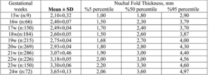
We calculated nuchal fold lenght %5, %50, %95 per-centile per week, which are shown in Table 1. We draw nuchal fold nomogram as seen in Figure 1. Er-ror bars have shown %95 confidence interval. It has been shown that there is a weak positive correlation between nuchal fold thickness and the gestational week (p: 0,001, r:0,18).
In 1625 analysed patients, 50 patients had CVS disea-ses and 32 patients had CNS diseadisea-ses. There was not a statistically significant relationship between nuchal fold thickness and CVS diseases (p= 0.98 and p<0.05). No statistically significant relationship was found
bet-Nuchal Fold Nomogram and Relationship With Heart and Central
67
Kaya ve ark. Kaya et al.
ween nuchal fold thickness and CNS diseases (p=0.55 and p<0.05). ( table.2)
Age may be a confounding factor for congenital CVS and CNS diseases. The patients were categorized above and below 35 years. There was no statistically significant relationship between CVS and CNS disea-ses in respect to category for 35 age (p=0.29, p=0.35 respectively and p<0.05). We also classified women above and below 40 years. We were not able to find statistically significant difference between CVS and CNS diseases in terms of category for 40 age (p=0.15, p=0.63 respectively and p<0.05).
Table 1. Age, gestational week, mean± standart deviati-on, min, max levels, %5, 50, 95 percentiles of Nuchal fold
Table 2. The relationship according to age, gestational week and nuchal fold thickness between patients with and without CNS diseases and CVS diseases
Discussion
Normal nuchal thickness significantly reduces the risk of Down syndrome and according to a study this may help reduce the number of amniocenteses done for abnormal triple screen results (7). The sensitivity of nuchal fold thickness for detection of Down syndro-me has been reported to be 42% to 43% with false-positive rates of 0.1% to 1.3%. Even when isolated, an abnormal nuchal fold is associated with a likeliho-od ratio (LR) of 11 to 49 for Down syndrome(5,7). We asked the question of what about in the absence of aneuploidy? We try to answer the question; should clinician investigate systemic diseases like cardiovas-cular and central nervous system in detail when de-tected increased nuchal fold thickness in fetus with normal karyotype.
It is clear that, in patients with karyotype abnorma-lity, association between nuchal fold thickness and systemic diseases. According to the study about nuc-hal oedema and related malformations by Nicolaides and Colleagues; in the fetuses with normal karyoty-pe, nuchal oedema or thickness may be a finding in a wide variety of fetal disorders, specially heart and than skeletal and craniospinal abnormalities. So it is emphasised that if increased nuchal fold thickness is an isolated abnormality, antenatal investigations should be performed with detailed sonografic scre-ening and echocardiography (8). Parents should be counselled about prognosis. Starting from this know-ledge, we investigated our data if there is a relations-hip between nuchal fold thickness and CVS diseases and CNS diseases.
The patients with normal karyotype, in addition to NT; a relationship has been shown between nuchal fold thickness and heart diseases (6,9). It has been recommended strongly that first-trimester fetuses with unexplained nuchal translucency elevation sho-uld have follow-up fetal echocardiography and pos-sibly postnatal evaluation for the presence of CHD. But there is limited data about second trimester. We pursued this study to search association between CVS, CNS diseases and thickened nuchal fold. We performed a nomogram of nuchal fold thickness of one of the cities in East Marmara Region in Turkey. In a study wrote from Khalil and Colleges, they said that in the absence of known major aneuploidy or genetic syndromes, fetuses with CVS were at incre-ased risk of brain abnormalities (10). Point of view, we wondered whether there is a togetherness of CVS and CNS abnormalities in the second trimester scre-ening in normal karyotype fetus. There was no statis-tically significant association about cooccurrence of CVS and CNS diseases. Also we did not find any
re-68
lationship between maternal advanced age and CNS or CVS diseases.
There was a limited data about the association bet-ween increased NFT and fetal CVS abnormalities so we thought that we should investigate the increased togetherness with CNS diseases and NFT because of positive correlation between CVS diseases and CNS diseases as above mentioned. As a result we could not establish a relationship between NTF and CNS diseases.
Similar to our study, many studies concluded that NFT increases with gestational age (11,12,13). It is known that nucal fold is a dynamic measurement, that incre-ases by gestational weeks. It has been thought that it is a continuous variable, it should be evaluated in the context of gestation-specific norms (9,13). Wit-hin this context, we gave the percentiles of every ges-tational week’s nuchal fold length in the table 2. Our outcomes are concordant with the previous studies. It is seen clearly this connection in figure 1.
Common view in literature, that is suitable to carry out detailed heart screening like fetal echo whom nuchal fold or nuchal translucency is measured as thick especially in case of abnormal karyotype. In the present study, we asked the question of what about normal karyotype fetus, should we be alert in terms of systemic disorders when we determined nuchal fold thickness? But we could not establish a relati-onship between nuchal fold thickness and CNS and CVS diseases.
In accordance with our results, Zelop and colleagu-es, found that; NFT did not appear to be increased in euploid fetuses with congenital cardiac disease. But there was a limitation about the patient counts (14). In order to clarify the relationship between increased NFT and CNS or CVS diseases, prospective randomi-sed and large scaled trails are needed.
References
1. Allan L, Benacerraf B, Copel JA et al. Isolated major congenital heart disease. Ultrasound Obstet Gynecol 2001;17:370-9.
2. Dolk H, Loane M, Garne E. The prevalence of congenital anomalies in Europe. Adv Exp Med Biol. 2010;686:349-64.
3. Engels AC, Joyeux L, Brantner C et al. Sonograp-hic detection of central nervous system defect s in the first trimester of pregnancy.PrenatalDiagno-sis2016,36,266–273.
4. Ritu Mogra, Nasser Alabbad, Jon Hyett. Increased nuchal translucency and congenital heart disease. Early Human Development 88 (2012) 261–267. 5. Benacerraf BR, Barss VA, and Laboda LA. A sonog-raphic sign for the detection in the second trimes-ter of the fetus with Down’s syndrome. Am J Obstet Gynecol 1985; 151: pp. 1078-107983
6. Ray O, Bahado-Singh, Minu Rowther et al. Midtri-mester nuchal thickness and the prediction of post-natal congenital heart defect. Am J Obstet Gynecol 2002;187:1250-3.
7. Ray O. Bahado-Singh, Israel Goldstein et al. Nor-mal nuchal thickness in the midtrimester indicates reduced risk of Down syndrome in pregnancies with abnormal triple-screen results. Am J Obstet Gyne-col. 1995 Oct;173(4):1106-10.
8. Nicolaides KH, Azal G, Snijders RJM, Gosden CM. Fe-tal nuchal edema: associated malformations and chro-mosomal defects. Fetal Diagn Ther 1992;7:123-31. 9. Ray O, Bahado-Singh, Ronald Wapner, et al. Eleva-ted first-trimester nuchal translucency increases the risk of congenital heart defects. American Journal of Obstetrics and Gynecology (2005) 192, 1357–61 10. Khalil A, Bennet S, Thilaganathan B, Paladini D, Griffiths P, Carvalho JS. Prevalence of prenatal bra-in abnormalities bra-in fetuses with congenital heart di-sease: a systematic review. Ultrasound Obstet Gyne-col. 2016 Sep;48(3):296-307.
11. Grandgean H, Sarramon MF. Sonographic measure-ment of nuchal skinfold thickness for detection for Down syndrome in the second-trimester fetus: a multicenter prospective study. Obstet Gynecol 1995; 85:103–106. 12. Watson WJ, Miller RC, Menard MK. Ultrasonog-raphic measurement offetal nuchal skin to screen for chromosomal abnormalities. Am J ObstetGynecol 1994; 170:583–586.
13. Borrell A, Costa D, Martinez JM, et al: Criteria for fetal nuchal thickness cut-off: a re-evaluation. Prenat Diagn 1997; 17: pp. 23-29
14. Carolyn Zelop, Lıllıan Kamınsky, Elısa Gıanferrarı et al. Nuchal Fold Thickness As A Marker For Conge-nıtal Cardıac Anomalıes. December 2003 Am J Obstet Gynecol Smfm Abstracts 638.
Nuchal Fold Nomogram and Relationship With Heart and Central