Synthesis of organic-inorganic hybrid nanoflowers using Trigonella foenum-graecum
seed extract and investigation of their anti-microbial activity
Cevahir ALTINKAYNAK1 Nilay ILDIZ2 Ayşe BALDEMİR3 Nalan ÖZDEMİR4 Vedat YILMAZ5 İsmail ÖÇSOY5
1
Department of Plant and Animal Production, Avanos Vocational School, Nevsehir Haci Bektas Veli University, Nevsehir, Turkey
2
Department of Pharmaceutical Microbiology, Faculty of Pharmacy, Erciyes University, Kayseri, Turkey 3
Department of Pharmaceutical Botany, Faculty of Pharmacy, Erciyes University, Kayseri, Turkey 4
Department of Chemistry, Faculty of Science, Erciyes University, Kayseri, Turkey 5
Department of Analytical Chemistry, Faculty of Pharmacy, Erciyes University, Kayseri, Turkey
Sorumlu Yazar/Corresponding Author: [email protected] ORCID: 0000-0003-0082-8521
Makale Bilgisi/Article Info Derim, 2019/36(2):159-167 doi:10.16882/derim.2019.549151
Araştırma Makalesi/Research Article Geliş Tarihi/Received: 04.04.2019 Kabul Tarihi/Accepted: 10.09.2019
Abstract
Herein we report a green method for the synthesis of organic-inorganic hybrid nanoflowers using a Trigonella
foenum-graecum L. (TF) (Fenugreek seeds) extracts as an organic part and copper ions acting as an inorganic
part. The organic-inorganic hybrid nanoflowers using TF seed extract (TF-Cu2+ hNFs) were characterized by SEM, XRD, EDX and FTIR. The morphology of the TF-Cu2+ hNFs was quite spherical and monodisperse with ∼18μm size. The TF-Cu2+ hNF exhibited the effective anti-bacterial activity against Enterococcus faecium,
Enterococcus faecalis, Staphylococcus aureus, Bacillus cereus, Salmonella typhi and Escherichia coli at
1-10 µg ml-1
concentrations except against Pseudomonas aeruginosa and Haemophilus influenza. However, both TF-Cu2+ hNFs and free TF extracts showed any antifungal activities against Candida albicans or Candida
glabrata. The study revealed that TF-Cu2+ hNFs could be used as a therapeutic agent for microbial infections and has the potential to overcome drug resistance.
Keywords: Biomaterials; Organic-inorganic hybrid nanoflowers; Copper; Anti-microbial activity
Organik-inorganik hibrit nano çiçeklerin çemen (Trigonella foenum-graecum L.) tohum ekstresi kullanılarak sentezi ve anti-mikrobiyal özelliklerinin araştırılması
Öz
Bu çalışmada organik-inorganik hibrit nano çiçek sentezi için organik bileşen olarak çemen (Trigonella
foenum-graecum L.) otu tohum ekstresi ve inorganik bileşen olarak Cu2+
iyonları kullanılarak yeşil bir üretim metodu raporlanmıştır. Çemen tohum ekstresi kullanılarak sentezlenmiş organik-inorganik hibrit nano çiçekler (TF-Cu2+ hNF); taramalı elektron mikroskobu (SEM), Enerji Dağılımlı Spektrometre (EDX), X-ışını kırınımı (XRD) ve Kızılötesi spektroskopisi (FT-IR) yöntemleri kullanılarak karakterize edilmiştir. TF-Cu2+ hNF’lerin elektron mikroskop görüntüsünde morfolojisi oldukça düzgün dağılımlı ve küresel formda olmak üzere yaklaşık 18 μm boyutunda gözlenmiştir. TF-Cu2+ hNF’ler; Pseudomonas aeruginosa and Haemophilus influenza dışında
Enterococcus faecium, Enterococcus faecalis, Staphylococcus aureus, Bacillus cereus, Salmonella typhi ve Escherichia coli türlerine karşı 1-10 µg ml-1 aralığında ve kullanılan antibiyotikler ile karşılaştırıldığında yüksek
düzeyde anti-mikrobiyal özellik göstermiştir. Ancak hem TF-Cu2+ hNF’ler hemde serbest TF ekstresi, Candida
albicans ve Candida glabrata türlerine karşı antifungal aktivite göstermemiştir. Yapılan bu çalışma çemen ekstresi
içeren organik-inorganik hibrit nano çiçeklerin test edilen patojen suşlar ile gelişen mikrobiyal enfeksiyonlar için terapötik bir ajan olarak kullanılabileceğini ve ilaç direncinin üstesinden gelme potansiyeline sahip olduğunu ortaya koymaktadır.
Anahtar Kelimeler: Biyomateryal; Organik-inorganik hibrit nano çiçekler; Bakır; Anti-mikrobiyal aktivite
1. Introduction
Various hybrid nanostructures with different morphologies are commonly used in the fields
of health care, cosmetic, biomedical, food, environment, health, medicine and biosensor (Ahmed et al., 2016; Lee et al., 2015). These hybrid nanostructures have been developed by
conventional synthesis techniques which are expensive and toxic. However, the development of green synthesis techniques has resulted in simple, cheap, rapid, stable and biocompatible procedures (Ahmed et al., 2016; Nadagouda et al., 2014).
The use of plant extracts has pointed out considerable attention within recent years in the synthesis of nanostructures. (Mittal et al., 2013). This is because plant extract is considered as an important protein source for hybrid nanostructures and plays a significant role in green nanotechnology as potential anti-microbial, and antioxidant agents (Phull et al., 2016). The TF plant, is widely cultivated throughout the world for medicinal uses due to its anti-cancer (Amin et al., 2005), and anti-microbial (Moradi et al., 2013; Aman et al., 2014) properties TF seeds contain 20-30% proteins consisting largely of albumins, globulins, glutelins and prolamines (Feyzi et
al., 2015), 45-60% carbohydrates, pyridine
alkaloids (0.2-0.38%), fixed oils (5-10%), choline (0.5%), flavonoids, free amino acids, tannic acid, Ca and Fe, saponins (0.6-1.7%), steroidal sapogenins, cholesterol, sitosterol, vitamins A, B1, C and nicotinic acid and volatile oils (0.015%) (Olli et al., 2006; Patel et al., 2013). TF extracts exhibited antibacterial effect on some negative and gram-positive bacteria at concentrations ranging from 250 to 1000 μg mL-1
have been shown by
Aman et al. (2014). Singh and co-workers have
stated that TF seed showed antibacterial activity at 500 μg mL−1 concentration againts B. cereus (Singh et al., 2014). Kamali and
El-Karim (2009) reported that various TF seed
extracts did not show antibacterial properties against bacteria. Various sizes of nanostructures have produced using plant extracts thus far (Karatoprak et al., 2017). As a new class of hybrid nanostructures, synthesis and characterization of flower-like hybrid nanostructures including organic (protein or enzyme) and inorganic parts were reported by Zare and co-workers for the first time (Ge et
al., 2012). They created novel hybrid
nanoflowers (hNFs) with much greater enzymatic activity and stability than those obtained with free enzymes. Some researchers have prepared various kinds of organic-inorganic nNFs and used them for different applications (Sun et al., 2014; Liang et
al., 2015; Huang et al., 2015; Somturk et al., 2015; Altinkaynak et al. 2016a; Altinkaynak et al., 2016b; Ariza-Avidad et al., 2016; Thawari et al., 2016; Zhang et al., 2016).
Inspired by the aforementioned works, we presented a green method for the production of organic-inorganic hNFs using an TF extracts as organic part and evaluated their comparative anti-microbial effect of TF-Cu2+ hNFs against some microorganisms.
2. Material and Methods 2.1. Materials
CuSO4.5.H2O, methanol, H3PO4, Bovine serum albumin (BSA), and other chemicals were bought from Sigma-Aldrich Products, MO, USA. Na2HPO4, KH2PO4, NaCl, KCl, HCl, NaOH and Coomassie G-250 were used for buffer solution. The chemicals used in the anti-microbial activity studies were obtained from Sigma-Aldrich, MO, USA except Mueller Hinton Broth (MHB) and Mueller Hinton Agar (MHA) (HiMedia, Mumbai, India), sterile disc (Oxoid, Hampshire, UK), and the standard strains were obtained from the ATCC, VA, USA; Enterococcus faecium (ATCC 8459), Bacillus cereus (ATCC 11778), Escherichia coli (ATCC 35218), Staphylococcus aureus (ATCC 25923), Enterococcus faecalis (ATCC 29212), Pseudomonas aeruginosa (ATCC 27853), Salmonella typhi (ATCC 14028), Haemophilus influenza (ATCC 49247), Candida albicans (ATCC 10231), and Candida glabrata (ATCC 90030). RPMI-1640 Medium with L-Glutamine, without sodium bicarbonate added with glucose (0.2%) at pH 0.165 molar morpholinepropanesulfonic acid (MOPS) and is buffered to be 7.0.
2.2. Preparation of TF extract
TF seeds registered as Gurarslan cultivar were harvested in field crops of Agricultural Faculty at Erciyes University. The seeds were kept at room temperature until the initiation of the experiments. TF was powdered using blender. 100 g powder was added to 500 mL flask including 250 mL methanol and incubated at room temperature (25°C) under stirring for one day. After incubation, solution was filtered twice Whatman No.1. The extracts were collected and evaporated under vacuum at 40°C.
2.3. Production of TF-Cu2+ hybrid nanoflowers
0.333 mL of 120 mM CuSO4.5.H2O solution was added to 50 mL of PBS, pH 7.4 with different concentration of TF dissolved extract (0.1 and 0.5 mg mL-1) as described in the literature (Ge et al., 2012; Altinkaynak et al, 2016a; Altinkaynak et al., 2016b; Somturk et al., 2015; Ariza-Avidad et al., 2016; Zhang et al., 2016; Thawari et al., 2016; Sun et al., 2014; Liang et al., 2015; Huang et al., 2015). The mixture was vigorously shaken for 30 s and kept without disturbing at +4°C for three days. The blue pellets were collected via centrifugation, and then washed 3 times. The obtained pellets were dried under vacuum at 50°C. The supernatant protein concentration was measured by the Bradford method (Bradford, 1976).
2.4. Characterization of TF-Cu2+ hybrid nanoflowers
The scanning electron microscopy (SEM) was used to generate the images of nanoflower on ZEISS EVO LS10 instrument (Oberkochen, Germany). The energy dispersive X-ray (EDX-ZEISS EVO LS10) technique was employed to analyze the elemental composition of nanoflower. The crystal structure of nanoflower was explained using the X-ray diffraction analysis (XRD-BRUKER AXS D8) (Karlsruhe, Germany). The Fourier Transform Infrared Spectroscopy (FTIR) spectra of nanoflowers were recorded to investigate its chemical structure using FTIR Spectrometer (Perkin Elmer 400 Spotlight 400 Imaging System, Waltham, USA).
2.5. Evaluation of anti-microbial activity The anti-microbial activities of TF extract and TF-Cu2+ hNFs compounds were tested by CLSI guidelines using minimum inhibitory concentration (MIC) and disc diffusion methods
(CLSI, 2012). The MIC assays were carried out
in triplicate assay at a dilution of the tested samples from 2000 𝜇g mL-1 to 125 𝜇g mL-1, and 100 𝜇g mL-1 to 0.01 𝜇g mL-1 for extracts and hNFs, respectively. Bacterial suspension (5×10⁵
CFU mL-1) was added in each well. Negative control comprised MHB and the positive control was suitable (Ampicillin
(100 mcg), Meropenem (10 mcg),
Ciprofloxacin (5 mcg), Bioanalyse/Turkey) for MHB + bacterial suspension and bacteria only. Plates were incubated at 37°C for 16 to 20 h. After incubation, plates were examined for bacterial growth. The MIC was calculated as lowest concentration of the samples inhibiting the visual growth of the microtitre plates. The experiments are done in triplicates. The anti-microbial behavior of the nanoflower compounds was assessed by Kirby Bauer disk diffusion method using MHA according to NCCLS guidelines at determined MIC concentrations for all tested microorganisms
(NCCLS, 2000). Because examined extract
concentrations had not effective. The bacterial cultures were sub-cultured in MHB and incubated for 14 h at 37°C. The bacterial cultures were sub-cultured in MHB then incubated for 14 h at 37°C. The sterile discs containing 30 𝜇g mL-1 samples were placed on MHA and plates were incubated after cultures were swabbed onto plates containing MHA. The zones of inhibition of each sample were calculated by measuring the diameter of the inhibition zone around the disc (in mm) including the disc diameter. Each experiment was performed in triplicates. Antifungal activities determined by using broth microdilution and disc diffusion method according to CLSI M27-A3, M44-A2 protocols
(CLSI, 2008; CLSI, 2009). Inoculum is
prepared in 0.85% NaCl to be 0.5 McFarland. The similar assay used for disc diffusion method by adding agar 1.5%. For each assay 30 𝜇g mL-1 sample used. Amphotericin B as positive control and DMSO as negative control were used. All experiments were performed in duplicate and repeated three times.
3. Results and Discussion
3.1. Production and characterization of TF-Cu2+ hNFs
In our work, we firstly used TF extract as an organic part to form the flower-like structures. The generation mechanism of the organic-inorganic hybrid nanoflowers was reported before (Ge et al., 2012; Altinkaynak et al, 2016a; Altinkaynak et al., 2016b; Somturk et al., 2016; Ariza-Avidad et al., 2016; Zhang et al., 2016; Thawari et al., 2016; Sun et al., 2014; Liang et al., 2015; Huang et al., 2015).
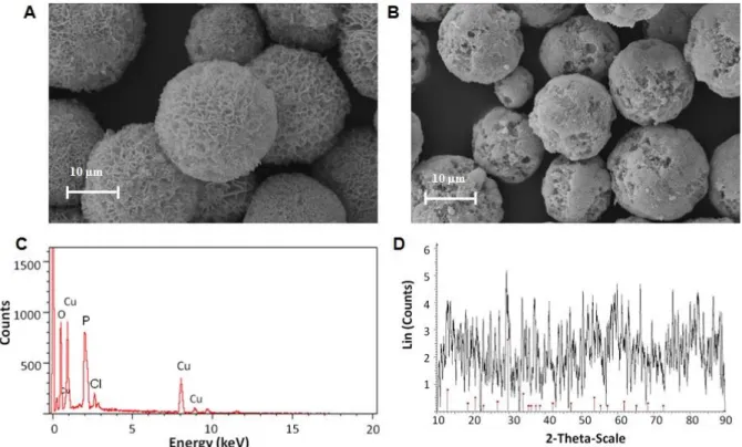
Figure 1. SEM images of TF-Cu2+ hNFs including (A) 0.1 mg mL-1 and (B) 0.5 mg mL-1 TF extract (C) EDX spectra of the hNFs (D) XRD patterns of hNFs in accord the peak position of the Cu3(PO4)2.3H2O (JCPDS card (00-022-0548, red line).
There are three steps in the mechanism: (a) nucleation and formation of primary crystals (b) to form nanoflowers through coordination of between the organic part and Cu2+ ions (c) complete formation of nanoflowers (Ge et al., 2012; Altinkaynak et al, 2016a; Altinkaynak et al., 2016b; Somturk et al., 2015; Ariza-Avidad et al., 2016; Zhang et al., 2016; Thawari et al., 2016; Sun et al., 2014; Liang et al., 2015;
Huang et al., 2015). The amine and diol groups
of molecules interact with copper phosphate complex to form TF-Cu2+ hNFs. To validate our design, Cu2+ ions and TF extract were mixed in PBS solution, after incubation for 3 days, blue precipitates were obtained. The impress of TF extract concentrations on the generation of the nanoflowers were investigated. As a result; the encapsulation yield (EY) of TF-Cu2+hNFs containing 0.1 and 0.5 mg mL-1 TF extract were determined as ~75% and ~86%, respectively. There could be a lower number of nucleation sites at lower TF extract concentration (0.1 mg mL-1). Concentrations of 0.02, 0.03, and 0.05 mg mL-1 were also tested but no nanoflower structure was formed at these concentrations (data are not shown). When the extract concentration was increased to
0.5 mg mL-1, a greater number of nucleation sites were available therefore the EY of TF-Cu2+ hNFs may increase. The different morphologies are demonstrated with SEM images in Fig. 1A and 1B. Interestingly, the morphology of TF-Cu2+hNF is very much uniform and monodisperse compared to protein-inorganic nanoflowers. When the concentration of extract was increased from 0.1 to 0.5 mg mL-1, the spherically shaped nanoflowers produced were more uniform and monodisperse. Although both concentrations of extract resulted in quite uniform and spherical NF (Fig. 1A and 1B), the NF had pores on its surface when extract (0.1 mg mL-1) was used (Fig. 1A). When extract (0.5 mg mL−1) was used, the pores on surface almost completely disappeared (Fig. 1B). Free extract molecules probably blocked the pores on the surface of the NF during growth process due to high concentration (Fig. 1B). The diameters of the hNFs formed with extract (0.1 mg mL-1) were measured as ∼22 μm in size. However, as the extract concentration increased (from 0.1 to 0.5 mg mL-1), the resulting nanostructures became smaller (~15-18 μm). This can be interpreted as showing that the number of nucleation sites may be
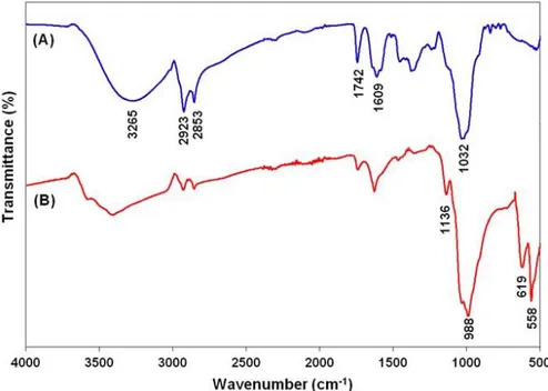
increased, resulting in small-sized NFs. In general, protein-inorganic hNFs including crude protein or enzyme etc. had an average diameter of ∼6-12 μm particle size. Interestingly, the morphology of TF-Cu2+ hNFs is much larger compared to that of protein-inorganic NFs. Based on these results, a TF extract concentration of 0.1 mg mL-1 was chosen for further evaluation. The structure of the hNFs was confirmed by EDX, XRD and FTIR. The EDX technique confirmed the presence of Cu2+ metals in the hNFs. Fig. 1C clearly demonstrated Cu2+ metal peaks on the EDX spectrum. The peak positions and relative intensities of the nanocrystals appeared on the XRD spectrum (Fig. 1D). Most of the diffraction peaks of Cu3 (PO4)2 nanocrystals in the hNFs were consistent with the JCPDS card number 00-022-0548. The strong peaks confirmed that the TF-Cu2+ hNFs were well crystallized. The chemical structures of extract and TF-Cu2+ hNFs were characterized using FT-IR for identification of functional groups. TF extract and TF-Cu2+ hNFs powder were dried at 70°C prior to typical sample preparation for FTIR. Simply, each powder was separately mixed with IR grade K Br and pressed into tablet form. The characteristic peaks of TF extract and TF-Cu2+ hNFs were analyzed on the FTIR spectra, respectively (Fig. 2A and 2B).
O P O groups showed very weak bending vibration at ∼532 cm−1, while the same vibration in very strong form appeared at ∼558 cm−1 (Yin et al., 2015; Wu et al., 2014). We assume that a high concentration of TF extract was packed in a nanoscale area of NF, which may have caused a very intense vibration peak. The vibration bands of the NH2 groups of TF extract and TF-Cu2+ hNFs appeared at ∼1609 cm−1 and ∼1626 cm−1, respectively (Wu et al., 2014; Yu et al., 2015). Finally, its stretching bands of it at 2800–3000 cm−1
were attributed to the CH3 and CH2 groups of TF extract and TF-Cu
2+ hNFs.
3.2. Determination of anti-microbial activities
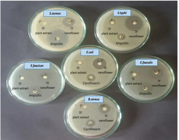
The anti-microbial activity of TF extract (2000-125 𝜇g mL-1) and TF-Cu2+ hNFs (100-0.01 𝜇g mL-1) was investigated against various pathogenic microorganisms. The diameter of inhibition zones (mm) around each well containing TF-Cu2+ hNFs is presented in Table 1 and imaged in Fig. 3. In this study, the anti-microbial properties of the liquid copper sulphate and phosphate buffer solution were also checked 0.8 mM and 10 mM concentrations, respectively and it was seen that these substances did not have significant activity against tested microorganisms.
Figure 2. (A) FTIR analysis of TF extract and (B) FTIR analysis of TF-Cu2+ hNFs formed with 0.1 mg mL-1 TF extract
Table 1. Anti-microbial activity of TF-Cu2+ hNFs
Microbial strains MIC (𝜇g mL-1) Inhibition zone (mm)
Escherichia coli ATCC 35218 10 24.625 ± 0.88
Salmonella typhi ATCC 14028 1 29.15 ± 1.62
Pseudomonas aeruginosa ATCC 27853 - NI
Staphylococcus aureus ATCC 25923 10 22.105 ± 0.15
Enterococcus faecium ATCC 8459 10 22.61 ± 0.86
Enterococcus faecalis ATCC 29212 10 22.115 ± 1.57
Candida albicans ATCC 10231 - NI
Candida glabrata ATCC 90030 - NI
Bacillus cereus ATCC 11778 10 20.6 ± 0.84
Haemophilus influenzae ATCC 49247 - NI
Values (mean ± SE) are average of three samples of hybrid nanoflower, analyzed in triplicate. NI: None Inhibition. TF extract was not effective any of all tested microorganisms (data not presented in Table 1).
Figure 3. Disc diffusion results image of TF extract, TF-Cu2+ hybrid nanoflower and drug (appropriate antibiotic disc)
The TF-Cu2+ hNFs were found to have the highest anti-microbial activity against S. typhi (29.15 ± 1.62), while the least anti-microbial activity was found against B. cereus (20.6 ± 0.84) by using disc diffusion method. TF-Cu2+ hNFs exhibited good antibacterial activity against both Gram-positive and Gram-negative microorganisms except P. aeruginosa and H.
influenza. In addition, TF extract was not effective against any of the tested microorganisms in the 2000-125 𝜇g mL-1 concentration range. Additionally, TF-Cu2+ hNFs and TF extract were not observed to display any antifungal activities against the tested yeasts. El-Kamali and El-Karim (2009) were reported TF seed extracts did not effective
against S. aureus-ATCC 25923, B. subtilis-NCTC 8236, E. coli-ATCC 25922, P. vulgaris-ATCC 6380, P. aeruginosa-vulgaris-ATCC 27853 and K. pneumonia-ATCC 1312. Singh et al. (2014) reported that TF seed extract; showed antibacterial activity at 500 μg mL−1
against B.cereus that only tested bacteria. We did not observe any effect against B.cereus. In contrast to our work, Aman et al. (2014) said that TF extracts had antibacterial effect on S.aureus, B.subtilis, E.faecalis, L.monocytogenes, E.coli, P.aeruginosa, S.typhi and S.dysenteriae at concentrations ranging from 250 to 1000 𝜇g mL -1
. In another work done by the same group, the snowball like hybrid nano structures comprising
Viburnum opulus L. extract called
“nanosnowball” (NSBs) were synthesized (Ildiz
et al., 2017). The NSBs exhibited effective
anti-microbial activity against E.coli, S.typhi, E.faecium, E.faecalis, B.cereus, S.aureus, except P.aeruginosa and H.influenza and C.albicans, C.glabrata, respectively. Also Baldemir et al. (2017) used Camellia sinensis (L.) Kuntze extracts incorporated Cu2+ ions nanoflowers and demonstrated their high catalytic activities and anti-microbial activities against against S.aureus (ATCC 25923), E.coli (ATCC 25922) and C.albicans (ATCC 90028) with broth microdilution and short time-kill assay. Subhapriya and Gomathipriya were reported that biosynthesis of TiO2 nanoparticles (TiO2NPs) using the aqueous leaf extract of
Trigonella foenum-graecum. These
nanoparticles that are to be 20–90 nm showed significant anti-microbial activity against Staphylococcus aureus, Enterococcus faecalis, Klebsiella pneumoniae, Streptococcus faecalis, Pseudomonas aeruginosa, Escherichia coli, Proteus vulgaris, Bacillus subtilis, Yersinia enterocolitica and fungus Candida albicans
(Subhapriya and Gomathipriya, 2018). In
contrast to our study, they synthesized nano-sized materials using titanium metal and extract from the leaf of Trigonella foenum-graecum.
Goyal et al. (2018) had been reported a simple
and eco-friendly method for the synthesis of silver nanoparticles using Trigonella foenum-graecum seed extract. They were investigated several parameters that dictated the biosynthesis of these nanoparticles such as reaction time, temperature, AgNO3 concentration, and amount of Trigonella
foenum-graecum seed extract.
Physicochemical characterization of these
nanoparticles was performed on Dynamic light scattering (DLS) field emission electron microscopy and energy distributor X-ray spectroscopy, X-ray diffraction and Fourier transform infrared spectroscopy. The size determination studies using DLS revealed of nanoparticles size between 95 and 110 nm. The antibacterial activities were studied against E. coli, P. vulgaris, P. aeruginosa and methicillin resistant Staphylococcus aureus (MRSA). The synthesized AgNPs were reported to show fewer efficacies against microorganisms as compared to the ones obtained via green synthesis. Also gram-positive microorganisms had shown more antibacterial activity than gram-negatives when compared our study.
The mechanism of TF-Cu2+ hNFs interaction with bacteria is not well known yet, but various hypotheses may be proposed to explain it. The positively charged copper ions play a vital role in the anti-microbial activity (Kruk et al., 2015). The bacterial cells may be accumulated inside the NFS’ petals due to the size differential and here the mechanism of anti-microbial activities may occur from the electrostatic attraction between NFs containing positively charged copper ions and negatively charged bacterial cells (Ahmed et al., 2016). Copper ions may be released slowly into the cells. Therefore, copper ions may lead to a break in the permeability of outer membrane and cause the leakage of cellular materials. Additionally, gram-positive and gram-negative bacteria have different membrane structures. The gram-positive bacteria cell wall has a thicker peptidoglycan layer that of gram-negative bacteria (Banerjee
et al., 2011). The layer of peptidoglycan
consisting of linear cross-linked polysaccharide chains is negatively charged, on the other hand, copper ions are positively charged. Although gram-positive bacteria are more sensitive to the anti-microbial activities of copper ions than gram-negative bacteria are, TF-Cu2+ hNFs showed the strongest anti-microbial activity against gram-negative bacteria rather than against gram-positive bacteria. C. albicans and C. glabrata were not affected by TF- Cu2+ hNFs. This difference may be due to cell wall differences because, fungi are eukaryotic cells and bacteria are prokaryotic. The differences in cell wall structure can be attributed to the abundance of the functional groups on the cell surface of the different microorganisms.
4. Conclusion
In summary, we reported a green synthesis method of organic-inorganic hybrid NFs by using Trigonella foenum graecum extract and Cu2+ ions. The obtained NFs were well dispersed, uniform and spherical. The TF extract amount affected the size of the NFs. The pores on the NFs’ surface almost completely disappeared when the TF extract amount was increased during the synthesis protocol, which is simple and economic. Our hNFs exhibited higher anti-microbial activity than TF extract against some Gram-positive and Gram-negative bacteria. Neither TF-Cu2+ hNFs nor TF extract demonstrated any anticandidal activities against C. albicans or C. glabrata. Hence, the TF-Cu2+ hNFs have potential applications due to their ability to overcome the drug resistance and can be used as a therapeutic agent in human health to overcome increased antibiotic resistance against pathogenic microorganisms.
Acknowledgements
The authors would like to thank PhD. Erman Beyzi from Agricultural Faculty, Erciyes University for the supply of Trigonella seeds. References
Altinkaynak, C., Yilmaz, I., Koksal, Z., Özdemir, H., Ocsoy, I., & Özdemir, N. (2016a). Preparation of lactoperoxidase incorporated hybrid nanoflower and its excellent activity and stability.
International Journal of Biological
Macromolecules, 84:402-409.
Altinkaynak, C., Tavlasoglu, S., Özdemir, N., & Ocsoy, I. (2016b). A new generation approach in enzyme immobilization: organic-inorganic hybrid nanoflowers with enhanced catalytic activity and stability. Enzyme and Microbial Technology, 93-94:105-112.
Ahmed, S., Ahmad, M., Swami, B.L., & Ikram, S. (2016). A review on plants extract mediated synthesis of silver nanoparticles for antimicrobial applications: A green expertise. Journal of
Advanced Research, 7(1):17–28.
Aman, S., Naim, A., Siddiqi, R., & Naz, S. (2014). Antimicrobial polyphenols from small tropical fruits, tea and spice oilseeds. Food Science and
Technology International, 20(4):241-51.
Amin, A., Alkaabi, A., Al-Falasi, S., & Daoud, S.A. (2005). Chemopreventive activities of Trigonella
foenum graecum (Fenugreek) against breast
cancer. Cell Biology International, 29:687-694. Ariza-Avidad, M., Salinas-Castillo, A., &
Capitán-Vallvey, L.F. (2016). A 3D mPAD based on a multi-enzyme organic–inorganic hybrid
nanoflower reactor. Biosensors and Bioelectronics, 77:51–55.
Baldemir, A., Kose, N.B., Ildız, N., İlgün, S., Yusufbeyoğlu, S., Yılmaz, V., & Ocsoy, I. (2017). Synthesis and characterization of green tea (Camellia sinensis (L.) Kuntze) extract and its major components-based nanoflowers: a new strategy to enhance antimicrobial activity. RSC Advances, 7:44303-44308.
Banerjee, M.,Sharma, S.,Chattopadhyay, A., & Ghosh, SS. (2011). Enhanced antibacterial activity of bimetallic gold-silver core-shell nanoparticles at low silver concentration.
Nanoscale, 3(11):5120-5125.
Bradford, M.M. (1976). A rapid and sensitive method for the quantitation of microgram quantities of protein utilizing the principle of dye binding.
Analytical Biochemistry, 72:248–254.
CLSI (2008). Reference method for broth dilution antifungal susceptibility testing of yeasts; approved standard-third ed. CLSI document M27-A3. Clinical and Laboratory Standards Institute, Wayne, USA.
CLSI (2009). Method for antifungal disk diffusion susceptibility testing of yeasts; approved guideline- second ed. CLSI document M44- A2. Clinical and Laboratory Standards Institute, Wayne, USA.
CLSI (2012). Performance standards for antimicrobial susceptibility testing. Twenty-second informational supplement ed. CLSI document M100-S22. Clinical and Laboratory Standards Institute, Wayne, USA.
El-Kamali, H.H., & El-Karim. E.M.A. (2009). Evaluation of antibacterial activity of some medicinal plants used in Sudanese traditional medicine for treatment of wound infections.
Academic Journal of Plant Sciences,
2(4):246-251.
Feyzi, S., Varidi, M., Zare, F., & Varidi, M.J. (2015). Fenugreek (Trigonella foenum graecum) seed protein isolate: extraction optimization, amino acid composition, thermo and functional properties, Journal of the Science of Food and
Agriculture, 95(15):3165-3176.
Ge, J., Lei, J., & Zare, R.N. (2012). Protein–inorganic hybrid nanoflowers, Nature Nanotechnology, 7:428-432.
Goyal, S., Gupta, N., Kumar, A., Chatterjee, S., & Nimesh, S. (2018). Antibacterial, anticancer and antioxidant potential of silver nanoparticles engineered using Trigonella foenum-graecum seed extract. IET nanobiotechnology, 12(4):526-533.
Huang, Y., Ran, X., Lin, Y., Ren, J., & Qu, X. (2015). Self-assembly of an organic-inorganic hybrid nanoflower as an efficient biomimetic catalyst for self-activated tandem reactions. Chemistry Communication, 51(21):4386-4389.
Ildiz, N., Baldemir, A., Altinkaynak, C., Özdemir, N., Yilmaz, V., & Ocsoy, I., (2017). Self-assembled
snowball-like hybrid nanostructures comprising
Viburnum opulus L. extract and metal ions for
antimicrobial and catalytic applications. Enzyme
and Microbial Technology, 102:60-66.
Karatoprak, G.Ş., Aydin, G., Altinsoy, B., Altinkaynak, C., Koşar, M., & Ocsoy, I. (2017). The effect of
Pelargonium endlicherianum Fenzl. root extracts
on formation of nanoparticles and their antimicrobial activities. Enzyme and Microbial
Technology, 97:21-26.
Kruk, T., Szczepanowicz, K., Stefanska, J., Socha, R.P., & Warszynski, P. (2015). Synthesis and antimicrobial activity of monodisperse copper nanoparticles. Colloids and surfaces B: Biointerfaces, 128:17-22.
Lee, S. W., Cheon, S.A., Kim, M.I., & Park, T.J.
(2015). Organic-inorganic hybrid
nanoflowers:types, characterictics, and future prospects. Journal of Nanobiotechnology, 13:54. Liang, L., Fei, X., Li, Y., Tian, J., Xu, L., Wang, X., &
Wang, Y. (2015). Hierarchical assembly of enzyme-inorganic composite materials with extremely high enzyme activity. RSC Advances, 5(117):96997-97002.
Mittal, AK.,Chisti, Y., & Banerjee, UC. (2013). Synthesis of metallic nanoparticles using plant extracts. Biotechnology Advances, 31(2):346-356.
Moradi Kor, N., Bagher Didarshetaban, M., & Saeid Pour, H.R. (2013). Fenugreek (Trigonella
foenum-graecum L.) as a valuable medicinal
plant. International journal of Advanced Biological
and Biomedical Research, 1(8):922-931.
Nadagouda, M.N., Iyanna, N., Lalley, J., Han, C., Dionysiou, DD., & Varma, R.S. (2014). Synthesis of silver and gold nanoparticles using antioxidants from blackberry, blueberry, pomegranate, and turmeric extracts. ACS Sustainable Chemistry & Engineering, 2(7):1717–
1723.
NCCLS (2000). Approved Standard: M7-A5. Methods for dilution antimicrobial susceptibility tests for bacteria that grow aerobically, 5th Ed., National Committee for Clinical Laboratory Standards, Wayne, USA.
Olli, S., & Kirti, P.B. (2006). Cloning, characterization and antifungal activity of defensin Tfgd1 from Trigonella foenum-graecum L., Journal of Biochemistry and Molecular Biology,
39(3):278-283.
Patel, D.K., & Dhanabal, S.P. (2013). Development and optimization of bioanalytical parameters for the standardization of Trigonella foenum-graecum, Journal of Acute Disease,
2(2):137-139.
Phull, A.R, Abbas, Q., Ali, A., Raza, H., Kim, S.J., Zia, M., & Haq, I. (2016). Antioxidant, cytotoxic
and antimicrobial activities of green synthesized silver nanoparticles from crude extract of
Bergenia ciliata. Future Journal of
Pharmaceutical Sciences, 2(1):31-36.
Singh, P., Vishwakarma, S.P., & Singh, R.L. (2014). Antioxidant, oxidative DNA damage protective and antimicrobial activities of the plant Trigonella
foenum‐graecum. Journal of the Science of Food and Agriculture, 94(12):2497-2504.
Somturk, B., Hancer, M., Ocsoy, I., & Özdemir, N. (2015). Synthesis of copper ion incorporated horseradish peroxidase-based hybrid nanoflowers for enhanced catalytic activity and stability. Dalton Transactions, 44:13845-13852. Somturk, B., Yilmaz, I., Altinkaynak, C., Karatepe, A.,
Ozdemir, N., & Ocsoy, I. (2016). Synthesis of urease hybrid nanoflowers and their enhanced catalytic properties. Enzyme and Microbial
Technology, 86:134-142.
Subhapriya, S. & Gomathipriya, P. (2018). Green synthesis of titanium dioxide (TiO2) nanoparticles by Trigonella foenum-graecum extract and its antimicrobial properties. Microbial Pathogenesis, 116:215-220.
Sun, J., Ge, J., Liu, W., Lan, M., Zhang, H., Wang, P., Wang, Y., & Niu, Z. (2014). Multi-enzyme co-embedded organic–inorganic hybrid nanoflowers: synthesis and application as a colorimetric sensor. Nanoscale, 6:255-262.
Thawari, A.G., & Rao, C.P. (2016) Peroxidase-like catalytic activity of copper-mediated protein– inorganic hybrid nanoflowers and nanofibers of β-lactoglobulin and α-lactalbumin: synthesis, spectral characterization, microscopic features, and catalytic activity. ACS Applied Material &
Interfaces, 8(16):10392–10402.
Wu, Z., Li, X., Li, F., Yue, H., He, C., Xie, F., & Wang, Z. (2014). Enantioselective transesterification of (R, S)-2-pentanol catalyzed by a new flower-like nanobioreactor. RSC
Advances, 4:33998-34002.
Yin, Y., Xiao, Y., Lin, G., Xiao, Q., Lin, Z., & Cai, Z. (2015). An enzyme-inorganic hybrid nanoflower based immobilized enzyme reactor with enhanced enzymatic activity. Jorunal of Materials
Chemistry B, 3:2295-2300.
Yu, Y., Fei, X., Tian, J., Xu, L., Wang, X., & Wang, Y. (2015). Self-assembled enzyme-inorganic hybrid nanoflowers and their application to enzyme purification. Colloids and Surfaces B Biointerfaces, 130:299-304.
Zhang, B., Li, P., Zhang, H., Li, X., Tian, L., Wang, H., Chen, X., Ali, N., Ali, Z., & Zhang, Q. (2016). Red-blood-cell-like BSA/Zn3(PO4)2 hybrid particles: Preparation and application to adsorption of heavy metal ions. Applied Surface