© TÜBİTAK
E-mail: [email protected] doi:10.3906/sag-0912-457
Eff ects of long-term passive smoking on the vascular
endothelial growth factor and apoptosis marker expression in
the retina and choroid: an experimental study
Sema DÜNDAR1, Fatih ÖZCURA2, İbrahim METEOĞLU3, Mehmet Erkut KARA4Aim: To investigate the eff ects of smoking, reported as a risk factor for age related macular degeneration (ARMD), on
the vascular endothelial growth factor (VEGF) expression and apoptosis in the retina and choroid of rats.
Materials and methods: Th is experimental study included 38 Sprague-Dawley rats. Th e rats were randomly assigned into 4 groups. Groups 1 (males) and 2 (females) were exposed to smoke beginning on day 21 aft er birth, whereas groups 3 (males) and 4 (females) were not exposed to smoke. At the end of the fourth month, the right eyes of all of the rats were enucleated. Immunohistochemical analysis was performed in the enucleated eyes in terms of the VEGF and apoptosis markers, namely caspase-3, Bcl-X, and p53. Comparisons between the groups were performed using the Mann-Whitney U test.
Results: Th e VEGF expression in the retina and choroid increased signifi cantly in rats exposed to smoking. Th is signifi cant diff erence did not change between the sexes. Th ere were no signifi cant diff erences in terms of Bcl-X and p53 expressions between the groups; however, the caspase-3 expression increased signifi cantly in the photoreceptor layer in rats exposed to smoking.
Conclusion: Our fi ndings show that smoking signifi cantly increases the VEGF expression in many of the retinal layers
and choroid. Smoking also increases caspase-3 expression only in the photoreceptor layer. According to these fi ndings, smoking may be a risk factor for retinal vascular disease such as exudative ARMD, via stimulating the VEGF.
Key words: Smoking, age related macular degeneration, vascular endothelial growth factor, apoptosis, retina, choroid
Uzun dönem pasif sigara maruziyetinin retina ve koroidde vasküler endotelyal
büyüme faktörü ve apoptozis markerları üzerine etkisi: Deneysel çalışma
Amaç: Yaşa bağlı makula dejenerasyonu (YBMD) için bir risk faktörü olduğu bildirilen sigaranın ratların retina ve
koroid dokusunda vasküler endotelyal büyüme faktörü (VEBF) ve apoptozis üzerine etkisini araştırmak.
Yöntem ve gereç: Çalışmaya 38 adet Sprague-Dawley rat alındı. Ratlar randomize olarak dört gruba ayrıldı. Grup
1 (erkek) ve 2 (dişi) doğumdan sonra 21. günden itibaren sigaraya maruz bırakılırken grup 3 (erkek) ve 4 (dişi) sigaraya maruz bırakılmadı. Dördüncü ayın sonunda tüm ratların sağ gözü enüklee edildi. Enüklee edilen gözlerde VEBF ve apoptozis markerları (kaspaz-3, Bcl-X, p53) yönünden immünohistokimyasal analiz yapıldı. Gruplar arası karşılaştırmalar Mann-Whitney U testi kullanılarak yapıldı.
Original Article
Received: 11.12.2009 – Accepted: 11.02.2011
1 Department of Ophthalmology, Faculty of Medicine, Adnan Menderes University, Aydın - TURKEY 2 Department of Ophthalmology, Faculty of Medicine, Dumlupınar University, Kütahya - TURKEY 3 Department of Pathology, Faculty of Medicine, Adnan Menderes University, Aydın - TURKEY 4 Department of Anatomy, Faculty of Veterinary, Adnan Menderes University, Aydın - TURKEY
Correspondence: Fatih ÖZCURA, Department of Ophthalmology, Faculty of Medicine, Dumlupınar University, Th e Central Campus, 43270 Kütahya - TURKEY E-mail: [email protected]
Introduction
It is known that smoking is a major risk factor for many disease groups that have fatal consequences, such as the heart, circulation, and respiration systems, as well as malign diseases (1). It has been reported that smoking is a risk factor for many important and common eye diseases such as cataract, glaucoma, age related macular degeneration (ARMD), Graves ophthalmopathy, and anterior ischemic optic neuropathy (1,2). ARMD is a major reason for legal blindness in western societies, especially for people over 65 years of age. It has 2 forms, one of which is non-exudative, characterized by the drusen and alterations of retinal pigment epithelium, while the other one is exudative, characterized by the formation of new vessels. Th e exudative form is seen less common than the other, yet causes serious loss of sight (1-3). It is known that smoking increases the risk of ARMD; however, the mechanism is still unclear (4-7).
Vascular endothelial growth factor (VEGF) is released from retinal pigment epithelium cells and it is of great importance in providing angiogenesis. In addition, it has an important role for pathologic angiogenesis. Spilsbury et al. have shown that the excessive production of VEGF in rats, via recombinant adenovirus vector, stimulates angiogenesis in retina and resulted in choroidalneovascularization (CNV) in their experimental study (8). Kliff en et al. reported that a signifi cantly increased expression of VEGF was found in postmortem human eyes with ARMD compared with the control macula (9).
Apoptosis has been identifi ed as the mechanism of cell death. It is an active process that has well described biochemical and morphological characteristics, including a lack of an infl ammatory response and DNA fragmentation caused by endonuclease activity
(10). It plays a major role in the development, homeostasis, healing of wounds, and pathophysiology of diseases in multi-cellular organisms. Besides the normal retinal growth, apoptosis also has a role in the development of some eye diseases, such as retinal detachment, retinitis pigmentosa, primary open angle glaucoma, and anterior ischemic optic neuropathy (10-12). While the caspase enzyme activity and p53 tumor suppressor gene activity increases in the cell that will undergo apoptosis, the level of Bcl-X molecule protecting the cell from apoptosis decreases (13-15). Hinton et al. reported that apoptosis related to choroidal neovascular membranes taken out by surgical intervention (11).
In this study, our aim was to analyze the eff ects of smoking, reported as a risk factor for ARMD, on VEGF, Bcl-X, caspase-3, and p53 expressions in the retina and choroid of rats.
Materials and methods
Th is study has been approved by the Animal Ethics Committee at Adnan Menderes University, Aydın, Turkey. Sprague-Dawley rats, 20 female and 10 male, were used in the study. Th e rats were placed into 28 × 28 × 16 cm polycarbonate cages, each of which had 2 females and 1 male. Th e rats were fed special feed brought from Gebze Best Yem Factory (İzmit, Turkey) and were provided with an unlimited water supply. Aft er mating, the male rats were taken out of the cages. Th e newborns were kept with their mothers until they were 3 weeks old.
Included in the study were 38 healthy newborn rats (19 males and 19 females). Group 1 (9 males) and Group 2 (10 females) were subjected to passive smoking 21 days aft er their birth. Group 3 (10 males) and Group 4 (9 females) were the control groups
Bulgular: Sigaraya maruz kalan ratların retina ve koroidinde VEBF anlamlı oranda arttı. Bu anlamlı fark cinsiyetler
arasında değişiklik göstermiyordu. Bcl-X ve p53 yönünden gruplar arasında anlamlı farklılık yoktu bununla birlikte sigaraya maruz kalan ratların fotoreseptör tabakasında kaspaz-3 anlamlı olarak yüksekti.
Sonuç: Bulgularımız sigaranın birçok retina tabakasında ve koroidde anlamlı olarak VEBF’yi artırdığını, ayrıca sadece
fotoreseptör tabakasında kaspaz-3’ü artırdığını gösterdi. Bu bulgulara göre sigara VEBF’yi sitümüle ederek eksüdatif YBMD gibi retina vasküler hastalıkları için risk faktörü olabilir.
Anahtar sözcükler: Sigara, yaşa bağlı makula dejenerasyonu, vasküler endotelyal büyüme faktörü, apoptozis, retina,
and were not subjected to smoking. For those rats exposed to passive smoking, their feed was taken out of their cages so that the smoke did not contaminate it and the rats would not receive nicotine orally; their feed was taken out of the cages for 2 h every day to air. Aft er the smoke exposure period was over, the rats were fed. On weekends, they were given unlimited feed. Th e rats assigned to be exposed to passive smoking were exposed to smoke in a unit for 120 min a day, 5 days a week, for 4 months. Th e smoke entered from one side of the unit and the air in the unit was circulated by an aspirator (the power of which could be adjusted). Th e brand name of the cigarette used in the study was Birinci (85 mm, Tekel, Turkey), which has a high level of nicotine. Th e amount of smoke exposure was gradually increased during the study. Th e animals were exposed to the smoke of 6 cigarettes in 120 min periods for 2 weeks, the smoke of 9 cigarettes for the next 2 weeks, and the smoke of 13 cigarettes for the following 12 weeks. Th e percentages of CO and CO2 were measured at certain intervals to evaluate the CO and CO2 levels created by the smoke and to test the reliability of the test environment (Sun Modular Gas Analyser 1200; England, UK). At the end of the month 4, all of the rats were killed and their right eyes were enucleated. Immunohistochemical analysis was performed in enucleated eyes.
Immunohistochemical evaluation
Placed on coated slides were 4 μm-thick sections from the formalin-fi xed, paraffi n-embedded tissue obtained. Immunostaining was performed using the avidin-biotin complex method. Aft er deparaffinization and dehydration, the sections were treated twice for 5 min in citrate buff er (0.01 mol/L, pH 6.0), in a microwave oven at 700 W. Th e slides were then cooled to room temperature for 1 h. Endogenous peroxidase activity was blocked by immersing the sections in 3% hydrogen peroxide in methanol for 30 min. Th e sections were then incubated with primary antibody for 1 h at room temperature. Biotinylated goat anti-rabbit secondary antibody was applied for 60 min at room temperature. Th e bound antibody was visualized with avidin-biotin-peroxidase complex (Zymed Histostain-Plus kit, Zymed, San Francisco, CA, USA, code no: 85-9843) for 1 h at room temperature. Th e color was developed
by 3,3’-diaminobenzidine tetrahydrochloride. Between steps, the slides were rinsed 3 times for 10 min in tris-buff ered saline (pH 7.6). Th e slides were counterstained lightly in Harris’ hematoxylin, and then were dehydrated and mounted. Th e antibodies used were as follows: VEGF, (Neomarkers, CA, USA, cat no: RB-222-R7), Bcl-X, (Neomarkers, CA, USA, cat no: MS-715-R7), Caspase 3 (Neomarkers, CA, USA, cat no: RB-1197-P0), and p53 (Neomarkers, CA, USA, cat no: MS-104-R7).
In the immunohistochemical staining for the positive control, various tissue samples were used. As the negative control, the primary antibody phase was skipped and the staining process was continued. Th e intensity of the staining (p53 nuclear, Caspase-3, Bcl-X, and VEGF cytoplasmic) was scored by the same investigator (IM) on a scale of 0 to 3 as follows: 0: absent, 1: weak and focal reaction, 2: moderate reaction, and 3: strong reaction.
Data were analyzed using SPSS for Windows (version 11.0, Chicago, IL, USA). Comparisons between groups were performed using the Mann-Whitney U test. P < 0.05 was considered statistically signifi cant.
Results
Primarily, the rats were evaluated in terms of whether or not they were exposed to smoking. Th en, to understand if smoking caused any diff erent eff ects on diff erent sexes, female and male rats were compared to other.
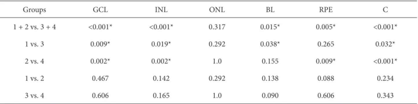
Table 1 shows the average and standard deviation of VEGF staining scores and Table 2 shows the statistical analyses results of these. VEGF expression increased signifi cantly in the ganglion cell layer (GCL), inner nuclear layer (INL), bacillary (photoreceptor) layer (BL), retinal pigment epithelium (RPE), and choroid (C) in rats exposed to smoking. When the analyses were subcategorized according to sexes, male rats exposed to smoking had a signifi cant diff erence in the GCL, INL, BL, and C compared to those not exposed to smoking. On the other hand, female rats exposed to smoking had a signifi cant diff erence in the GCL, INL, RPE, and C compared to those not exposed to smoking. When the male and the female rats exposed to smoking (Group 1 vs. 2) and those not exposed to
smoking (Group 3 vs. 4) were compared, there was no signifi cant diff erence in any of the layers in terms of VEGF expression.
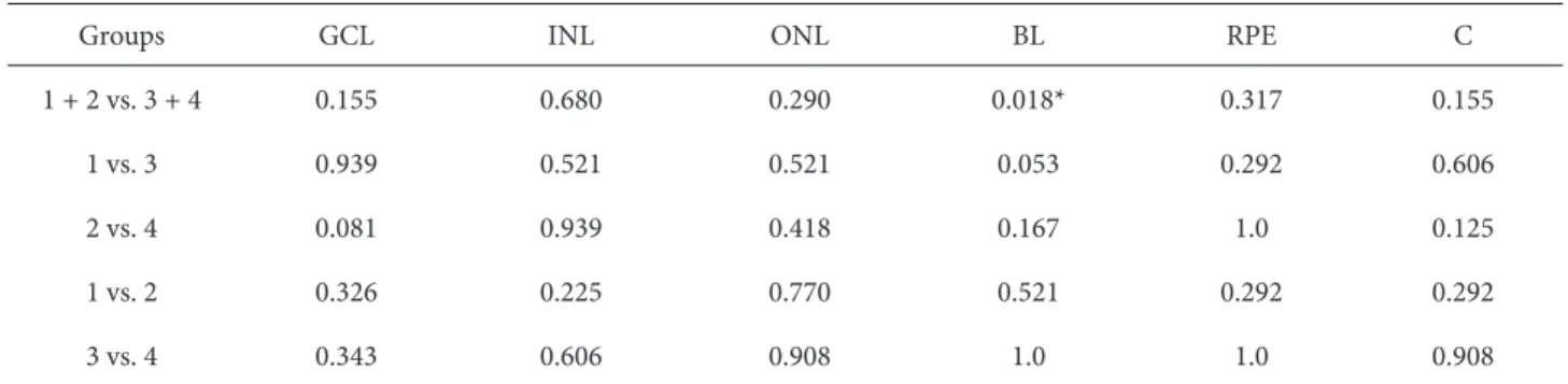
Table 3 shows the average and standard deviation of the caspase-3 staining scores and Table 4 shows the statistical analyses results of these. Th e caspase-3
expression was signifi cantly increased only in the BL of the rats exposed to smoking. Th ere was no signifi cant diff erence in any of the retinal layers and the choroid between the female and male rats exposed to smoking and those not exposed to smoking. Figure shows the immunohistochemical staining pattern of VEGF and caspase-3.
Table 1. Th e average staining scores of the VEGF in the retina layers.
Groups GCL INL ONL BL RPE C
1 (n: 9) 1.22 ± 0.97 1.33 ± 0.87 0.11 ± 0.33 0.56 ± 0.53 0.44 ± 0.53 0.78 ± 0.83 2 (n: 10) 1.50 ± 0.97 1.90 ± 1.10 0 1.00 ± 0.67 1.20 ± 1.03 1.10 ± 0.32 3 (n: 10) 0.20 ± 0.42 0.40 ± 0.52 0 0.10 ± 0.32 0.20 ± 0.42 0.10 ± 0.32
4 (n: 9) 0.11 ± 0.33 0.11 ± 0.33 0 0.56 ± 0.73 0.11 ± 0.33 0
VEGF: vascular endothelial growth factor, GCL: ganglion cell layer, INL: inner nuclear layer, ONL: outer nuclear layer, BL: bacillary (rod and cone) layer, RPE: retinal pigment epithelium, C: choroid.
Table 2. Th e statistical analysis results of the VEGF staining in the retina layers.
Groups GCL INL ONL BL RPE C
1 + 2 vs. 3 + 4 <0.001* <0.001* 0.317 0.015* 0.005* <0.001*
1 vs. 3 0.009* 0.019* 0.292 0.038* 0.265 0.032*
2 vs. 4 0.002* 0.002* 1.0 0.155 0.009* <0.001*
1 vs. 2 0.467 0.142 0.292 0.138 0.088 0.234
3 vs. 4 0.606 0.165 1.0 0.090 0.606 0.343
VEGF: vascular endothelial growth factor, GCL: ganglion cell layer, INL: inner nuclear layer, ONL: outer nuclear layer, BL: bacillary (photoreceptor) layer, RPE: retinal pigment epithelium, C: choroid. *P < 0.05.
Table 3. Th e average staining scores of caspase-3 in the retina layers.
Groups GCL INL ONL BL RPE C
1 (n: 9) 0.11 ± 0.33 0.33 ± 0.50 0.33 ± 0.50 0.33 ± 0.50 0.11 ± 0.33 0.11 ± 0.33 2 (n: 10) 0.30 ± 0.48 0.10 ± 0.32 0.40 ± 0.52 0.20 ± 0.42 0 0
3 (n: 10) 0.10 ± 0.32 0.20 ± 0.42 0.20 ± 0.42 0 0 0.20 ± 0.42
4 (n: 9) 0 0.11 ± 0.33 0.22 ± 0.44 0 0 0.22 ± 0.44
GCL: ganglion cell layer, INL: inner nuclear layer, ONL: outer nuclear layer, BL: bacillary (photoreceptor) layer, RPE: retinal pigment epithelium, C: choroid.
GCL: ganglion cell layer, INL: inner nuclear layer, ONL: outer nuclear layer, BL: bacillary (photoreceptor) layer, RPE: retinal pigment epithelium, C: choroid. *P < 0.05.
Groups GCL INL ONL BL RPE C
1 + 2 vs. 3 + 4 0.155 0.680 0.290 0.018* 0.317 0.155
1 vs. 3 0.939 0.521 0.521 0.053 0.292 0.606
2 vs. 4 0.081 0.939 0.418 0.167 1.0 0.125
1 vs. 2 0.326 0.225 0.770 0.521 0.292 0.292
3 vs. 4 0.343 0.606 0.908 1.0 1.0 0.908
Table 4. Th e statistical analysis results of caspase-3 staining in the retina layers.
A B C D E F Figure. Anti-VEGF; A) Score 1 (anti-VEGF, ×100), B) Score 2 (anti-VEGF, ×200), C) Score 3 (anti-VEGF, ×400).
Anti-caspase 3 staining pattern; D) Score 1 (anti-caspase 3, ×200), E) Score 2 (anti-caspase 3, ×200), F) Score 3 (anti-caspase 3, ×400).
GCL: ganglion cell layer, IPL: inner plexiform layer, INL: inner nuclear layer,
Th ere was no signifi cant diff erence in terms of the Bcl-X and p53 expressions between the groups.
Discussion
ARMD is a multi-factorial disease. Many environmental factors have been analyzed and it has been found that smoking and ARMD has a consistent parallelism (16,17). Khan et al. compared 435 cases, with ARMD defi ned as the presence of geographic atrophy (GA) or choroidal neovascularization (CNV), and 280 control cases. Th ey have demonstrated a strong association between the risk of both GA and CNV, especially in people who smoke more than 40 packs a year. Th ey also showed an increased risk for ARMD in non-smokers exposed to passive smoking (18).
In this study we demonstrated that VEGF expression increased signifi cantly in the GCL, INL, BL, RPE, and C in rats exposed to smoking experimentally. Th is signifi cant diff erence did not change between the sexes. We did not determine any signifi cant changes in the Bcl-X and p53 expressions; however, the caspase-3 expression increased signifi cantly only in the BL of rats exposed to smoking.
VEGF, released from retinal pigment epithelium, is required for physiological vasculogenesis, but overexpression of VEGF induces the pathological neovascularization. It is well known that VEGF has an important role in the pathogenesis of retinal vascular diseases such as CNV and diabetic retinopathy. Increased expression of VEGF has been demonstrated in CNV removed from patients and in experimentally induced CNV, and the blockade of VEGF causes dramatic inhibition of CNV as well (19). Similar to CNV, an increased expression of VEGF has been established in diabetic retinopathy (20) and nowadays anti-VEGF treatments have been proven to be a successful protocol in treatment of diabetic retinopathy and CNV (21).
Th e expression of VEGF was shown to increase in the event of hypoxia related to ischemia (22,23). Smoking may trigger atherosclerosis in the retinal and choroidal circulation, and therefore may cause hypoxia. Because the plasma HDL cholesterol level decreases in smokers, while total cholesterol level, LDL cholesterol level, platelet adhesiveness, and fi brinogen increase (24-26), it is not a surprise that these eff ects cause hypoxia and ischemia in the choroidal circulation and increase the expression of VEGF.
Apoptosis is an important mechanism for the natural development of the body and for the continuation of homeostasis. In addition, it also has a role in the limitations of the pathologic processes (13). Hinton et al. investigated several frozen sections from 10 surgically excised CNV, which were stained by the TUNEL method, and found that many of the TUNEL-positive apoptotic cells were stromal retinal pigment epithelial cells (11). Dunaief et al. studied postmortem retinas with ARMD (geographic atrophy or exudative ARMD) and normal retinas by TUNEL and immunocytochemistry. Th ey found that maculas with ARMD had signifi cant increases in TUNEL-positive cells in the inner C, RPE, photoreceptors, and INL, compared with normal retinas (27). In this study, we determined that the Bcl-X and p53 expressions were not diff erent between groups; however, the caspase-3 expression increased signifi cantly only in the BL of rats exposed to smoking. We studied some apoptosis markers but we did not evaluate apoptosis via the TUNEL method in this study. Th is may be thought as a defi ciency in our study, because many of the cellular mechanisms have a role in the apoptosis pathway.
In conclusion, our fi ndings show that smoking signifi cantly increases VEGF expression in many of the retinal layers and choroid. Smoking also increases the caspase-3 expression only in the photoreceptor layer. According to these fi ndings, smoking may be a risk factor for retinal vascular disease such as exudative ARMD via stimulating VEGF.
References
1. Cheng AC, Pang CP, Leung AT, Chua JK, Fan DS, Lam DS. Th e association between cigarette smoking and ocular diseases. Hong Kong Med J 2000; 6: 195-202.
2. Solberg Y, Rosner M, Belkin M. Th e association between cigarette smoking and ocular diseases. Surv Ophthalmol 1998; 42: 535-47.
3. Suner IJ, Espinosa-Heidmann DG, Marin-Castano ME, Hernandez EP, Pereira-Simon S, Cousins SW. Nicotine increases size and severity of experimental choroidal neovascularization. Invest Ophthalmol Vis Sci 2004; 45: 311-7.
4. Vingerling JR, Hofman A, Grobbee DE, de Jong PT. Age-related macular degeneration and smoking. Th e Rotterdam Study. Arch Ophthalmol 1996; 114: 1193-6.
5. Delcourt C, Diaz JL, Ponton-Sanchez A, Papoz L. Smoking and age-related macular degeneration. Th e POLA Study. Arch Ophthalmol 1998; 116: 1031-5.
6. Klein R, Klein BE, Moss SE. Relation of smoking to the incidence of age-related maculopathy. Th e Beaver Dam Eye Study. Am J Epidemiol 1998; 147: 103-10.
7. Clemons TE, Milton RC, Klein R, Seddon JM, Ferris FL. Age-Related Eye Disease Study Research Group. Risk factors for the incidence of Advanced Age-Related Macular Degeneration in the Age-Related Eye Disease Study (AREDS) AREDS report no. 19. Ophthalmology 2005; 112: 533-9.
8. Spilsbury K, Garrett KL, Shen WY, Constable IJ, Rakoczy PE. Overexpression of vascular endothelial growth factor (VEGF) in the retinal pigment epithelium leads to the development of choroidal neovascularization. Am J Pathol 2000; 157: 135-44. 9. Kliff en M, Sharma HS, Mooy CM, Kerkvliet S, de Jong PT.
Increased expression of angiogenic growth factors in age-related maculopathy. Br J Ophthalmol 1997; 81: 154-62. 10. Barber AJ, Lieth E, Khin SA, Antonetti DA, Buchanan
AG, Gardner TW. Neural apoptosis in the retina during experimental and human diabetes. Early onset and eff ect of insulin. J Clin Invest 1998; 102: 783-91.
11. Hinton DR, He S, Lopez PF. Apoptosis in surgically excised choroidal neovascular membranes in age-related macular degeneration. Arch Ophthalmol 1998; 116: 203-9.
12. Chang CJ, Lai WW, Edward DP, Tso MO. Apoptotic photoreceptor cell death aft er traumatic retinal detachment in humans. Arch Ophthalmol 1995; 113: 880-6.
13. Wilson SE. Stimulus-specifi c and cell type-specifi c cascades: emerging principles relating to control of apoptosis in the eye. Exp Eye Res 1999; 69: 255-66.
14. Podesta F, Romeo G, Liu WH, Krajewski S, Reed JC, Gerhardinger C, Lorenzi M. Bax is increased in the retina of diabetic subjects and is associated with pericyte apoptosis in vivo and in vitro. Am J Pathol 2000; 156: 1025-32.
15. Yoshizawa K, Nambu H, Yang J, Oishi Y, Senzaki H, Shikata N, et al. Mechanisms of photoreceptor cell apoptosis induced by N-methyl-N-nitrosourea in Sprague-Dawley rats. Lab Invest 1999; 79: 1359-67.
16. Kahn HA, Leibowitz HM, Ganley JP, Kini MM, Colton T, Nickerson RS, Dawber TR. Th e Framingham Eye Study. II. Association of ophthalmic pathology with single variables previously measured in the Framingham Heart Study. Am J Epidemiol 1977; 106: 33-41.
17. Smith W, Assink J, Klein R, Mitchell P, Klaver CC, Klein BE, et al. Risk factors for age-related macular degeneration: Pooled fi ndings from three continents. Ophthalmology 2001; 108: 697-704.
18. Khan JC, Th urlby DA, Shahid H, Clayton DG, Yates JR, Bradley M, et al. Smoking and age related macular degeneration: the number of pack years of cigarette smoking is a major determinant of risk for both geographic atrophy and choroidal neovascularisation. Br J Ophthalmol 2006; 90: 75-80.
19. Kwak N, Okamoto N, Wood JM, Campochiaro PA. VEGF is major stimulator in model of choroidal neovascularization. Invest Ophthalmol Vis Sci 2000; 41: 3158-64.
20. Crawford TN, Alfaro DV, Kerrison JB, Jablon EP. Diabetic retinopathy and angiogenesis. Curr Diabetes Rev 2009; 5: 8-13. 21. Eyetech Study Group. Preclinical and phase 1A clinical
evaluation of an anti-VEGF pegylated aptamer (EYE001) for the treatment of exudative age-related macular degeneration. Retina 2002; 22: 143-52.
22. Shweiki D, Itin A, Soff er D, Keshet E. Vascular endothelial growth factor induced by hypoxia may mediate hypoxia-initiated angiogenesis. Nature 1992; 359: 843-5.
23. Shima DT, Adamis AP, Ferrara N, Yeo KT, Yeo TK, Allende R et al. Hypoxic induction of endothelial cell growth factors in retinal cells: identifi cation and characterization of vascular endothelial growth factor (VEGF) as the mitogen. Mol Med 1995; 1: 182-93.
24. Ogston D, Bennett NB, Ogston CM. Th e infl uence of cigarette smoking on the plasma fi brinogen concentration. Atherosclerosis 1970; 11: 349-52.
25. Hawkins RI. Smoking, platelets and thrombosis. Nature 1972; 236: 450-2.
26. Craig WY, Palomaki GE, Haddow JE. Cigarette smoking and serum lipid and lipoprotein concentrations: an analysis of published data. BMJ 1989; 298: 784-8.
27. Dunaief JL, Dentchev T, Ying GS, Milam AH. Th e role of apoptosis in age-related macular degeneration. Arch Ophthalmol 2002; 120: 1435-42.