See discussions, stats, and author profiles for this publication at: https://www.researchgate.net/publication/265050776
Regulation of Glutathione S-Transferase Mu with type 1 diabetes and its
regulation with antioxidants
Article in Türk Biyokimya Dergisi / Turkish Journal of Biochemistry · April 2013
DOI: 10.5505/tjb.2013.96720 CITATIONS 3 READS 231 3 authors, including: Gökhan Sadi
Karamanoglu Mehmetbey Üniversitesi 54PUBLICATIONS 439CITATIONS
SEE PROFILE
Tulin Guray
Middle East Technical University 28PUBLICATIONS 423CITATIONS
SEE PROFILE
All content following this page was uploaded by Gökhan Sadi on 07 May 2015.
Regulation of Glutathione S-Transferase Mu with type 1
diabetes and its regulation with antioxidants
[Glutatyon S-Transferaz Mu izoziminin Tip 1 diyabet ve antioksidanlar ile
düzenlenmesi]
Research Article [Araştırma Makalesi]
Türk Biyokimya Dergisi [Turkish Journal of Biochemistry–Turk J Biochem] 2013; 38 (1) ; 92–100
Yayın tarihi 15 Nisan, 2013 © TurkJBiochem.com [Published online 15 April, 2013]
TÜ R K BİY OKİMYA DERNEĞİ DER G İSİ TÜ R K BİY OKİMYA DERNEĞİ DER G İSİ 1976 TÜ R K BİY OKİMYA DERNEĞİ DER G İSİ TÜ R K BİY OKİMY A DERNEĞ İ D ERG İS İ 1976 ORJİNAL 1. ÖRNEK 2. ÖRNEK Gökhan Sadi1,
Deniz İrtem Kartal2,
Tülin Güray3
1Karamanoglu Mehmetbey University, Kamil
Ozdag Science Faculty, Department of Biology, Karaman, Turkey
2Department of Biochemistry, Graduate School
of Natural And Applied Sciences, Middle East Technichal University, Ankara, Turkey
3Department of Biological Sciences, Middle East
Technichal University, Ankara, Turkey
Yazışma Adresi
[Correspondence Address]
Dr. Gökhan Sadi
Karamanoglu Mehmetbey University, Kamil Ozdag Science Faculty, Department of Biology, Karaman, Turkey
Tel. 0338 2262000/3824 Fax. 0338 2262150 E-mail. [email protected]
Registered: 16 August 2012; Accepted: 21 October 2012 [Kayıt Tarihi: 16 Ağustos 2012; Kabul tarihi: 21 Ekim 2012]
ABSTRACT
Objective: Increased oxidative stress is now related with the pathogenesis and the chronic comp-lications associated with the disease, diabetes mellitus. While roles of oxidative stress in diabetic complications are widely studied, the molecular mechanisms playing role in the regulations of detoxification enzymes in the presence of antioxidants have not been clearly established because of the complexity of the pathways.
Methods: Diabetes was induced by single dose streptozotocin application and rats were treated with 50mg/kg antioxidants DL-α-lipoic acid (LA), vitamin C (VC) and combination of both for three weeks. Lipid peroxidation, protein oxidation and reduced glutathione levels were measured and Glutathione S-Transferase Mu (GST-Mu) isozyme activity was determined by using enzyme specific substrate. mRNA and protein expressions were evaluated by RT-PCR and western blot methods respectively.
Results: In diabetic animals the products of lipid peroxidation and protein oxidation reactions were all found to be elevated significantly (p<0.05) and supplementing the animals either individually or in combination with two powerful antioxidants restored this effect. GST-Mu activities were also significantly decreased in diabetic group compared to controls and RT-PCR and Western blot analysis results showed that this decrease in activity is regulated at the level of gene expression, as both mRNA and protein expressions were suppressed. All antioxidant treatments did not reverse the effect of diabetes at the gene expression level and interestingly protein expressions were further suppressed indicating a post-translational regulation by antioxidants.
Conclusion: Presence of oxidative stress and the effects of antioxidants in diabetic rats were con-firmed and it was found that diabetes leads to GST-Mu to be down regulated at mRNA expressi-on levels. Future post-translatiexpressi-onal modificatiexpressi-on studies will help to determine the mechanism or combination of mechanisms underlying the damaging effects of oxidative stress observed in streptozotocin induced diabetic rats.
Key Words: GST-Mu, gene expression, lipid peroxidation, protein carbonylation, diabetes mellitus Conflict of Interest: The authors declare that they do not have any conflict of interest.
ÖZET
Amaç: Günümüzde artan oksidatif stresin, diyabet hastalığı ile ilişkili kronik komp-likasyonlar ve diyabetin patogenezi ile ilişkisi olduğu bilinmektedir. Oksida-tif stresin diyabetin komplikasyonlarındaki görevleri çok çalışılmış olmasına rağ-men, detoksifikasyon enzimlerinin diyabet ve antioksidan uygulamaları ile moleküler olarak nasıl düzenlendiği, hücre içi yolakların karmaşıklığı nedeniyle açıkça belirlenememiştir. Yöntemler: Diyabetin hayvansal modeli, sıçanlarda tek doz streptozotocin uygula-ması ile oluşturulmuştur. Sıçanlar üç hafta boyunca DL-α-lipoik asit (LA), vitamin C (VC) ve her ikisinin kombinasyonu ile muamele edilmiş, bütün gurupların karaci-ğer dokularında lipit peroksidasyonu, protein oksidasyonu ve indirge GSH düzeyleri ölçülmüştür. Glutatyon S-Transferaz Mu (GST-Mu) izozim aktivitesi enzime özel sübstrat kul-lanılarak, mRNA ve protein ekspresyonu RT-PCR ve Western blot teknikleri ile belirlenmiştir. Bulgular: Diyabetik hayvanlarda lipid peroksidasyonu ve protein oksidasyonu ürünlerinin istati-sitksel olarak yükseldiği (p<0.05) ve LA ve VC gibi iki güçlü antioksidanın tekil veya birlikte uy-gulamasının bu yükselmeyi normale getirdiği bulunmuştur. GST-Mu enzim aktivitesinin diyabetik gurupta anlamlı ölçüde azaldığı ve bu azalmanın nedeninin, RT-PCR ve Western blot analizleri ile elde edilen düşük mRNA ve protein ekspresyon seviyelerine dayanarak, enzimin gen ekspresyonu düzeyinde gerçekleştiği ortaya çıkarılmıştır. Antioksidan uygulamalarının gen ekspresyon düzey-lerini değiştirmediği, fakat protein ekspresyonlarını ilave olarak düşürdüğü bulunmuş, bu durum GST-Mu izoziminin antioksidanlar ile post-translasyonel olarak düzenlenmesi ile açıklanmıştır. Sonuç: Çalışma ile diyabette oksidatif stresin ve antioksidan uygulamalarının etkileri ortaya çı-karılmış, GST-Mu enziminin diyabet ile aşağı yönde düzenlendiği ve düzenlenmenin gen eks-presyonu düzeyinde gerçekleştiği bulunmuştur. İleri post-translasyonel modifikasyon çalışmaları, streptozotocin ile oluşturulmuş diyabetik sıçanlarda oksidatif stresin detoksifikasyon enzimleri-nin düzenlenme mekanizmalarını ortaya çıkaracaktır.
Anahtar Kelimeler: GST-Mu, gen ekspresyonu, lipid peroksidasyonu, protein karbonilasyonu, diabetes mellitus
Çıkar Çatışması: Yazarlar çıkar çatışması bulunmadığını beyan ederler. doi: 10.5505/tjb.2013.96720
Turk J Biochem, 2013; 38 (1) ; 92–100. 93 Sadi et al.
Introduction
Oxidative stress is defined as inequality between the productions of reactive oxygen species (ROS) and the organism’s antioxidant defense potential. Cells and tissu-es control the levels of reactive oxygen specitissu-es with the help of enzymatic and non-enzymatic cellular defense mechanisms. Enzymatic systems include antioxidant enzymes such as catalase (CAT: EC 1.11.1.6), superoxide dismutase (SOD: EC 1.15.1.1) and glutathione peroxidase (GPx: EC 1.11.1.9), whereas non-enzymatic ROS regulati-on is achieved by scavengers including glutathiregulati-one (GSH), α-lipoic acid (LA), vitamin C (VC) and α-tocopherol [1,2]. LA and VC are non-enzymatic ROS scavengers which can alleviate intracellular ROS stress. LA and its reduced form dihydrolipoic acid (DHLA) are strong ROS scaven-gers as a result of their interactions with various radicals and oxidized form of other radicals promoting the antio-xidant effects of other antioantio-xidants like VC. Another pro-tective mechanism against oxidative stress is Glutathione S-transferases (GST: EC 2.5.1.18) which catalyze the con-jugation of glutathione to a wide range of electrophiles. GSTs also exhibit differential response to a wide variety of chemicals and oxidative stress in the normal and pat-hophysiological conditions such as diabetes mellitus [3-5]. It has been confirmed that GSTs are also regulated both in vivo and in vitro by reactive oxygen species (ROS) such
as superoxides, H2O2 and by the products of membrane
lipid peroxidation [6,7]. Among the different isoenzymes of GSTs, the cytosolic Mu isoenzyme is predominantly present in the liver and function in the elimination of di-verse array of foreign compounds as well as wide variety of products of oxidative stress. Even though, alpha class GST isoenzyme have been shown to possess highest ac-tivity towards the lipid peroxidation end product which is 4-hydroxynonenal, GST Mu and Pi also display imp-ressive catalytic efficiency toward this end product [8,9]. Therefore, GST-Mu family is critical in the protection of cells from reactive oxygen species since the total pool of GST-Mu which is quite large in liver cells might contri-bute to the process of detoxification of the products of the oxidative stress. The aim of this study was to demonstrate how the regulation of the expression of GST-Mu under oxidative stress conditions is taking place. Therefore this study may provide new insights for analyzing cellular mechanism for protection of liver cells against oxidative damage observed in diabetes.
Materials and Methods
Materials
Streptozotocin, DL-6,8-Thioctic acid (a-lipoic acid), as-corbic acid (vitamin C), reduced glutathione (GSH), Bovi-ne serum albumin (BSA), Folin-Ciocalteu Phenol Reagent, tris (hydroxymethyl)aminomethane (Tris), Ethylenediami-netetraacetic acid (EDTA), 5,5’-dithiobis-(2-nitrobenzoic acid (DTNB), Phenylmethylsulfonyfloride (PMSF), Dit-hiothreitol (DTT), 5-Bromo-4-chloro-3-indolyl
phospha-te (BCIP), Nitro blue phospha-tetrazolium (NBT) were purchased from Sigma (St. Louis, MO, USA). All other chemicals used were of the highest analytical grade available, and the buffers were prepared by using sterile distilled water.
Induction of diabetes and animal treatments
Male Wistar rats which were at the same age and weight (200-250gr) were used as experimental animals. All the experimental protocols were performed according to ru-les of local ethical committee. Animals were fed with a standard diet and diabetes was induced with single in-jection of streptozotocin (STZ) (55mg/kg) dissolved in 0,05M citrate buffer (pH: 4.5) to the IP cavity of animals. One week after STZ injection, blood glucose levels were checked with Accu-check-go blood glucose analyzer (Roche, Foster City, California, USA) and the animals ha-ving blood glucose concentration higher than 200 mg/dl were taken as diabetic. Then, LA (50mg/kg/day) in physi-ological saline, VC (50mg/kg/day) in physiphysi-ological saline, and combination of them (50+50 mg/kg/day) were given with daily IP injection until the end of four weeks diabe-tic growing period. After the diabetes and antioxidants application, experimental test and control groups were as follows: control (n=9), diabetic (n=9), diabetic group gi-ven VC (D+VC) (n=12), diabetic group gigi-ven LA (D+LA) (n=8), diabetic group given combination of VC and LA (D+LA+VC) (n=7). Animals were decapitated after four weeks of diabetes and three weeks of antioxidant treat-ment and then liver tissues were collected, quick frozen in liquid nitrogen, and kept at -85°C for subsequent bioc-hemical analysis.Tissue homogenization
Liver tissues were homogenized in 1,15% (w/v) KCl, 5mM EDTA, 0,2mM PMSF, 0,2mM DTT, in 25 mM phosphate buffer, pH 7,4 using teflon-glass homogeni-zer and homogenates were centrifuged at 1.500g. Super-natants were sampled for the determination of enzyme activities, reduced glutathione (GSH), lipid peroxidati-on (MDA), protein carbperoxidati-onylatiperoxidati-on, and for western blot analysis. Protein contents of the homogenates were deter-mined according to Lowry method [10].
Total RNA isolation and RT-PCR studies
Total RNAs were isolated from the liver tissues bygu-anidine isothiocyanide method[11] and cDNA synthe-sis by using 1 μg total RNA were carried out by using
Revert AidTM cDNA synthesis kit according to
manu-facturer protocols (Fermentas; Burlington, Canada). Multiplex RT-PCR was performed in order to amplify GST-Mu mRNA and the internal control gene (β-actin) simultaneously as described previously [12]. The pri-mer pairs used for amplification of cDNAs of GST-Mu were 5′-AGAAGCAGAAGCCAGAGTTC (forward) and 5′-GGGGTGAGGTTGAGGAGATG (reverse) and β-actin were 5′-CCTGCTTGCTGATCCACA (forward) and 5′-CTGACCGAGCGTGGCTAC (reverse).
Western Blot anlaysis
For the western blot analysis of GST-Mu, 20 μg of total cytosolic protein were separated by SDS-PAGE and elect-roblotted onto PVDF membrane [13]. Then, membranes were incubated with primary antibody solutions of GST-Mu (1:2000- Anti-GST-GST-Mu Rabbit IgG, Abcam: Camb-ridge, USA), and GAPDH (1:2000- Anti-GAPDH Rabbit IgG, Santa Cruz, USA) for 2 hours with constant shaking. GAPDH was used as internal standard for the normaliza-tion. Afterwards, alkaline phosphatase (AP) conjugated secondary antibodies (1:10.000) were applied; proteins were visualized by BCIP/NBT substrate solution. Then, membranes were scanned and the intensities of the bands corresponding to antioxidant enzymes and GAPDH were measured with the aid of Image J software [14].
Activity measurements
GST-Mu Class isozyme activity was determined accor-ding to the method of Habig and coworkers [15] with small modifications. Enzyme activity was calculated as the amount of thioether (nmol) formed by 1 mg total
protein in one minute by using 0,0085 µM.cm-1 as an
ex-tinction coefficient of thioether that is formed by GSTs.
Determination of oxidative biomarkers
GSH determination was carried out based on the oxi-dation of reduced GSH by 5,5’-dithiobis-(2-nitrobenzoic acid), [DTNB] to produce pale yellow color that gives its maximum absorbance at 412 nm [16]. The results were expressed as nmol GSH per mg protein. MDA concent-rations were determined by using extinction coefficient
of colored complex as 1,56 x 10⁵ (M.cm)-1 and expressed
as nmol MDA for one mg of protein containing super-natant [17]. A spectrophotometric assay [18] was used for the quantification of protein carbonyl groups as a biomarker of oxidative damage. Carbonyl contents of the proteins were calculated by using a molar absorption
coefficient of 2,2x10⁴ lt/mol.cm.
Statistical Analysis
Data were given as mean ± standard error of mean (SEM). SPSS 15.0 statistical software (IBM Corporati-on, Armonk, NY, USA) was used for the calculations of statistical significance between groups that was deter-mined by one-way ANOVA with appropriate post hoc test (Tukey’s Honestly Significant Difference). Probabi-lity of 0,05 as the level of significance was set in the data analysis and the comparisons.
Results
Biomarkers of oxidative stress: GSH, MDA
and protein carbonyl concentrations
Increasing evidence in both experimental and clinical studies suggests that oxidative stress plays a major role in the pathogenesis of diabetes mellitus. We studied the oxidative stress caused by diabetes in liver and
determi-ne the extent of oxidative damage by quantifying the oxi-dation products of the lipids and proteins by measuring the MDA content and protein carbonylation, respectively. As an important oxidative stress marker, GSH
concent-rations were also determined. Figure 1a demonstrates how GSH levels were modified with diabetes and anti-oxidant treatments. According to the results, diabetes reduced GSH concentrations below the levels of control groups and the decrease was found statistically signifi-cant (p<0,05). Even though, individual LA (α-lipoic acid) and VC (vitamin C) treatments brought the diabetic GSH state toward the control levels, their combination had no significant effect on GSH concentration.
One of the end products of lipid peroxidation reactions is malonedialdehyde (MDA) which was found to be ele-vated significantly (p<0,05) in diabetic animals. Supp-lementing the animals with antioxidants either alone or in combination reduced the effect of diabetes and the MDA values reached almost to control values as shown in Figure 1b. Similarly protein carbonyl contents, the indicator of radical induced oxidative protein damage, were found to be higher in diabetics compared to cont-rols. About 30% elevation was observed in diabetes which was also statistically significant (p<0,005). Both LA and VC treatments and their combination reduced the increased protein carbonyl contents observed in di-abetes toward the control values, as shown in Figure 1c. The antioxidant treatments either alone or in
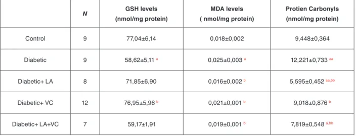
combinati-on seem to prevent at least some of the ccombinati-onsequences of oxidative stress which can promote the development of complications of diabetes mellitus. Table 1 summarizes the overall changes in the reduced glutathione concent-rations, and the degree of lipid peroxidation and protein carbonylation in control, diabetic, and the antioxidant supplemented diabetic rat liver tissues.
Regulation of GST-Mu activity in diabetes
and the effect of antioxidants
As the activity of GST Mu is related to cellular defense against oxidative stress, regulation of its expression is an important factor in cellular response to a wide vari-ety of oxidants and antioxidants. Previous studies have shown that altered GST isoenzyme expression has been implicated in oxidative stress. In this study GST-Mu iso-enzyme was selected as an indicator of defense against oxidative stress. GST-Mu mRNA expressions were de-termined with multiplex RT-PCR. Figure 2 shows the agarose gel electrophoresis bands of the RT-PCR pro-ducts obtained from multiplex amplification of GST-Mu and β-actin mRNAs.
After densitometric analysis of the gels with ImageJ soft-ware, the ratios of the densities of GST-Mu and β-actin genes were calculated and the results are given as bar diagrams in Figure 4a. According to the results, diabetes caused a significant decrease in mRNA expressions of GST-Mu, and this reduction was statistically significant (p<0,05). Supplementing the rats with either antioxidant
Turk J Biochem, 2013; 38 (1) ; 92–100. 95 Sadi et al. Figure 1: Liver tissue (a) GSH concentrations, (b) MDA contents and (c) protein carbonyl contents of control, diabetic, diabetic animals supplemented with α-lipoic acid (D+LA), diabetic animals supplemented with vitamin C (D+VC), and diabetic animals supplemented with both α-lipoic acid and vitamin C (D+LA+VC).
a represents significance at p<0,05 as compared with control groups. aa represents significance at p<0,005 as compared with control groups. b represents significance at p<0,05 as compared with untreated diabetic groups. bb represents significance at p<0,005 as compared with untreated diabetic groups
Table 1. Summary of overall changes in tissue GSH levels, MDA (final product of lipid peroxidation) levels and protein carbonylation levels in control, diabetic, LA supplemented diabetic, VC supplemented diabetic and LA+VC supplemented diabetic rat liver tissues.
N GSH levels (nmol/mg protein) MDA levels ( nmol/mg protein) Protien Carbonyls (nmol/mg protein) Control 9 77,04±6,14 0,018±0,002 9,448±0,364 Diabetic 9 58,62±5,11 a 0,025±0,003 a 12,221±0,733 aa Diabetic+ LA 8 71,85±6,90 0,016±0,002 b 5,595±0,452 aa,bb Diabetic+ VC 12 76,95±5,96 b 0,021±0,001 b 9,018±0,876 b
Diabetic+ LA+VC 7 59,17±1,91 0,019±0,001 b 7,819±0,548 a,bb
Data were expressed as mean ± Standard Error of Mean (S.E.M)
a-represents significance at p<0,05, aa-represents significance at p<0,005 as compared with control groups. b-represents significance at p<0,05,bb-represents significance at p<0,005 as compared with untreated diabetic groups.
Figure 2: Multiplex RT-PCR products of GST-Mu and β-actin genes amplified by using cDNAs synthesized from 1 µg total RNAs isolated from corresponding tissues. 500bp β-actin 450bp GST-Mu bands are clearly observed in control, diabetic, LA treated diabetic (D+LA), VC treated diabetic (D+VC) and LA+VC treated diabetic (D+LA+VC) groups.
Figure 3: Western-blot analysis of GST-Mu and GAPDH proteins from corresponding tissues of individual rats belonging control, diabetic, LA treated diabetic (D+LA), VC treated diabetic (D+VC) and LA+VC treated diabetic (D+LA+VC) groups. On the membranes lower band represents 25kD GST-M1 protein and upper band represents 36kD GAPDH protein. 20 µg total proteins were loaded in each well and separated by 12% separating gel.
Turk J Biochem, 2013; 38 (1) ; 92–100. 97 Sadi et al. Figure 4: Bar diagrams of the results of GST-Mu (a) mRNA expressions, (b) protein expressions, and (c) the activities in control, diabetic, diabetic animals supplemented with α-lipoic acid (D+LA) and diabetic animals supplemented with vitamin C (D+VC), and diabetic animals supplemented with both α-lipoic acid and vitamin C (D+LA+VC).
a represents significance at p<0,05 as compared with control groups. aa represents significance at p<0,005 as compared with control groups. b represents significance at p<0,05 as compared with untreated diabetic groups.
alone or in combination, did not change the mRNA exp-ressions of GST-Mu in diabetic state.
For the determination of GST-Mu protein expression in tissues, Western-blot analysis was carried out by using rabbit anti GST-Mu IgG and the membranes were co-immunostained also by using rabbit anti GAPDH IgG. Figure 3 shows the results of multiplex Western-blot analysis of GST-Mu and GAPDH protein in control, diabetic, LA, VC and LA+VC treated diabetic rat liver tissues
The results showed that, there was a marked decrease in the relative protein expression of GST-Mu (p<0,005) in diabetic animals and neither antioxidants showed a res-toring effect. Instead relative protein expressions were reduced further with antioxidant applications as shown in Figure 4b.
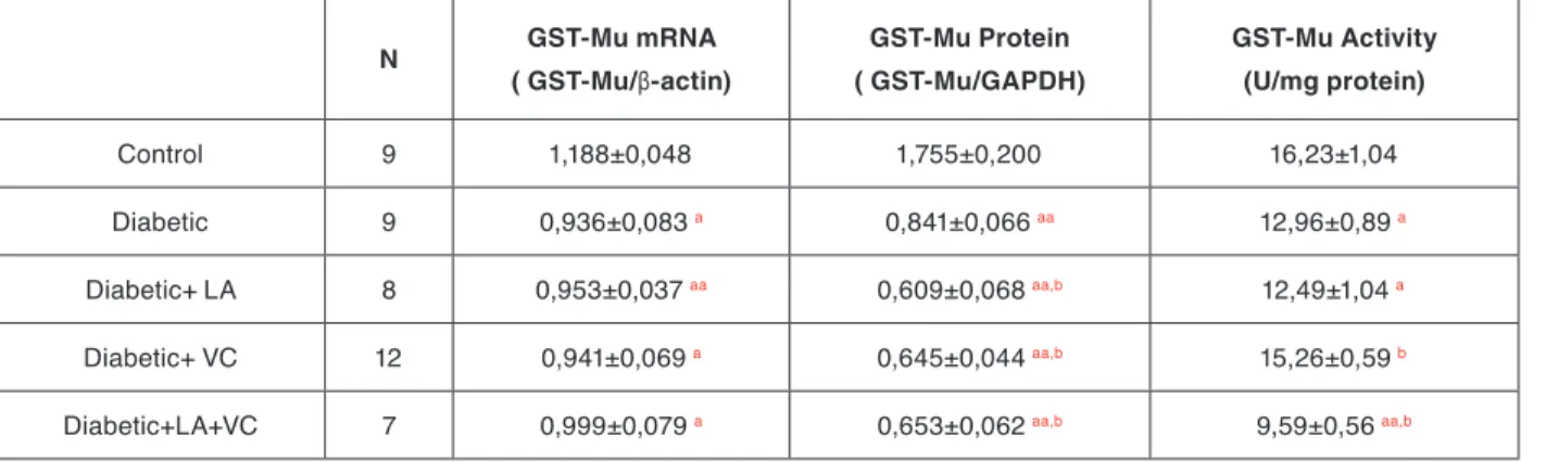
GST-Mu activities were measured in all groups by using DCNB as a specific substrate for that isoenzyme. Diabe-tes leads to a significant reduction in the activities of the enzyme (p<0,05) when compared to the control group. Although LA did not change GST-Mu activities, VC tre-atment caused a significant increase in the activities but still below the control values. Interestingly, when two antioxidants were given together, they reduced the GST-Mu activities further. Effects of diabetes and the antioxi-dant treatment on GST-Mu activities are shown in Figu-re 4c. Table 2 summarizes overall changes in the mRNA expressions, protein amounts and enzyme activities of GST-Mu isoenzyme in control, diabetic and antioxidant supplemented diabetic rat liver tissues.
Discussion
Diabetes mellitus is a group of metabolic diseases cha-racterized by high blood glucose levels that could result from the defects in insulin secretion, or action, or both. Increased oxidative stress is playing an important role in the etiology and pathogenesis of the chronic compli-cations associated with this disease. Numerous studies
proposed that, inhibition of oxidative stress with antio-xidants might be an effective strategy for reducing the complications of diabetes. The beneficial effects of an-tioxidants in the treatment of diabetes have been shown by several investigators [19-23]. Although it is clear that oxidative stress plays a major role in diabetic complica-tions, the molecular mechanisms of the effects of antio-xidants especially on antioxidant and detoxification enz-ymes have not been clearly established. In the present study, we investigated the role of GST Mu expressions on the enzymatic activity and the role of strong antioxi-dants, LA and VC on the expressions and activity of the enzyme.
At first, we have showed the presence of oxidative stress during the diabetes in the liver tissues by measuring the extent of oxidative damage, namely by quantifying the oxidation products of the lipids and proteins, by measu-ring the MDA content and protein carbonylation levels respectively. Furthermore, the concentration of the ma-jor cellular antioxidant, reduced glutathione (GSH) was also quantified in diabetes and the effects of antioxidant treatments were evaluated in both control and diabetic rat liver tissues. Glucose oxidation, nonenzymatic glyca-tion of proteins, and the subsequent oxidative degrada-tion of glycated proteins observed in diabetics leads to inevitable productions of free radicals which in turn reduces the antioxidant defense mechanisms simultane-ously leading to lipid peroxidation and hence damage the cells.
Reduced glutathione (GSH) is one of the most important biomolecules towards the toxicity induced by chemicals and free radicals and it is involved in the removal of re-active intermediates in the presence of GPx and GSTs with hidroperoxidation reactions. GSH has also free ra-dical scavenging activity and repair the rara-dical induced biological injuries. Previous studies have shown that, hepatic GSH concentration of STZ induced diabetic rats was significantly lower when compared with the normal rats [20,21,24]. According to our results, diabetes redu-Table 2. Summary of overall changes in the mRNA expressions, protein amounts and enzyme activities of GST-Mu isoform in control, diabetic and antioxidant supplemented diabetic rat liver tissues.
N GST-Mu mRNA ( GST-Mu/β-actin) GST-Mu Protein ( GST-Mu/GAPDH) GST-Mu Activity (U/mg protein) Control 9 1,188±0,048 1,755±0,200 16,23±1,04 Diabetic 9 0,936±0,083 a 0,841±0,066 aa 12,96±0,89 a Diabetic+ LA 8 0,953±0,037 aa 0,609±0,068 aa,b 12,49±1,04 a Diabetic+ VC 12 0,941±0,069 a 0,645±0,044 aa,b 15,26±0,59 b
Diabetic+LA+VC 7 0,999±0,079 a 0,653±0,062 aa,b 9,59±0,56 aa,b
Data were expressed as mean ± Standard Error of Mean (S.E.M)
a-represents significance at p<0,05, aa represents significance at p<0,005 as compared with control groups. b-represents significance at p<0,05
Turk J Biochem, 2013; 38 (1) ; 92–100. 99 Sadi et al. ced the GSH concentrations below the levels observed
in control groups. Even though, individual LA and VC application brought the diabetic GSH state toward the control levels, their combined application yielded no sig-nificant changes in GSH concentration. The decrease in GSH in liver during diabetes most probably is the result of its increased utilization due to oxidative stress and the addition of antioxidants (LA or VC) slow down the excess usage of GSH. Since, LA has the ability to cor-rect deficient thiol status of cells by increasing de novo synthesis of GSH and hence increased the GSH levels in diabetic animals [25].
Oxidative damage to unsaturated lipid molecules in a cell is a free radical induced phenomena leading to a process called lipid peroxidation. Even though low le-vels of lipid peroxides are present in a healthy cell, as the free radical concentration increases or the antioxidant system fails chain oxidation reactions of lipids results in enhanced lipid peroxidation. So, it is an important mar-ker of early and reversible tissue damage caused by di-abetes mellitus and decreased antioxidant defense capa-city of the tissue. Lipid peroxidation also cause protein damage and inactivation of membrane bound enzymes either through direct attack by free radicals or through chemical modification by its end products which are MDA and 4-hydroxynonenal (4-HNE) [26]. According to our results, lipid peroxidation levels in the liver tis-sues of diabetic rats were significantly higher compared to controls and these findings are in agreement with the results of other related studies [23,27,28]. This marked increase in the lipid peroxidation rates in diabetic tissues suggests an accumulation of oxygen free radicals which can be due to either increased production and/or dec-reased elimination. Furthermore, application of antioxi-dants either alone or in combination reduced the levels towards the control values, most probably by decreasing the cellular redox potential.
The oxidative modification of proteins has been shown to play an important role in a number of human diseases [26]. Protein carbonyl content is actually a general in-dicator and the most commonly used marker of protein oxidation [29,30]. Accumulation of protein carbonyls has been observed in Alzheimer’s disease, diabetes, inflam-matory bowel disease, and arthritis [27,30]. Our results showed that, the carbonyl content of proteins in diabetic groups were higher compared to control groups, most probably, due to exposure of proteins to reactive oxy-gen species which could lead to modifications in ami-no acid side chains resulting in altered structure and/or functions. Other studies also showed that, the increase in the number of carbonyl groups within a protein corre-lated well with diabetes mellitus [27,31]. Application of LA and VC, alone or in combination decreased the level toward the control values. Effect of LA on the protein carbonylation was more prominent then the effect of VC alone. It reduced the protein carbonylation even below the control values.
Conjugating the glutathione to a wide range of electrop-hiles, Glutathione S-transferases also reveal diverse res-ponse to lots of chemicals and oxidative stress in the nor-mal and disease conditions as diabetes mellitus [32]. Our data also demonstrated that, excessive oxidative stress occurred in the liver due to diabetes mellitus, resulted in the down regulation of the expression GST-Mu gene. We have previously reported that, the other main antio-xidant enzymes such as SOD and CAT gene expressi-ons were also suppressed in diabetes [12]. The decrease in the mRNA expressions of GST-Mu also reflected in both the protein expressions and the activities, indica-ting the transcriptional regulation of the gene. The re-duction in the mRNA of GST-Mu isoenzyme in diabetes could be due to oxidation of transcriptional factors (e.g. Nrf2) responsible for the initiation machinery of antioxi-dant enzyme transcription process found in antioxiantioxi-dant response elements. The decrease could also be due to the decrease in the half lives of mRNAs since increased oxidative stress may lead to destabilization of mRNA [33]. Supplementing the diabetic animals with both LA and VC did not restore the gene and protein expressions of GST Mu. Furthermore, the level of protein synthe-sis were decreased drastically well below the diabetic values. But, especially VC treatment by itself increased GST Mu activities almost to control values through post translational mechanisms. Combinational effect of the two antioxidants was not so powerful even though it is known that LA also enhances the antioxidant properti-es of other antioxidants like VC in both aqueous and in hydrophobic environments [12].
In conclusion, we have found that GST Mu could be a marker enzyme in order to examine the effect of oxida-tive stress caused by diabetes. The results showed that oxidative stress dependent reduced cellular concentra-tions of glutathione, and increased oxidation-modified protein and lipid contents down regulated the gene and protein expressions of GST Mu. Antioxidants can resto-re the enzyme activities partially not through transcrip-tional regulation but by post translatranscrip-tional mechanisms.
Acknowledgements
The work cited in this article was included in the author’s Ph.D and M.S studies. The financial support provided by grants from Middle East Technical University (BAP-08-11-DPT2002K120510-TB3) and Karamanoglu Meh-metbey University (BAP-25-M-11) are gratefully ack-nowledged. We also thank to Prof.Dr. Ökkeş Yılmaz for providing the animals.
Ethical Issues
All experiments were carried out with the approval of lo-cal animal use committee (Fırat University, 02.08.2008 with 12/56 number). The procedures involving animals and their care are conformed to the institutional guide-lines.
Turk J Biochem, 2013; 38 (1) ; 92–100. 100 Sadi et al.
Conflict of Interest
The authors declare that they do not have any conflict of interest.
References
[1] Baynes JW, Thorpe SR. The role of oxidative stress in diabetic complications. Curr Opin Endocrinol 1996; 3: 277-284 [2] Mates JM, Perez-Gomez C, De Castro IN. Antioxidant enzymes
and human diseases. Clin Biochem 1999; 32(8): 595-603 [3] Palsamy P, Subramanian S. Resveratrol protects diabetic kidney
by attenuating hyperglycemia-mediated oxidative stress and re-nal inflammatory cytokines via Nrf2–Keap1 sigre-naling. Biochi-mica et Biophysica Acta 2011; 1812: 719-731
[4] Raza H. Dual localization of glutathione S-transferase in the cytosol and mitochondria: implications in oxidative stress, toxi-city and disease. FEBS J 2011; 278: 4243-4251
[5] Raza H, Ahmed I, John A. Tissue specific expression and im-munohistochemical localization of glutathione S-transferase in streptozotocin induced diabetic rats: Modulation by Momordica charantia (karela) extract. Life Sci 2004; 74(12): 1503-1511 [6] Awasthi YC, Zimniak P, Singhal SS, Awasthi S. Physiological
role of glutathione S-transferases in protection mechanism aga-inst lipid peroxidation: A commentary. Biochem Arch 1995; 11: 47-54
[7] Mari M, Cederbaum A. Induction of catalase alpha and mic-rosomal glutathione S-transferase in CYP 2E1 overexpressing HepG2 cells and protection against short-term oxidative stress. Hepatology 2001; 33: 652-661
[8] Knapen MFCM, Zusterzeel PLM, Peters WHM, Steegers EA. Glutathione and glutathione-related enzymes in reproduction - A review. Eur J Obstet Gynecol Reprod Biol 1999; 82(2): 171-184 [9] Yang Y, Cheng JZ, Singhal SS, Saini M, Pandya U, et al.. Role of glutathione S-transferases in protection against lipid peroxidati-on. Overexpression of hGSTA2-2 in K562 cells protects against hydrogen peroxide-induced apoptosis and inhibits JNK and cas-pase 3 activation. J Biol Chem 2001; 276(22): 19220-19230 [10] Lowry OH, Rosebrough NJ, Farr AL, Randall RJ. Protein
mea-surement with the folin phenol reagent. J Biol Chem 1951; 193: 265-275
[11] Chomczynski P, Sacchi N. Single-step method of RNA isolation by acid guanidinium thiocyanate-phenol-chloroform extraction. Anal Biochem 1987; 162: 156-159
[12] Sadi G, Yilmaz O, Guray T. Effect of vitamin C and li-poic acid on streptozotocin-induced diabetes gene expression: mRNA and protein expressions of Cu-ZnSOD and catalase. Mol Cell Biochem 2008; 309(1-2): 109-116
[13] Towbin H, Staehelin T, Gordon J. Electrophoretic transfer of proteins from polyacrylamide gels to nitrocellulose sheets: procedure and some applications. Proc Natl Acad Sci U.S.A. 1979; 76(9): 4350-4354
[14] Rasband WS. ImageJ, U. S. National Institutes of Health, Bet-hesda, Maryland, USA: http://rsb.info.nih.gov/ij/ (Last accessed: October 2011)
[15] Habig WH, Pabst MJ, Jakoby WB. Glutathione-S-transferases: the first enzymatic step in mercapturic acid forma-tion. J Biol Chem 1974; 249: 7130.-7139
[16] Sedlak J, Lindsay RH. Estimation of total, protein-bound, and nonprotein sulfhydryl groups in tissues with Ellman’s rea-gent. Anal Biochem 1968; 25: 192-205
[17] Jain SK, Levine SN. Elevated lipid peroxidation and vita-min E quinine levels in heart ventricles of streptozoticin-treated
diabetic rats. Free Radic Biol Med 1995; 18: 337-341
[18] Levine RL, Garland D, Oliver CN, Amici A, Climent I, et al. De-termination of carbonyl content in oxidatively modified proteins. Method Enzymol 1990; 186: 464-478
[19] Cay M, Nazıroglu M, Simsek H, Aydilek N, Aksakal M et al. Effects of intraperitoneally administered vitamin C on anti-oxidative defense mechanism in rats with diabetes induced by streptozotocin. Res Exp Med 2001; 200(3): 205-213
[20] Dinçer Y, Telci A, Kayalı R, Yılmaz IA, Çakatay U, et al. Effect of α-Lipoic acid on lipid peroxidation and antioxidant enzyme activities in diabetic rats. Clin Exp Pharm Phy 2002; 29: 281-284
[21] Maritim AC, Sanders RA, Watkins JB. Effects of alpha-lipoic acid on biomarkers of oxidative stress in streptozotocin-induced diabetic rats. J Nutr Biochem 2003; 14(5): 288-294
[22] Sadi G, Guray T. Gene expressions of Mn-SOD and GPx-1 in streptozotocin induced diabetes: effect of antioxidants. Mol Cell Biochem 2009; 327: 127-134
[23] Seven A, Güzel S, Seymen O, Civelek S, Bolayırlı M, et al. Ef-fects of vitamin E supplementation on oxidative stress in strep-tozotocin induced diabetic rats: Investigation of liver and plas-ma. Yonsei Med J 2004; 45(4): 703-710
[24] Pari L, Latha M. Antidiabetic effect of scoparia dulcus: effect on lipid peroxidation in streptozotocin diabetes. Gen Physiol Bi-ophys 2005; 24: 13-26
[25] Han D, Handelman G, Marcocci L, Sen CK, Roy S, et al. Lipoic acid increases de novo synthesis of cellular glutathione by imp-roving cystine utilization. Biofactors 1997; 6: 321-338 [26] Halliwell B, Gutteridge J. Free Radicals in Biology and
Medici-ne 2007; Oxford University Press, USA
[27] Bhor V M, Raghuram N, Sivakami S. Oxidative damage and altered antioxidant enzyme activities in the small intestine of streptozotocin-induced diabetic rats. Int J Biochem Cell B 2004; 36: 89-97
[28] Kakkar R, Mantha SV, Radhi J, Prasad K, Kalra J. Increased oxidative stress in rat liver and pancreas during progression of streptozotocin-induced diabetes. Clin Sci 1998; 94(6): 623-632 [29] Berlett BS, Stadtman ER. Protein oxidation in aging, disease,
and oxidative stress. J Biol Chem 1997; 272(33): 20313-20316 [30] Chevion M, Berenshtein E, Stadtman ER. Human studies
rela-ted to protein oxidation: protein carbonyl content as a marker of damage. Free Radic Res 2000; 33: 99-108
[31] Limaye PV, Raghuram N, Sivakami S. Oxidative stress and gene expression of antioxidant enzymes in the renal cortex of strep-tozotocin-induced diabetic rats. Mol Cell Biochem 2003; 243: 147-152
[32] Raza H, Ahmed I, John A, Sharma AK. Modulation of xenobiotic metabolism and oxidative stress in chronic streptozo-tocin-induced diabetic rats fed with Momordica charantia fruit extract. J Biochem Mol Toxic 2000; 14(3): 131-139
[33] Navarro MMM, Roca RC, Belli G, Martinez JG, Navarro JM, et al. Comprehensive Transcriptional Analysis of the Oxida-tive Response in Yeast. J Biol Chem 2008; 283(26): 17908-17918